How do bilaterally equivalent structures in the forebrain become left-right asymmetric?

How do bilaterally equivalent structures in the forebrain become left-right asymmetric?

This publication summary relates to the paper:

Cachd1 is a novel Frizzled- and LRP6-interacting protein required for neurons to acquire left-right asymmetric character. (Powell et al., 2022)

bioRxiv preprint doi: https://doi.org/10.1101/2022.05.16.492129;


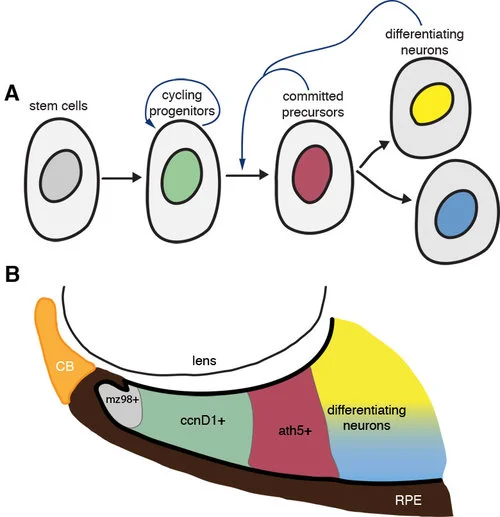
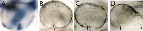
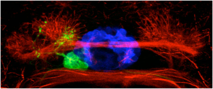
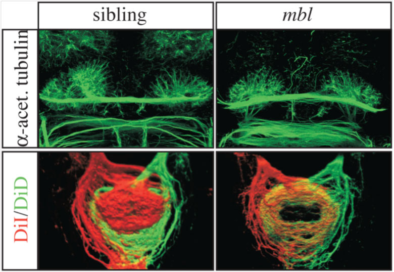
The human brain is divided into left and right hemispheres that superficially look like mirror copies of each other. However, there are differences between the sides that ultimately affect the types of neural circuits formed, the function of those circuits, and our overall behaviour. Many of these lateralised differences are also found in other animals and appear to have been conserved in evolution. One example of such conservation is the habenulae: two small brain nuclei, one present on either side, that act as a relay station to integrate sensory information and then pass a signal on to other circuits that control some forms of motor behaviour. We study the habenulae in zebrafish because there are easy to observe left-right asymmetries in the translucent embryo, such as size and shape, gene expression and circuit inputs and outputs (see figure below, A-C: compare wildtype panels on the left with mutant panels on the right). How those differences arise, and what affect they have on behaviour, is still not very well understood. In the Wilson lab, we look at how asymmetries are established in the brain during embryonic development by exploring what genes are involved in the process, what effect those genes have on the structure of the habenulae, and finally, how the zebrafish behave.

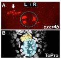
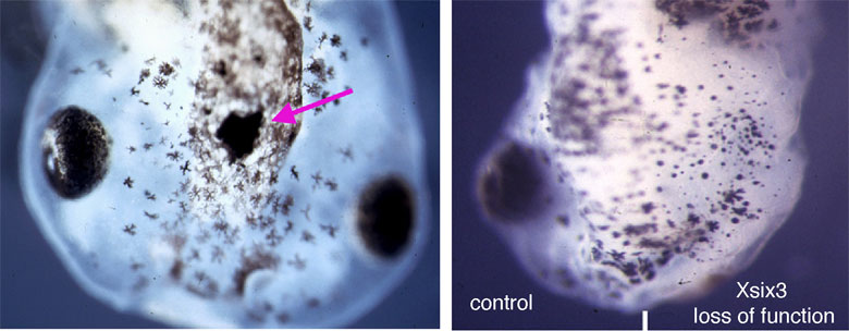
We have recently discovered that a gene called cachd1 plays a very important early role in establishing the difference between the left and right habenulae: when it is mutated, the right habenula develops into an almost mirror copy of the left habenula (see figure below, A and B). We have learnt that cachd1 does this, at least in part, by controlling the time at which the habenula neurons are born. Normally, more neurons are born early on the left-hand side than the right, and this affects the character of the mature habenulae (see figure below, D, left panel). When cachd1 does not function, more neurons are born early on both sides (see figure below, D, right panel), leading to mature habenula that look alike, so-called “double left” habenulae. This in turn affects the type of sensory circuits that can connect to the habenula, and how the habenulae connect to downstream circuits (see figure below, C).

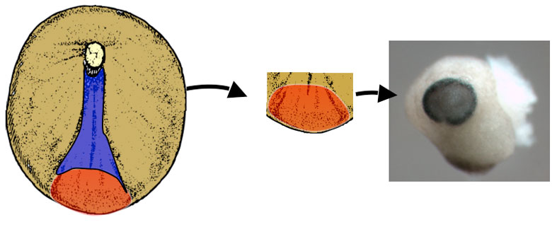
We believe that cachd1 is able to control the timing of neurons by modulating a key developmental signalling pathway called the Wnt pathway. Wnt is a secreted protein that simultaneously binds to two receptor proteins (Frizzled and LRP6) on the surface of cells. This interaction sets off a chain reaction of events inside the cell, passing information to the nucleus allowing the cell to react to its environment. Wnt signalling is involved in lots of different developmental processes in the embryo, typically (but not exclusively) to do with controlling the proliferation of cells. Similar to the Wnt protein, Cachd1 protein is able to physically bind to the Frizzled and LRP6 receptors (see figure below, E), potentially competing with the signal to alter the output of the pathway; in this case, the time at which a habenula neuron is born. Consistent with this, mutation of lrp6 also causes “double left” habenulae to develop (see figure below, F).

Interestingly, cachd1 isn’t limited to either side of the brain in the embryo, so how does it control asymmetry? We believe that the function of cachd1 is suppressed by a signal from the parapineal – a small population of neurons that migrates to the left-hand side and contacts the left habenula (see [Ingrid’s paper summary]). When the parapineal is ablated, or cannot function properly due to mutation, the left habenula develops as a mirror copy of the right habenula, so-called “double right” habenulae (see figure below, G, left panel). However, when the parapineal is ablated in embryos lacking cachd1 function, “double left” habenulae develop (see figure below, G, right panel). This means that the parapineal must inhibit cachd1 in order for the left habenula to form in the embryo.

This is an important piece of the puzzle, but lots remain to be discovered. We are particularly interested in learning about the consequences of the cachd1 mutation on behaviour, as well as developing a better understanding of how the Cachd1 protein affects Wnt signalling and what the identity of the antagonising parapineal signal is. Furthermore, our discovery of the function of cachd1 will help us uncover more genes that play roles in determining left-right asymmetry.

Loss of function of cachd1 disrupts asymmetry of the habenulae. Mutation of cachd1 (right panels A-D, G; compare to wildtype, left panels) affects asymmetry of gene expression (A) and neuron composition (B; neurons in red, Cachd1 protein in cyan) of the habenulae (dashed box in A), as well as connections of the habenula neurons to other brain regions (C; left side neurons in magenta, right side neurons in green). Neurons are born early in cachd1 mutant embryos on both sides (D), with an increased proportion of left-character neurons on the right (magenta). When neurons are born in the habenulae depends (in part) on Wnt signalling through Frizzled (FZD) and Lrp6 co-receptors. Cachd1 protein can bind to these simultaneously, shown in the crystal structure of the complex formed by the three proteins (E). Mutation of lrp6 also affects asymmetry in the habenula (F), consistent with its interaction with Cachd1. When the parapineal is ablated in a wildtype embryo, both left and right habenula express a gene characteristic of the right habenula (G, left panel, green; pineal gland shown in white); this suggests that a signal from the parapineal is needed to break asymmetry. When it is ablated in the cachd1 mutant however, “double left” habenulae form; this suggests that cachd1 is needed to form the right habenulae, and must be suppressed by the parapineal signal on the left.


For the whole story of cachd1 check out our preprint on biorxiv.

By Gareth Powell

The power of the parapineal in brain asymmetry development

The power of the parapineal in brain asymmetry development

A summary of the paper “ Sox1a mediates the ability of the parapineal to impart habenular left-right asymmetry” eLife 2019;8:e47376 DOI: 10.7554/eLife.47376

The transparent larval zebrafish is excellent for studying brain development, allowing whole-embryo imaging and relatively simple genetic manipulation. Using these advantages, we study how the left and right side of the brain become different from each other (i.e. asymmetric) during embryogenesis.

The pineal is midline brain structure that contributes to the regulation of circadian rhythms in vertebrates. In zebrafish, the pineal is accompanied by an exclusively left-sided parapineal nucleus, function of which is not quite clear. It is known, however, that during embryonic development the parapineal emerges from the pineal and migrates to the left side (Figure 1). On this journey the parapineal cells instruct adjacent habenular neurons on the left side of the epithalamus (dorsal part of the thalamus) to become different from equivalent neurons on the right with regards to molecular properties and neuronal connectivity. In other words, the parapineal determines the development of left-right asymmetries in the habenulae. Eliminating or changing the side of the parapineal therefore leads to loss or reversal of habenular asymmetries, respectively (Figure 1).

Figure 1: The parapineal emerges from the pineal during zebrafish embryogenesis, as shown by confocal images of a transgenic line with GFP in the pineal complex (top pictures). The pineal complex lies at the midline of the epithalamus, flanked by le…

Figure 1: The parapineal emerges from the pineal during zebrafish embryogenesis, as shown by confocal images of a transgenic line with GFP in the pineal complex (top pictures). The pineal complex lies at the midline of the epithalamus, flanked by left (green on the scheme) and right (magenta on the scheme) habenulae. The habenulae are innervated by the parapineal and themselves send neuronal projections to the midbrain interpeduncular nucleus (IPN). The side of the parapineal, on the left in wild-type (wt), determines which habenula acquires a left-type identity projecting to the dorsal part of the IPN (dIPN).

We show that during embryogenesis the parapineal instructs the cells in the left habenula to initiate neurogenesis earlier compared to the right habenula. The parapineal is also required at later stages for the establishment neuronal connectivity specific to the left habenula.

Upon precise laser-ablation of the embryonic parapineal cells prior to their migration to the left side, both habenulae exhibit right habenula characteristics at larval stages. A similar phenotype can be observed upon mutating the sox1a gene, normally active in the parapineal. In sox1a mutants the parapineal forms properly, but the habenulae develop as if there was no parapineal. Finally, transplanting only a few parapineal cells to the right side of the embryonic brain induces left-sided character in neurons within the right habenula. The results of these experiments are schematically summarised in Figure 2.

Figure 2: One of the mechanisms leading to left-right asymmetries in the zebrafish habenulae is the different timing of neurogenesis on each side. In sox1a-/- mutants and upon parapineal ablation the early onset of neurogenesis in the left habenula …

Figure 2: One of the mechanisms leading to left-right asymmetries in the zebrafish habenulae is the different timing of neurogenesis on each side. In sox1a-/- mutants and upon parapineal ablation the early onset of neurogenesis in the left habenula is delayed and the habenulae acquire a double-right character. Hence, in the absence of the left-sided parapineal or upon loss of sox1a-/- in the parapineal the left habenula becomes identical to the right habenula. Conversely, transplantation of external parapineal cells to the right side induces left-habenula characteristics in the right habenula.

The development of the zebrafish epithalamus serves as a great example of a genetically determined mechanism for brain asymmetry formation. The emergence of a parapineal nucleus in the pineal complex during evolution has in turn led to asymmetries in the habenulae, ensuring that appropriate lateralised character is propagated within left and right-sided circuitry. As we and others have previously shown, the asymmetric neuroanatomy translates into asymmetric function of the habenulae and lateralised behaviour of the fish. Hence, from studies in zebrafish a clearer picture is forming on how and why brain asymmetries emerge in largely­ bilaterally symmetric animals.

By Ingrid Lekk

Sizing the eyes- a study of the mechanisms by which the developing eye copes with deleterious genetic mutations

Sizing the eyes- a study of the mechanisms by which the developing eye copes with deleterious genetic mutations

A summary of the article “Compensatory growth renders Tcf7l1a dispensable for eye formation despite its requirement in eye field specification” Elife. 2019 Feb 19;8. pii: e40093. doi: 10.7554/eLife.40093.


Although some genetic mutations lead to congenital abnormalities and/or disease, DNA sequencing projects have shown that compromised function of many genes has no obvious effect.  Indeed, people that carry such mutations live an apparently normal life that is not impacted by altered gene function.  This suggests that genetic pathways and developmental processes are robust and able to cope when challenged by altered gene function.  An important goal in biomedicine is to identify the compensatory mechanisms that enable such mutations to be carried without consequence  

Zebrafish embryos are well suited to explore the link between genetic mutations (genotype) and developmental outcome (phenotype).  In a paper recently published in eLife (elifesciences.org/articles/40093), we studied zebrafish carrying a mutation in a gene called tcf7l1a that encodes a protein functioning in the Wnt pathway, one of the most critical pathways by which cells signal to each other during development.  The Wnt pathway is particularly important for development of the eyes.  Zebrafish embryos with the tcf7l1a mutation initially have tiny forming eyes, but within a day or two, these eyes undergo compensatory growth to reach a size comparable to that in unaffected individuals. We found that the smaller eyes delayed the onset of differentiation, enabling them to continue growing until they reached the right size. How does the eye know what the right size is? We propose that there is a mechanism by which eye size is intrinsically assessed and this informs retinal cells when to stop dividing and start differentiating into neurons. This mechanism may also explain how it is that left and right eyes develop independently and yet still grow to the same size.

 Although compensatory growth could restore eye size when tcf7l1a function was absent, we speculated that developing eyes in these mutants may be very sensitive to the effect of additional genetic mutations.  Indeed, we found additional interacting mutations that completely impaired eye development such that no eyes were formed, and others in which the developing eyes were unable to compensate their growth and remained small.

 These results contribute to a better understanding of the origin of developmental eye pathologies such as microphthalmia and anophthalmia, in which people are born with small eyes or no eyes at all.

Read the digest of this paper on Elife or treat yourself to the full text here.

Key Publications

Young RM, Hawkins TA, Cavodeassi F, Stickney HL, Schwarz Q, Lawrence LM, Wierzbicki C, Cheng BY, Luo J, Ambrosio EM, Klosner A, Sealy IM, Rowell J, Trivedi CA, Bianco IH, Allende ML, Busch-Nentwich EM, Gestri G, Wilson SW.
Compensatory growth renders Tcf7l1a dispensable for eye formation despite its requirement in eye field specification.
Elife. 2019 Feb 19;8. pii: e40093. doi: 10.7554/eLife.40093.

Sizing the eyes-a study of the mechanisms by which the developing eye copes with deleterious genetic mutations

Brain asymmetry improves processing of sensory information

Many of us are aware that the left and right sides of the brain are believed to have slightly different roles in cognition and in regulating behavior. However, we don't know whether these asymmetries actually matter for the efficient functioning of the brain.

Unravelling the mysteries of brain diversity

Brain diversity has puzzled scientists for centuries. But, what do we mean by 'brain diversity'? If one compares brains from many different species of vertebrates, soon one realises how different they look. This diversity in brain form or morphology is extraordinary not only for brains from very separate groups (e. g. mammals vs. fishes), but also within the same group.

A gene and a population of cells important for shaping the eye

Back to publication summaries


A GENE AND A POPULATION OF CELLS IMPORTANT FOR SHAPING THE EYE

Gaia Gestri and Steve Wilson
Lmx1b coordinates ocular FGF signaling to regulate patterning and morphogenesis of multiple eye tissues in zebrafish

LMX1B is a gene that when mutated in humans causes Nail-Patella Syndrome (NPS), a condition with many different features including limb, joint and renal (kidney) defects and disrupted function of the central nervous system (brain and spinal cord) and eyes. Approximately 50% of patients develop elevated intraocular pressure and glaucoma. LMX1B encodes a LIM-homeodomain transcription factor - a protein that functions of a kind of genetic switch to regulate the activity of many other genes. Individuals can exhibit the condition if they lack function of just of their two copies of the gene (one from mother and one from father). Although the identity of the causative gene is known, how defective function of the gene leads to the wide range of abnormalities observed in patients is poorly understood. In this study, a collaboration with Brian Link, we studied the function of the zebrafish version of the LMX1B gene to elucidate how it might function during embryonic development of the eye.

Figure 1: A. The lmx1b gene (in blue) is expressed in cells that surround the eye and in an open groove in the retina, the choroid fissure (indicated by the black line in B), that is present in the eye early in development. Eyes in embryos with comp…

Figure 1: A. The lmx1b gene (in blue) is expressed in cells that surround the eye and in an open groove in the retina, the choroid fissure (indicated by the black line in B), that is present in the eye early in development. Eyes in embryos with compromised Lmx1b function exhibit varying degrees of coloboma (the condition where the choroid fissure doesn't properly close).

The eyes form during embryogenesis as outgrowths from the developing brain - these outgrowths are called optic vesicles and they will give rise to the retina and the pigmented cells at the back of the eye (for more information, see our research pages on eye development www.ucl.ac.uk/zebrafish-group/research/eye.php). Other groups of cells contribute to other eye structures such as the lens, blood vessels and protective coat of the eyeball. How all of these different groups of cells interact in a highly coordinated way during eye formation is poorly understood. In this study, we show that the lmx1b gene is expressed in highly motile cells that migrate around the optic vesicles as they are forming (Figure 1A and Movies 1 and 2). To test the function of the gene in these cells, we removed its function using anti-sense reagents that prevent formation of Lmx1b protein. This resulted in the motile cells failing to properly migrate around the eye and indeed many of these cells subsequently died (Movie 2). This disruption to the migratory cells was correlated with a failure in the optic vesicle to under the tissue movements that enable the eye to take on its spherical shape. Indeed, we often observed that eye exhibited coloboma, a condition in which two lips of the forming eye fail to fuse together in the ventral (lower) part of the eye (Figure 1B-D). These results strongly suggest (but have not proven) that the migratory cells are required for the optic vesicle to form the spherical eye. If correct, how might this happen? We suspect that the migratory cells may secrete proteins that help the nascent eye cells to undergo tissue re-shaping but this will require further research to resolve.

We also found that retinal cells are incorrectly patterned when Lmx1b function is disrupted - this would lead to defects in visual map formation when connections are made between the eye and the brain. We do not know if this problem is a consequence of the death of the motile cells around the eye or to some other action of the Lmx1b gene.

Many of the deficits we describe in fish are more severe than observed in human patients - this is most likely due to the fact that our experiments deplete Lmx1b function more severely than in patients. If the function of the gene is severely compromised in humans, it would lead to early lethality as we observe in fish. Our studies in fish will continue to elucidate how genes function and cells and tissues interact during vertebrate eye formation and will help us to understand human diseases, conditions and syndromes in which these processes are disrupted.

Other links
UK Nail Patella Syndrome charity website
Wikepedia
NIH Genetics page

If you have any further questions, please contact Gaia Gestri or Steve Wilson

This study was a collaboration with Brian Link and our work received financial support from

The complex choreography of eye formation

Back to publication summaries
 

the complex choreography of eye development

Florencia Cavodeassi and Stephen Wilson, October 2009

Original paper reference
Dynamic Coupling of Pattern Formation and Morphogenesis in the Developing Vertebrate Retina

If we are to interpret correctly what we see, a precise representation of the visual input received the retina must be formed in our brain. This "visual map" forms during embryogenesis, when retinal neurons (nerve cells) project long processes (axons) to the brain to establish the wiring between the eye and those parts of the brain that process visual information. Each retinal axon targets a very specific area of the superior colliculus (or optic tectum), a region of the brain that interprets visual information, thus generating a map of connections that reproduces the external visual field in the brain. For the visual map to be accurate, the neurons in the retina must be able to interpret where in the retina they are located, and who are their neighbours. We know quite a lot about the genetic "codes" that give the retinal cells their identity but much less about how the retinal cells acquire this knowledge of who they are and where they must go.

Until now, it has been particularly challenging to resolve the mechanisms that instruct positional information in the retina, since this process happens in parallel to the extensive remodelling (or morphogenesis) of tissues that occur during formation of the eye. Using a combination of imaging techniques and genetic manipulations, in this study, we describe how the processes of morphogenesis and allocation of retinal identity are coordinated during maturation of the optic cup.

In a previous study, our collaborators Alexander Picker and Michael Brand proposed that a combination of secreted Fibroblast Growth Factor (Fgf) proteins are required by the eye cells to 'know' where they are. The study determined that retinal cells acquire their identity during a very early stage of eye development, when the nascent eye (optic vesicle/cup) is just beginning to emerge from the brain. This presented a puzzle - Fgfs work through being secreted from one group of cells and received and interpreted by nearby cells - however, during the stage when Fgfs appear to function in the eye, the nascent eye tissues are undergoing complex rearrangements. How then, could this process of morphogenesis be temporally and spatially coordinated with the localised action of Fgfs on retinal cells? This study found that as the prospective eye cells undergo various movements, it brings them transiently into proximity with other tissues that produce Fgfs which can act consequently act upon their temporarily neighbouring nascent retinal cells.

Figure 1: cartoon illustrating the reorganisation of the nasal-temporal axis during maturation of the optic cup. The source of Fgf signals is shown in blue, the future nasal region of the retina is shown in green and the future temporal region in re…

Figure 1: cartoon illustrating the reorganisation of the nasal-temporal axis during maturation of the optic cup. The source of Fgf signals is shown in blue, the future nasal region of the retina is shown in green and the future temporal region in red. The olfactory placode (the future nasal epithelium, dark blue) is one source of Fgfs and the other is the telencephalic region of the forebrain (pale blue).

The retina is divided into two axes - a nasal to temporal axis (nose to ear) and a dorsal to ventral axis (up to down) and Fgf promotes nasal identity. One surprising finding was that Fgfs are produced by tissues dorsal to retina at early stages (Figure 1) suggesting that nasal retina is initially located dorsally in the nascent eye and that the eye somehow rotates by 90 degrees during subsequent stages of development. But how is this shift of the axis effected?

To answer this question required labelling subsets of retinal cells with green fluorescent protein and tracking them in live zebrafish embryos. In this way, it was possible to follow the complex reorganisation movements that transform the initial dorsal-ventral axis of the optic vesicle into the nasal-temporal axis of the mature retina. During this process, the dorsal, future nasal cells of the retina become compacted, the future temporal cells move from ventrally to join the nasal cells in the forming retina and simultaneously the whole optic vesicle rotates to bring the dorsal domain to its final location anteriorly in the eye. Remarkably, this reorganisation is coordinated with a similar shift of the source of Fgfs, which act as choreographers of some aspects of the complex morphogenetic process. In this way, one set of signals (the Fgfs) instructs positional information in the retina and simultaneously ensures that the axis is maintained during the subsequent maturation of the eye.

The study also revealed that many aspects of morphogenesis proceed very well in the absence of Fgfs indicating that other pathways must be involved. We hope that future collaborations will help to identify these additionally choreographers of eye formation.

If you have any further questions, please contact Florencia Cavodeassi or Steve Wilson. Alexander Picker and Michael Brand would also be very happy to receive any questions on the study.

This study was a collaboration with Alexander Picker and Michael Brand and our contribution to the research received financial support from the MRC and Wellcome Trust.

Back to publication summaries


The left brain leads the right in making neurons

Myriam Roussigne and Steve Wilson
Nodal signalling imposes left-right asymmetry upon neurogenesis in the habenular nuclei

The left and right sides of our brain mediate different cognitive and sensory functions; for instance, the left hemisphere is dominant in processing aspects of language and tool use while the right hemisphere handles visuospatial attention and interpretation of emotion. This feature of the brain is known as functional lateralisation and is thought to improve our cognitive performance as the specialization of each hemisphere broadens the repertoire of tasks that can be undertaken. Brain lateralisation is not unique to humans and underlies asymmetric behaviour in all vertebrates, a common example being visual lateralization, which is manifest as an eye preference that is dependent on what is being looked at.

Functional lateralisation of the brain is a consequence of structural differences between the left and right sides of particular brain regions. One of the best characterised neuroanatomical asymmetries is found in a region of the brain called the epithalamus and in the last ten years, studies of developing zebrafish have led the way in elucidating the mechanisms by which these epithalamic asymmetries develop. The zebrafish epithalamus consists of several groups of neuron (neuronal nuclei) - the pineal complex and a pair of habenular nuclei which display left-right (LR) asymmetries in the proportion of different subtypes of neurons and in their connections (see the publication summary entitled "Brain asymmetry at the level of single cells"). The pineal complex is composed of the symmetrically positioned epiphysis and a left-sided parapineal nucleus that migrates from the midline and connects to the left habenula. The left-sided migration of the parapineal nucleus is dependent upon the activity of a protein called Fgf8 expressed bilaterally in the presumptive habenulae (see the publication summary entitled "Fgf8 signalling breaks symmetry in the brain"). As it migrates, the parapineal sends signals to the left habenula promoting "left-sided" development by these neurons. If the parapineal is removed, the asymmetry between the left and right habenula is considerably reduced.

The sidedness/laterality of parapineal migration and habenulae asymmetries are always concordant (both on the same side) and dependent on the earlier left-sided signalling by Nodal proteins. Importantly, in the absence of one-sided Nodal signalling, neuroanatomical asymmetries develop but with a randomised sidedness: the parapineal migrates to the left or to right side of the epithalamus with an equal probability and the pattern of habenular asymmetry is either normal or mirror reversed correlating with parapineal position. Thus, Nodal signalling is not required for the establishment of structural asymmetry per se but is responsible of the directionality/laterality of asymmetries.

Figure 1: Temporal LR asymmetry in habenular neurogenesis. Images looking down onto the midline of a zebrafish embryo brain. A : cxcr4b expressing neurons appear earlier in the left habenula (arrow) than in the right. The epiphysis is indicated by a…

Figure 1: Temporal LR asymmetry in habenular neurogenesis. Images looking down onto the midline of a zebrafish embryo brain. A : cxcr4b expressing neurons appear earlier in the left habenula (arrow) than in the right. The epiphysis is indicated by a dotted blue circle. A’ Staining of all cells in the same brain allows detection of the parapineal (yellow shading) and pineal (blue shading).

The manner in which Nodal signaling imposes left bias in the brain is not known. To address this issue, we explored the involvement of Nodal signalling in promoting the production (neurogenesis) of habenular neurons. We found that a novel marker of habenular precursors/neurons called cxcr4b appears earlier in the left habenula than in the right. This is consistent with previous results from our colleague Hitoshi Okamoto's team showing temporal LR asymmetries in the generation of neuronal sub-types between left and right habenulae. Asymmetry in habenular neurogenesis can be detected before the migration of the parapineal and, indeed, we found that it is independent of the parapineal. In contrast, when we remove the L/R bias in Nodal signaling, habenular neurons appear at the same time in both habenular nuclei. Thus, the activity of Nodal signals in the left epithalamus can directly drive an asymmetry in habenular neurogenesis and this is the first example for a role for Nodal signalling in promoting an asymmetry per se rather than in directing laterality in the brain.

Figure 2. Nodal signalling promotes early asymmetric neurogenesis Images looking down onto the midline of embryonic zebrafish brains showing neurons in green. A and C show normal brains with LR asymmetric neurons (arrows). B and D show brains that d…

Figure 2. Nodal signalling promotes early asymmetric neurogenesis Images looking down onto the midline of embryonic zebrafish brains showing neurons in green. A and C show normal brains with LR asymmetric neurons (arrows). B and D show brains that don’t have asymmetric Nodal signalling and have LR symmetric neurons (small arrows). The epiphysis is indicated by a dotted blue circle.

The roles/consequences of this Nodal dependent LR asymmetry in habenular neurogenesis are not clear yet but it could be a way by which Nodal biases the orientation of parapineal migration and subsequent habenular asymmetries. This model is consistent with previous data suggesting that the left habenula may provide cues that influence the orientation of parapineal migration. This early asymmetric neurogenesis could also partly influence the later identity of habenular neurons and could consequently be responsible for subtle differences that remain between the left and right habenulae after removal of the parapineal nucleus (see the publication summary entitled "Brain asymmetry at the level of single cells").

How might an early neurogenesis in the left habenula influence the orientation of migration of the parapineal? As we have shown that the protein Fgf8 is required for parapineal migration (see the publication summary entitled "Fgf8 signalling breaks symmetry in the brain"), it will be important to determine if Nodal signalling biases levels of Fgf8 activity, for instance by promoting a L/R asymmetry in the number of fgf8 expressing habenular cells. An alternative possibility is that both asymmetric neurogenesis and directed parapineal migration might be independent consequences of a Nodal-dependent asymmetry in Fgf activity. Our ongoing research is addressing the interactions between Nodal and Fgf signalling pathways in the epithamus.

If you have any further questions, please contact Myriam Roussigne or Steve Wilson

This study was a collaboration with Patrick Blader and our work received financial support from the Wellcome Trust and FEBS.

A zebrafish model of the human Oral-facial-digital syndrome

Leila Romio and Steve Wilson

Original paper reference
Convergent extension movements and ciliary function are mediated by ofd1, a zebrafish orthologue of the human oral-facial-digital type 1 syndrome gene

Oral-facial-digital syndrome type 1 (OFD1) is a severe condition that occurs in 1:50,000-250,000 live births. The disease is caused by a defect in a gene called ofd1 that is carried on the X chromosome. We know that Ofd1 protein has a crucial role in development, because XY males inheriting the mutation have no Ofd1 and die before birth, whereas heterozygous XX females (who carry one mutant and one working copy of the gene) are born with several congenital defects: malformation of the face and mouth, abnormalities of the digits and malformation of the central nervous system. OFD1 syndrome often features polycystic kidneys, which is the main cause of death among patients and that can only be treated effectively by kidney transplant. Ofd genes are present in all vertebrate animals and this means that one can potentially model the disease in animals in which it is easier to study why developmental events go wrong than it is in humans. In this study, we elucidated the function of ofd1 during development by depleting Ofd1 protein during zebrafish embryogenesis, using morpholino (Mo) antisense reagents that inhibit the activity of the gene.

Ofd1 protein localises to basal bodies - these are structures inside cells from which arise cilia - tail-like projections from the cell surface. Recent studies have highlighted that many genes mutated in complex genetic syndromes produce proteins that localise in cilia or at their root. Cilia are present on almost every cell type in vertebrates, and can be motile or non-motile. Motile cilia are found on various structures during development including the embryonic node in mouse and Kupffer's vesicle (KV) in zebrafish. These are structures in which active motile cilia generate fluid flow towards the left side of the body and this is important for establishing the left-right placement of organs like the heart and liver. Cilia also mediate cerebrospinal fluid movement and respiratory tract mucous clearance. A class of cilia called primary cilia are generally not actively motile but are important for the transmission of signals from the outside to the inside of cells. For example those located on mammalian renal (kidney) epithelial cells sense renal tubular flow by bending, thus instigating signalling which maintains epithelial differentiation.


Figure 1. Cilia movement is disrupted when Ofd1 doesn't function: the yellow line shows the path of a single bead inside Kupffers' vesicle, A is a normal embryo, while B is an embryo where Ofd1 function is disrupted. The plot shows the difference in…

Figure 1. Cilia movement is disrupted when Ofd1 doesn't function: the yellow line shows the path of a single bead inside Kupffers' vesicle, A is a normal embryo, while B is an embryo where Ofd1 function is disrupted. The plot shows the difference in bead speed between normal and affected embryos

We have studied how zebrafish development is affected by partial depletion of Ofd1 protein, as happens in female patients. The main findings of our study are: Ofd1 depletion disrupts cilia motility. Although cilia are present they don't function correctly when Ofd1 function is disrupted. This is demonstrated by an experiment where we injected very tiny beads into Kuppfers' vesicle (a vesicular structure covered by motile cilia) and observed that cilia movement is disrupted in ofd1-depleted embryos compared to normal embryos (Figure 1). The consequence of this disruption is that some of the affected embryos show altered left-right patterning, that is, asymmetric positioned organs (the heart for example) can be found on either side of the body (Figure 2).

Figure 2. Heart laterality: A shows the normal postion of the heart (blue), B is an example of a laterality defect where the heart loops in the opposite direction. The plot shows the proportion of right/medial/left positioned hearts in normal embryo…

Figure 2. Heart laterality: A shows the normal postion of the heart (blue), B is an example of a laterality defect where the heart loops in the opposite direction. The plot shows the proportion of right/medial/left positioned hearts in normal embryos and embryos with disrupted Ofd1 function.

Ofd1 has a role in movements by which cells elongate the embryo during early development. Cell movements during the gastrulation phase of embryonic development are regulated by the so-called Wnt/PCP signalling pathway. When Ofd1 is not functioning correctly, these movements are disrupted and embryos are shorter than normal. We showed ofd1 has genetic interactions with two genes of the Wnt/PCP pathway, vangl2 and wnt11 (Figure 3). This means that mutations in two genes leads to a more severe developmental defect than in either alone.

Figure 3. This picture shows how ofd1 genetically interacts with vangl2 in gastrulation cell movements, resulting in shorter larvae: partial loss of ofd1 doesn't affect embryo length, but when there is also disruption to the vangl2 gene, this result…

Figure 3. This picture shows how ofd1 genetically interacts with vangl2 in gastrulation cell movements, resulting in shorter larvae: partial loss of ofd1 doesn't affect embryo length, but when there is also disruption to the vangl2 gene, this results in much shorter embryos.

Ofd1 is required for fusion of the two primordia that normally merge at the midline to form the kidney. Embryos lacking functional ofd1 often have split kidneys due to a failure in migration of the cells that from this structure (Figure 4). We speculate this might be related to the other defects we observe in cell migrations during early development.

Figure 4. The pronephroic glomerulus (the precursor of the kidney) forms from two primordia that migrate towards the midline and fuse. In ofd1 compromised embryos, this process is impaired, and we observed the same phenomenon in mutant fish called t…

Figure 4. The pronephroic glomerulus (the precursor of the kidney) forms from two primordia that migrate towards the midline and fuse. In ofd1 compromised embryos, this process is impaired, and we observed the same phenomenon in mutant fish called trilobite (tri) that carry a mutation in vangl2. We speculate that this could be another consequence of disrupting Wnt/Pcp dependent cell migrations.

Overall, our study has helped to elucidate why mutations in ofd1 can cause severe problems in humans by demonstrating critical roles for the gene in cilia function and in cell movements during embryogenesis.

Further reading

This study was a collaboration between the groups of Derek Stemple at the Sanger Centre, Adrian Woolf at the Institute of Child Health at UCL and Steve Wilson also at UCL. Most of the support for the work was provided by the Wellcome Trust.

Fgf8 signalling breaks symmetry in the brain

Jenny Regan and Steve Wilson

Original paper reference:
An Fgf8-dependent bi-stable cell migratory event establishes CNS asymmetry

Left-right asymmetry is a universal feature of the central nervous system (CNS) and is fundamental to proper brain function. In this study, we sought to answer a question about which virtually nothing was known: "How is symmetry broken in the vertebrate brain?"

The zebrafish brain shows differences between left and right sides in terms of structure, organization and connectivity of nerve cells (neurons). These features have helped to make the zebrafish a focus for studies of brain asymmetry. We have previously shown that the consistent development of brain asymmetries in one direction (laterality or handedness) is dependent on left-sided activity of a Nodal-family signalling protein. Crucially, if Nodal signalling occurs on both sides of the brain or is absent, brain asymmetries still develop, but are randomised, such that normal brain laterality and reversed brain laterality are equally likely outcomes. Therefore, whilst consistent laterality relies on Nodal signalling, development of an asymmetric brain per se does not, and must be dependent on other signals. To uncover the signalling pathways required to break symmetry in the brain, we looked for lines of zebrafish carrying genetic mutations that prevent the development of brain asymmetry.


Images of brains of normal (wild-type, WT left) and ace/fgf8 mutant (right) zebrafish in which all cells are labelled with a red nuclear marker (TOPRO-3) and parapineal neurons with an additional green marker (green fluorescent protein). The parapin…

Images of brains of normal (wild-type, WT left) and ace/fgf8 mutant (right) zebrafish in which all cells are labelled with a red nuclear marker (TOPRO-3) and parapineal neurons with an additional green marker (green fluorescent protein). The parapineal neurons are on the left in the wild-type brain but stuck at the middle in the fgf8 mutant.

We discovered that fish carrying a mutation in the fgf8 gene, called acerebellar (ace), have symmetric brains. Although all relevant brain structures are specified in the mutant, they are unable to develop asymmetrically. This is first evident in the failure of a small group of neural cells, the parapineal, to migrate in a stereotypical leftward arc away from the midline. This leftward migration normally initiates a cascade of events that leads to elaboration of brain asymmetries and culminates in the establishment of asymmetric brain circuits. We found that in ace embryos, the parapineal remains at the midline and never migrates, and later-developing brain structures remain symmetric.

When we looked to see where the fgf8 gene is expressed in normal embryos, surprisingly, we found it on both sides of the brain adjacent to the parapineal, as are some genes turned on in response to Fgf signalling. Supporting the idea that Fgf signals act upon the parapineal, several genes functioning in the Fgf-pathway, including the Fgf-receptor FgfR4, are expressed specifically in this structure.

In order to determine whether signalling by Fgf8 is required for the parapineal nucleus to move leftward from the midline, we provided ace brains with a localised source of Fgf8. This was able to rescue the migration of the parapineal nucleus in ace mutants, however, migration was usually to the left, irrespective of the location of the source of Fgf8. This led us to suspect that another signal acts together with Fgf8 to influence the direction of migration. Indeed, we found that the leftward bias in Fgf-dependent migration is due to left-sided Nodal signalling. In situations where the strong Nodal bias is removed, the Fgf8 source can determine the direction of brain laterality, possibly by acting as an attractant to parapineal cells.

This and other data allowed us to produce a model for generation of brain asymmetry, where left and right sides of the brain compete to attract the parapineal via Fgf8-signalling, initiating a cascade of asymmetric development on the winning side. In normal brains (Panel A below), Nodal signalling strongly biases Fgf-dependent migration to the left. However, Nodal in the absence of Fgf8 is not sufficient to promote migration (Panel C). If Nodal is taken away, the side of the brain that wins the competition is probably that which has stochastically slightly higher levels of Fgf8 (Panel B). Indeed, if we experimentally provide Fgf8 on one side in such situations, that side that wins the competition. The study shows that the combined action of Fgf and Nodal signals ensures the establishment of brain asymmetries with consistent laterality, and suggests that mechanisms to generate asymmetry and direct laterality can be uncoupled and may have evolved sequentially.

Schematic representing Fgf8 and Nodal signalling (top) and resulting brain asymmetry (bottom) in normal (wild-type) embryos (A), in embryos with unbiased Nodal signals (B) and in fgf8 mutants (ace, C). Lh, left habenula; Rh, right habenula. The habe…

Schematic representing Fgf8 and Nodal signalling (top) and resulting brain asymmetry (bottom) in normal (wild-type) embryos (A), in embryos with unbiased Nodal signals (B) and in fgf8 mutants (ace, C). Lh, left habenula; Rh, right habenula. The habenulae are paired structures that produce Fgf8 signals and elaborate asymmetries themselves in response to parapineal migration.

For further reporting and discussion of this work


If you would like to read more about our asymmetry research, please visit our asymmetry research web pages. If you have any more questions about this work, please contact Jenny or Steve Wilson

Our work on this project was primarily funded by the Wellcome Trust

Brain asymmetry at the level of single cells

Isaac Bianco and Stephen Wilson
Original paper reference:
Brain asymmetry is encoded at the level of axon terminal morphology

We use the left and right sides of our brains in different ways and this lateralisation of neural processing is observed throughout the animal kingdom. For instance, in humans, many aspects of the perception and generation of language occur predominantly in the left cerebral hemisphere and in many vertebrate species the left and right visual fields are specialised for processing different types of visual scenes. Presumably, these asymmetries in processing function are associated with left-right differences in the underlying neural architecture wherein the two sides of the brain are specialized for particular computational tasks. Although neuroanatomical asymmetries have been described at the level of differences in overall size, gross connectivity between brain regions and in the types of neurotransmitters used by neurons in equivalent regions on the left and right sides of the brain, very little is known about asymmetries in the microarchitecture of defined neuronal circuits that have distinct information processing roles. One reason for this is that such differences are likely to be relatively subtle. For instance, there could be differences in dendritic and/or axonal arbors - cellular processes by which neurons receive or transmit information to or from other neurons in the circuit, or in the synaptic connections that are formed on the arbors.

In this study, we resolved asymmetry at single-neuron resolution in a structure in the larval zebrafish brain called the habenulo-interpeduncular pathway and identified a previously unrecognized strategy by which neural circuits on the left and right sides of the brain can become distinct. We focused on this particular pathway because asymmetries between the left and right habenular nuclei are amongst the most conserved and conspicuous in the vertebrate brain. Furthermore, we had previously determined that the left and right habenulae establish laterotopic connections with their target, the interpeduncular nucleus (IPN), which lies at the midline of the ventral midbrain: whilst most left habenular axons innervate the dorsal part of the IPN, the majority of right habenular axons connect to the ventral IPN (Aizawa et al, 2005)

In collaboration with Jon Clarke (now at King's College London), we optimised a focal electroporation technique that allows nucleic acids or dyes to be introduced into individual cells in living zebrafish embryos. By expressing a membrane-tethered fluorescent protein, GFP, we were able to visualize the entire morphology of single habenular projection neurons in the intact brain (Fig 1A).

Figure 1 : Two subtypes of habenular projection neuron revealed by focal electroporation. (A) This panel shows an intact fish brain in which the entire morphology of a single right-sided habenular neuron is revealed following electroporation with a construct driving expression of GFP that localizes to the membrane of the neuron. (B and movie 1) An example of a L-typical axonal arbor, formed by most left habenular neurons and only a few right-sided cells. (C and movie 2) An example of a R-typical arbor elaborated by the majority of right habenular neurons. All panels show dorsal views, anterior top Scale bar in (A): 100µm.


Figure 1 : Two subtypes of habenular projection neuron revealed by focal electroporation. (A) This panel shows an intact fish brain in which the entire morphology of a single right-sided habenular neuron is revealed following electroporation with a …

Figure 1 : Two subtypes of habenular projection neuron revealed by focal electroporation. (A) This panel shows an intact fish brain in which the entire morphology of a single right-sided habenular neuron is revealed following electroporation with a construct driving expression of GFP that localizes to the membrane of the neuron. (B and movie 1) An example of a L-typical axonal arbor, formed by most left habenular neurons and only a few right-sided cells. (C and movie 2) An example of a R-typical arbor elaborated by the majority of right habenular neurons. All panels show dorsal views, anterior top Scale bar in (A): 100µm.

Movies 1- Movies 2


This led to the identification of two sub-types of habenular projection neuron which have axon terminal arbors with very distinct morphologies. The vast majority of left-sided habenular neurons elaborate axon arbors of one type, which we have called "L-typical" (Fig 1B). These arbors are shaped like a domed crown, localize to the dorsal part of the IPN and extend over a relatively large depth. Such arbors are also formed by a small number of right-sided cells. However, most right habenular neurons instead elaborate "R-typical arbors" which take the form of a flattened spiral and innervate the ventral IPN (Fig 1C). This axon arbor sub-type is also formed by a minority of left habenular cells.

Thus we have identified two neuronal subtypes that display distinct morphologies and connectivity and that are generated on both sides of the brain but in very different numbers. This represents a novel strategy for lateralisation of neuronal circuitry: the same (or very similar) circuit components are generated on both sides of the brain, but differences in the relative ratios of these shared components results in left-right specialization of circuit microarchitecture (Fig 2).

Figure 2: Models for lateralization of neural circuits. (A) In a simple 'scaling' model, equivalent regions on the left and right sides of the brain have the same composition and differ only in overall size. Cortical territories and language-associa…

Figure 2: Models for lateralization of neural circuits. (A) In a simple 'scaling' model, equivalent regions on the left and right sides of the brain have the same composition and differ only in overall size. Cortical territories and language-associated fibre tracts appear to show this mode of lateralization. (B) Unique types of neuron or patterns of connectivity might be specified on one side of the brain (indicated by the unique red neurons on the left in this schematic). For example, the parapineal nucleus of neurons is located on the left side of the brain and innervates the left habenula with no equivalent neurons on the right. (C) The same circuitry components are produced on both sides of the brain but in different relative ratios so as to produce unique, lateralized circuits. This is the model we propose for habenulo-interpeduncular circuitry.

How do these different neuronal subtypes contribute to asymmetric processing in the habenulo-interpeduncular pathway? To try to shed light on this question we again turned to focal electroporation and examined the morphology of neurons in the target IPN. We found that whilst some post-synaptic neurons have their dendritic arbors completely confined to either the dorsal or ventral regions of the IPN neuropil (and as such will receive either L-typical or R-typical inputs), others types have dendritic arbors in both sub-regions. This suggests the IPN could integrate inputs from both sides of the brain whilst also containing circuits that preferentially relay left-dominant or right-dominant information to downstream targets.

To understand how asymmetric circuitry develops in the vertebrate brain, we examined the role of the parapineal, a small cluster of neurons that migrates towards and innervates the left habenula and is important for the expression of many of the molecular-genetic asymmetries between the left and right habenulae (see publications page for Concha 2003). In larvae in which the parapineal was removed by laser ablation, both habenulae predominantly innervated the ventral IPN, yielding a connectivity pattern that is superficially 'double right-sided'. However, examination of single neuron morphologies showed that subtle left-rght differences persist in parapineal-ablated embryos, suggesting that other, as yet unidentified developmental signals act to lateralise this highly conserved circuit.

Additional links

If you would like to read more about our asymmetry research, please visit our asymmetry research web pages. If you have any questions about this work, please contact Isaac (i.bianco@ucl.ac.uk) or Steve (s.wilson@ucl.ac.uk).

This project was funded by the Wellcome Trust and an European Communities grant entitled "Evolution and Development of Cognitive, Behavioural and Neural Lateralisation"

From body to brain asymmetry - distinguishing left from right in the developing brain

Matthias Carl and Steve Wilson
Original paper reference:
Wnt/Axin1/β-catenin signaling regulates asymmetric Nodal activation, elaboration, and concordance of CNS asymmetries

The vertebrate brain is an immensely complex structure which exhibits numerous neuroanatomical and functional asymmetries. In humans, some of these asymmetries are evident as biases in hand preference and from the different deficits that arise as a consequence of strokes on left or right sides of the brain. However, it is very difficult to study brain development in humans and so we use animal model systems to study the genetic and cellular mechanisms that underlie brain asymmetry.

Due in part to its small size, relative simplicity and transparency, the zebrafish embryo is particulary well suited to study the genetic pathways that allow the correct establishment of brain asymmetries. In the fish brain, several structures develop prominent asymmetries that are easy to visualise. In the dorsal part of the brain, so called parapineal neurons (nerve cells) are found exclusively on the left side of the brain and also the bilaterally positioned clusters of habenular neurons exhibit asymmetric features (figure 1).

Figure 1. An image of asymmetric neurons in the larval fish brain. The blue neurons are at the midline of the brain, the green parapineal neurons are on the left and the red habenular neurons are differently organised on the left and the right.

Figure 1. An image of asymmetric neurons in the larval fish brain. The blue neurons are at the midline of the brain, the green parapineal neurons are on the left and the red habenular neurons are differently organised on the left and the right.

In the search for genes that underlie the laterality/handedness of these brain asymmetries, we showed the Nodal signalling pathway plays an important and evolutionary conserved role. Nodal is one of a small number of genetic pathways that allow cells to communicate with each other during embryonic development. Some genes in this signalling cascade are active during a short period of time only on the left side of the brain and determine the subsequent directionality of the migration of parapineal cells from the midline towards the left side of the brain. Subsequent to this, further signals from the parapineal neurons are important to elaborate asymmetries in the habenulae. These various interactions eventually lead to the development of asymmetric neuronal circuits and behaviour.

Figure 2. Images of brains of normal (sibling on the left) and masterblind/Axin1 (mbl on the right) mutant embryos. The habenulae in the top panel are asymmetric in the normal brain but symmetric in the mutant. (Bottom panel) These panels shows the …

Figure 2. Images of brains of normal (sibling on the left) and masterblind/Axin1 (mbl on the right) mutant embryos. The habenulae in the top panel are asymmetric in the normal brain but symmetric in the mutant. (Bottom panel) These panels shows the connections made by the left habenula neurons (red) and the right habenula neurons (green). Left and right connections are segregated in the normal embryo but intermixed in the mutant consistent with the brain having symmetric double right character.

Although Nodal signalling in the left brain determines the direction of brain asymmetry, this begs the question of how Nodal signalling is restricted to the left side of the brain in the first place? In our search for genes which influence asymmetric Nodal signalling, we found a zebrafish mutant in which the asymmetry of Nodal is lost and the brains develop symmetrically, as if they are double right-sided (figure 2). These masterblind mutants carry a mutation in Axin1, a gene which acts to turn down the activity of another signalling cascade, the Wnt/β-catenin pathway. We were able to show that Axin1 mediated signaling during very early stages of embryonic development in the future brain is required to establish Nodal gene asymmetry. Too much Wnt/β-catenin signalling at these stages (as happens in the mutant) leads to activation of Nodal signalling on both sides of the brain.

These and other results led us to propose a rather complicated and counter-intuitive model of how Nodal comes to be on the left. A first step is that both sides of the brain are primed to express Nodal but the pathway is switched off on both sides (this step involves the Wnt pathway). Later in development, a signal from the left side of the body signals to the brain to remove the 'off-switch' and consequently Nodal is activated only in the left brain.

So we take a step backwards and now ask why does only the left side of the body send the signal to the brain. It turns out that the Nodal pathway is involved again. It is required to determine the asymmetries in our internal organs like the heart, liver and lungs and in doing this, the pathway appears to have been co-opted to signal left from right in the brain.

Our studies also revealed a later role for Wnt signalling in the communication between the asymmetric brain structures and this is a topic that we are currently following up.

Here's a link to our research description pages.

A related study from Lila Solnica-Krezel's group was published together with ours
Inbal A, Kim SH, Shin J, Solnica-Krezel L. (2007)
Six3 represses nodal activity to establish early brain asymmetry in zebrafish
Neuron 55:407-15

And both studies are reviewed in
Sagasti A (2007)
Three ways to make two sides: genetic models of asymmetric nervous system development
Neuron. 55:345-51

If you have any more questions about this work, please contact Matthias or Steve Wilson

Our work on this project was primarily supported by the BBSRC and Wellcome Trust with some additional funding from the EU.

Little RNAs - big roles?

Marika Kapsimali and Steve Wilson
Original paper reference
MicroRNAs show a wide diversity of expression profiles in the developing and mature central nervous system

Our genome contains the instructions, in the form of genes, for making tens of thousands of proteins that are the building blocks and machinery of all the cells in our body. The genome is composed of DNA and each gene is a stretch of DNA that encodes a matching RNA that is then used as a template for building proteins. In recent years, it has become evident that not all RNAs that are encoded by the genome are used to make proteins. There are lots of very small RNA molecules called microRNAs or miRNAs that are thought to regulate whether other, larger, RNAs are made into proteins or are degraded. In this study, we aimed to examine the expression of lots of miRNAs in the developing and mature brain to find out which groups of neural cells might be using each of the different miRNAs. We reasoned that a detailed analysis of miRNA expression in different brain areas would be a useful resource for studies in the future that try to address the functions of the various miRNAs

Previous work in Ronald Plasterk's lab (Science 2005 309:310-1.) had identified a considerable number of miRNAs expressed in the zebrafish brain. For our study, we chose 38 that are conserved in vertebrates and have brain specific expression. A method called in situ hybridization was already established to detect their expression (Nat Methods. 2006 3:27-9.). The principle is to incubate cells with a labelled modified nucleic acid (Locked Nucleic Acid, LNA) that is complementary in sequence to the mature miRNA - if the miRNA is present in the cell, the complimentary LNA will bind to it and as the LNA can be visualised (the blue staining in the pictures below), one can determine which cells express the miRNA (the blue ones) and the ones that don't (pink in the pictures). Once the data on miRNA expression were gathered, we identified the brain areas where they expressed with the help of available neuroanatomical atlases (zfin.org).


Images of sections through the eyes showing a variety of expression profiles for different miRNAs. miRNA expression is detected by in situ hybridization (blue) and the sections are counterstained with nuclear red stain to visualize the cell nuclei. …

Images of sections through the eyes showing a variety of expression profiles for different miRNAs. miRNA expression is detected by in situ hybridization (blue) and the sections are counterstained with nuclear red stain to visualize the cell nuclei. let-7b is expressed in proliferating cells (arrow, CMZ-ciliary marginal zone), mir-124 in the all differentiated retina cells, mir-183 in photoreceptors (Ph) and mir-181b in a subset of differentiating cells (INL-inner and GCL-ganglion cell layers).

The results show that miRNAs have a wide variety of different expression profiles in neural cells including: expression in proliferative cells and/or their differentiated progeny, regionally restricted expression, cell-type specific expression, constitutive expression in mature neurons. This suggests several modes of miRNA action in the neural cells. For example, cell-type specific miRNAs may modulate the spatial and/or temporal regulation of target mRNA translation within mature neuronal cells. Overall this survey provides an important resource for future functional studies of miRNAs in the brain.

Other links
Faculty of 1000 assessment

Scientific reviews
Bartel DP. Cell 2009 136:215-33. MicroRNAs: target recognition and regulatory functions.
Asli NS and colleagues. Curr Mol Med. 2008 8:698-710. MicroRNAs in organogenesis and disease.
Kosik KS. Nat Rev Neurosci. 2006 7:911-20.The neuronal microRNA system.

Big eyes/small eyes: identifying signals that regulate eye formation

Florencia Cavodeassi and Steve Wilson
Original paper reference:
Early stages of zebrafish eye formation require the coordinated activity of Wnt11, Fz5 and the Wnt/β-catenin pathway

You might not realise it but your eyes are in fact part of your brain. Early in embryonic development, the eyes arise as outgrowths from the forming brain. How does a subset of brain cells become destined to form the eyes and how do these cells then undergo the complex movements that must occur during eye formation? In this study, we addressed the role of genes functioning in the Wnt pathway, a cell-to-cell signalling pathway, in the regulation of early steps of eye formation.

The primordium of the mature eyes is specified as a single "eye-field" of cells at very early stages of embryonic development. At this stage, the nervous system is just a simple sheet of cells called the neural plate. As the neural plate folds up to form the brain, the eye-field splits in two and evaginates from the brain to form left and right eyes.

In zebrafish embryos, the eye field and other regions of the prospective brain can be readily visualised within the neural plate by the overlapping expression of various genes encoding transcription factors. The eye field is surrounded anteriorly by the prospective telencephalon, and posteriorly by the prospective diencephalon (Figure 1).

Figure 1: (A) Organisation of brain territories at neural plate stage. The groups of cells giving rise to each territory are shown in different colors. (B-D) The same colour code is used to show the relative positions of the different territories in…

Figure 1: (A) Organisation of brain territories at neural plate stage. The groups of cells giving rise to each territory are shown in different colors. (B-D) The same colour code is used to show the relative positions of the different territories in embryonic brains at a later stage of development, from lateral (B), dorsal (C) and ventral (D) views. Modified from Cavodeassi et al., (2009) Squire LR (ed.) Encyclopedia of Neuroscience, vol. 4, 321-325

The Wnt signalling pathway has been well studied and work done by our group and others, suggested a model whereby a gradient of Wnt activity specifies different regional fates within the anterior neural plate. The model suggested high levels of Wnt activity specify diencephalic fates and lower levels of Wnts specify gradually more anterior fates, such as the eye field and the telencephalon. In this study, we re-examined the role of the Wnt pathway during eye field specification, and found that it is more complex than expected. Wnt proteins can activate several different signalling cascades: simplistically, Wnt/β-catenin signalling leads to changes in gene expression and the identity of cells (like brain versus eye) whereas Wnt/PCP signalling leads to changes in cell shape and movements.

By doing a series of experiments where we activated or switched off the Wnt pathway locally within the anterior neural plate, we found that these two branches of the pathway have very different effects on eye formation (Figure 2). High levels of Wnt/β-catenin signalling tells cells to become diencephalic brain and blocks eye formation. In contrast, Wnt/PCP activity within the eye field promotes eye formation, and it does so, at least partially, by antagonising the Wnt/β-catenin pathway. Each branch of the Wnt pathway appears to be activated by a different combination of Wnts and their receptors (called Fzs) in the nascent forebrain. Thus, Wnt8b and Fz8a activate the Wnt/β-catenin pathway, while Wnt11 and Fz5 activate the non-canonical pathway.

Figure 2: Manipulation of two branches of the Wnt pathway has opposite effects on eye formation. Dorsal views of embryos with transplants of cells (labelled in brown) activating different branches of the Wnt pathway. Wnt8b/βcatenin activity blocks e…

Figure 2: Manipulation of two branches of the Wnt pathway has opposite effects on eye formation. Dorsal views of embryos with transplants of cells (labelled in brown) activating different branches of the Wnt pathway. Wnt8b/βcatenin activity blocks eye formation (asterisk in B), while Wnt11/PCP activity leads to the formation of bigger, misshapen eyes (asterisk in C). A control transplant expressing GFP does not have any effect on eye development (A). The optic vesicles are labelled by the expression of the rx2 gene (blue). Anterior is to the left.

Six ways to make a brain

Gaia Gestri and Steve Wilson
Original paper reference:
Six3 functions in anterior neural plate specification by promoting cell proliferation and inhibiting Bmp4 expression

During development of the embryo, cells make decisions about the tissues they will contribute to and how they will differentiate. The outer layer of the embryo is called ectoderm and cells in this layer make the nervous system (brain and spinal cord) and the skin as well as migratory cells called neural crest. How do the ectoderm cells know which of these tissues to form? Within cells there are genes that encode proteins called transcription factors that act as molecular switches turning on different developmental programmes that control cell identity. In this study, we explored how one such transcription factor, called Six3, promotes the ability of cells to form eyes and brain.

Six3 is sometimes called a 'master regulator' for eye and forebrain development in that it is necessary and sufficient for the eyes and brain to form. What this means is that in gain of function experiments (in which the embryos express more Six3 protein), embryos show brain enlargement and ectopic eye structures (see the arrow pointing to 'the third eye' in Fig.1) while in loss of function experiments (where the Six3 protein is absent or not functioning) embryos show reduction of the most anterior part of the brain and no eyes (Fig. 1, right).

Figure 1. Pictures of the heads of frog tadpoles the black blobs are the forming eyes. In the tadpole on the left there is a small extra eye on top of the head when there is too much Six3 protein and the tadpole on the right is missing an eye due to…

Figure 1. Pictures of the heads of frog tadpoles the black blobs are the forming eyes. In the tadpole on the left there is a small extra eye on top of the head when there is too much Six3 protein and the tadpole on the right is missing an eye due to lacking Six3 protein on one side of its brain.

So how might Six3 control brain and eye development? With this in mind, we asked when and how Six3 exerts it function taking advantage of both the frog, Xenopus laevis, and the zebrafish as model systems for our experiments.

The processes that regulate brain development start at the early neural plate stage, this is much earlier than the first morphological appearance of the eye and the brain. The neural plate is a sheet of cells that will form both the brain and the eyes and at first, it is continuous with a sheet of cells called the epidermis that will form the skin. It is the anterior part of the neural plate, ANP, (Fig. 2 red) that gives rise to the brain and eyes (Fig. 2).

Figure 2. The schematic shows a very young frog embryo where the cells that are going to form the nervous system are coloured in red and blue. The red cells are those that will develop into the brain and eyes (right photo)

Figure 2. The schematic shows a very young frog embryo where the cells that are going to form the nervous system are coloured in red and blue. The red cells are those that will develop into the brain and eyes (right photo)

Given that the ANP has the capacity to form the brain and eyes, then the key genes that control eye and brain development must already be active in the ANP. These ANP genes must positively confer brain/eye identity as well making sure that the cells don't acquire the identity of the surrounding tissues such as the posterior neural plate (blue in Fig. 2), that will give rise to the spinal cord, and the surrounding epidermis that will give rise to the skin (brown in Fig 2). In our study we found that Six3 plays key roles in both specification and maintenance of the brain/eye promoting properties of the ANP.

Figure 3. Schematic and two photos of the anterior neural plate (blue in the schematic and white in the two photos. The neural plate is surrounded by epidermis (yellow in the schematic and blue in the photos). The little brown dots in the photos are…

Figure 3. Schematic and two photos of the anterior neural plate (blue in the schematic and white in the two photos. The neural plate is surrounded by epidermis (yellow in the schematic and blue in the photos). The little brown dots in the photos are individual cells that either have a lot of Six3 (middle), or lack Six3 (right)

One of the properties of the ANP is that it is more highly proliferative compared with the posterior neural plate and we found that Six3, expressed in this territory, promotes proliferation and represses the production of neurons.

The ANP is surrounded by the epidermis, (Fig. 3) that will later form the skin. The epidermis produces a signal, called Bmp, that has the ability to inhibit expression of ANP genes. We found that Six3 switches off Bmp expression (Fig. 3 middle) ensuring the maintenance of the ANP as a Bmp-free domain. This is really important given that if Six3 function is lost, Bmp signalling encroaches into the ANP (Fig. 3 right) and doing so, leads to embryos with severely reduced anterior brain and eyes and expanded epidermis (Fig. 1 right panel).

In humans, mutations in Six3 can lead to severe congenital abnormalities of the brain and eyes, and this study has helped to elucidate the function of this gene, one of the most important genes for brain and eye development.

This study was done as a collaboration between Guiseppina Barsacchi's lab in Pisa and Steve Wilson's lab here at UCL.