

Acetylcholinesterase
ABOUT THIS LABELLING
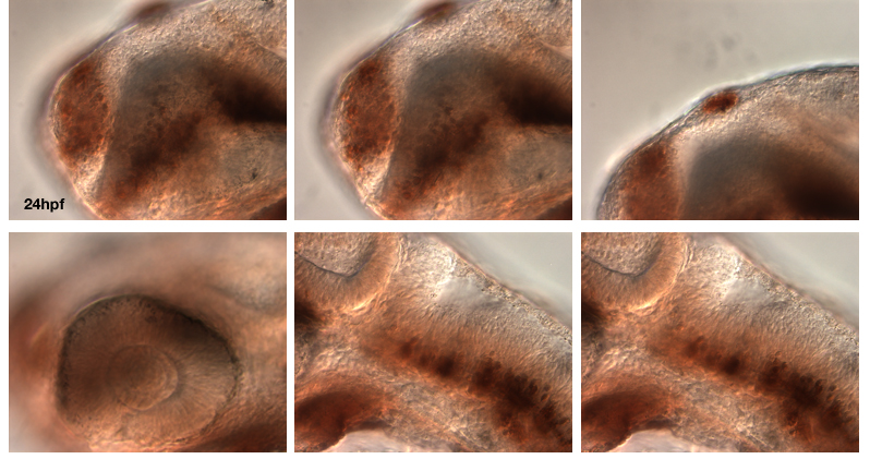
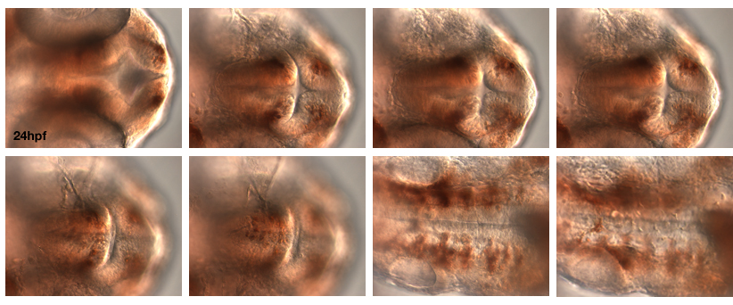
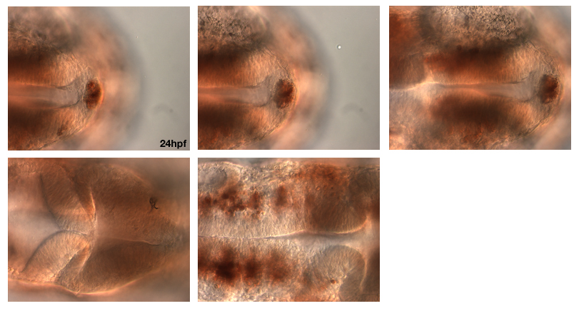
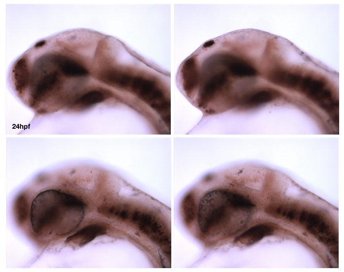
AChE is believed to be present transiently in neurons, including non-cholinergic ones, soon after they complete their final mitosis, and therefore has been used as a marker of differentiation (Wilson et al., 1990).
METHOD FROM Wilson et al., 1990.
We used the method of Karnovsky and Roots (1964). 1-day embryos were fixed in 4% formalin buffered with either maleate (pH6.0, 0.1M) or Pipes (disodium salt - Sigma, pH6.95, O.lM-plus 2mM-EGTA [JV,/V,/V',A''-tetraacetic acid, Sigma], and lmM-MgSO4). Embryos prepared as whole mounts (n=18) were incubated for 2.5 h in maleate buffer (pH6.0) with 5mM-sodium citrate, 3mM-copper sulphate, 0.5mM-potassium ferricyanide and 0.5mgml~' substrate (acetylthiocholine iodide).
Select images
All produced by the boss: Steve Wilson.
External Links:
ZFIN:
LABELS THESE BRAIN STRUCTURES:
differentiated neurons, cholinergic neurons.
KEY PUBLICATIONS
Wilson et al., Wilson, S.W., Ross, L.S., Parrett, T., and Easter, S.S. Jr. (1990)
The development of a simple scaffold of axon tracts in the brain of the embryonic zebrafish, Brachydanio rerio.
Development (Cambridge, England). 108:121-145.
KARNOVSKY, M. J. AND ROOTS, L. (1964).
A direct coloring thiocholine method for cholinesterases.
J. Histocliem. Cytochem. 12, 219-221.