

Synaptic Vesicle 2 (SV2)
ABOUT THIS ANTIBODY
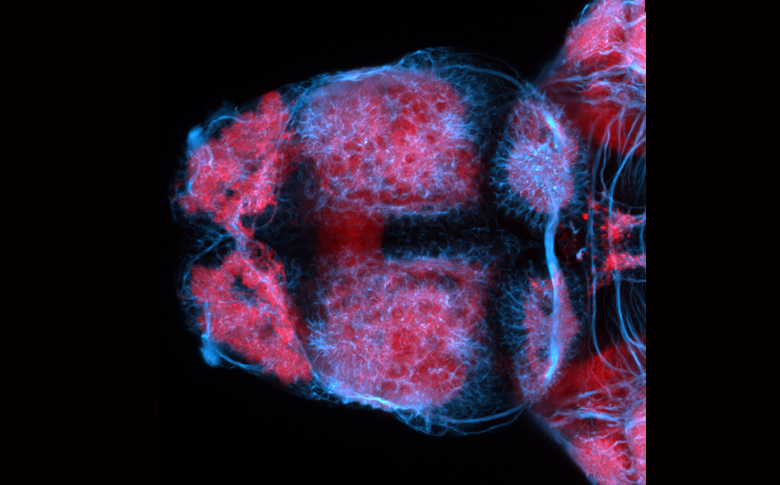
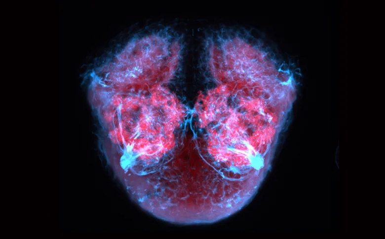
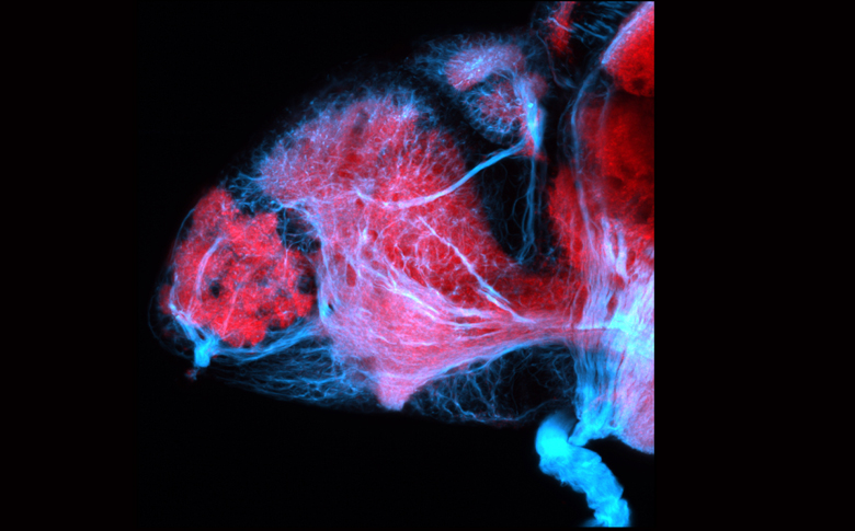
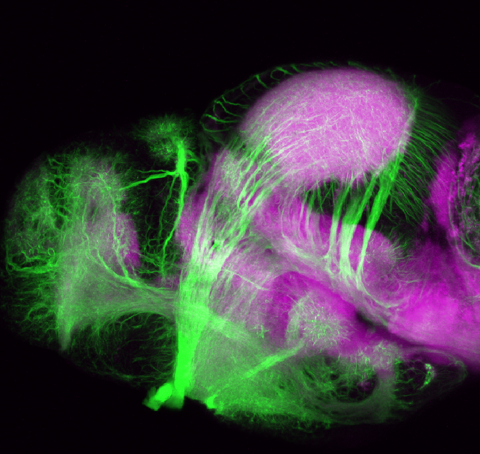
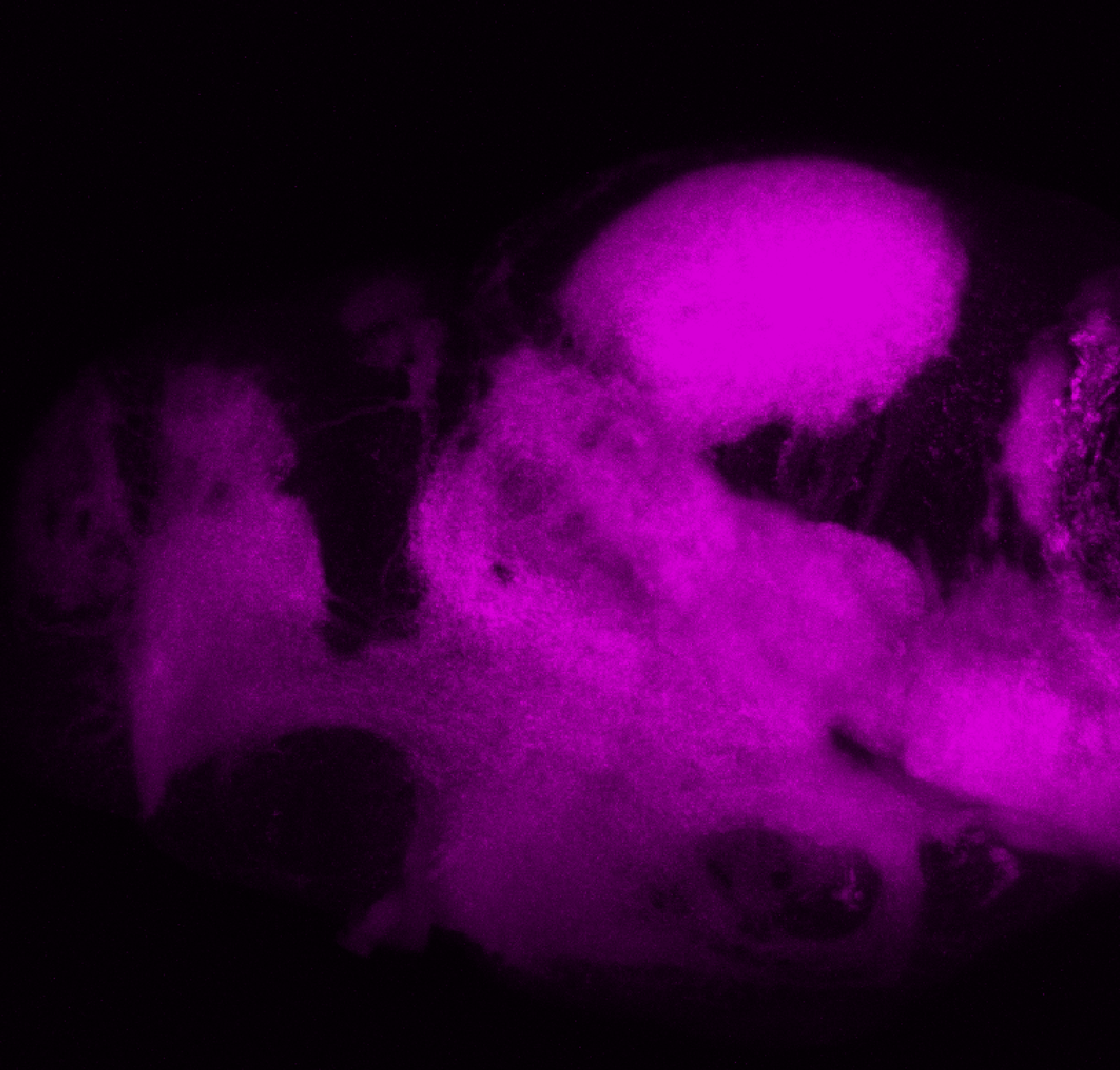
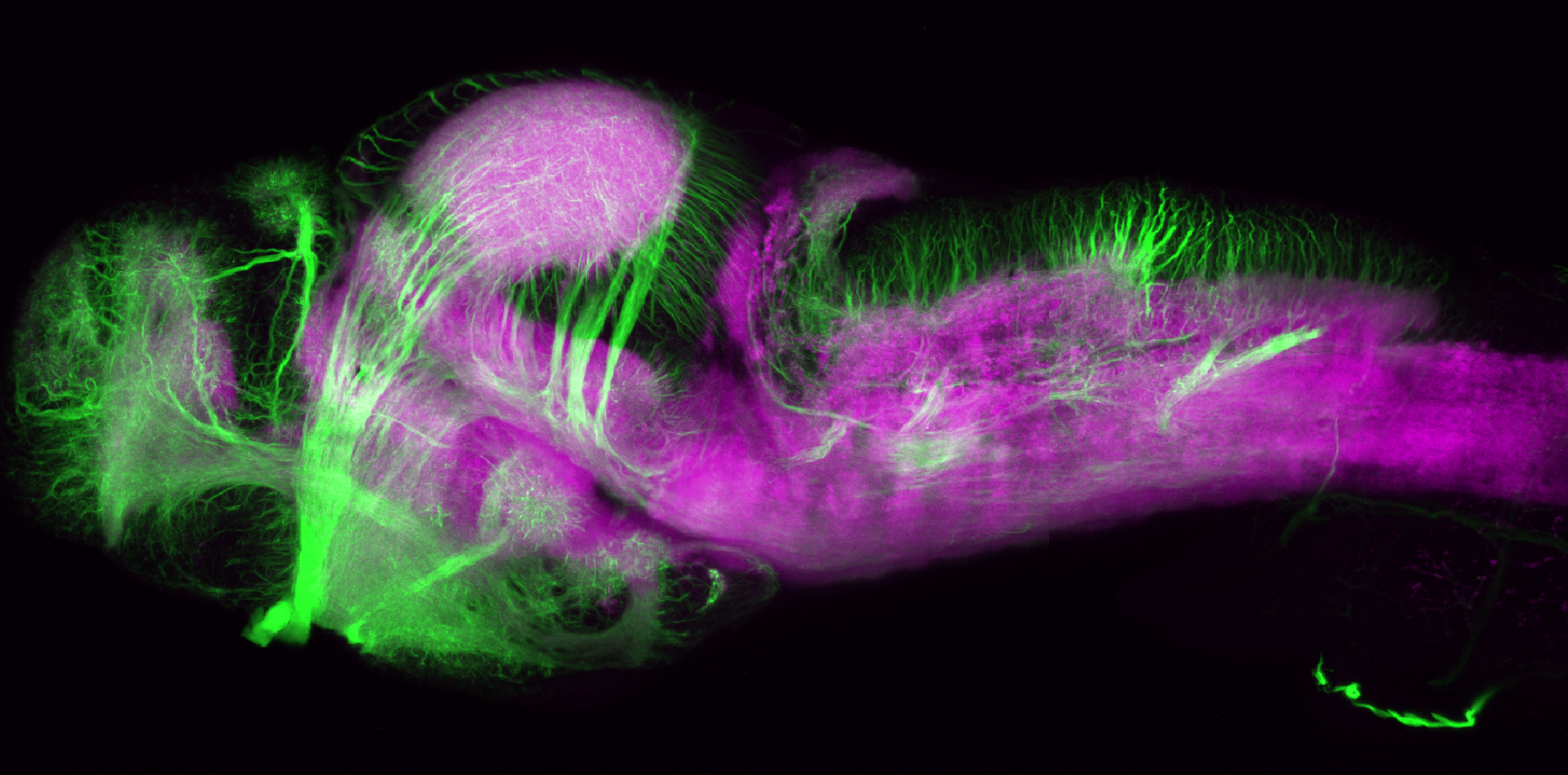
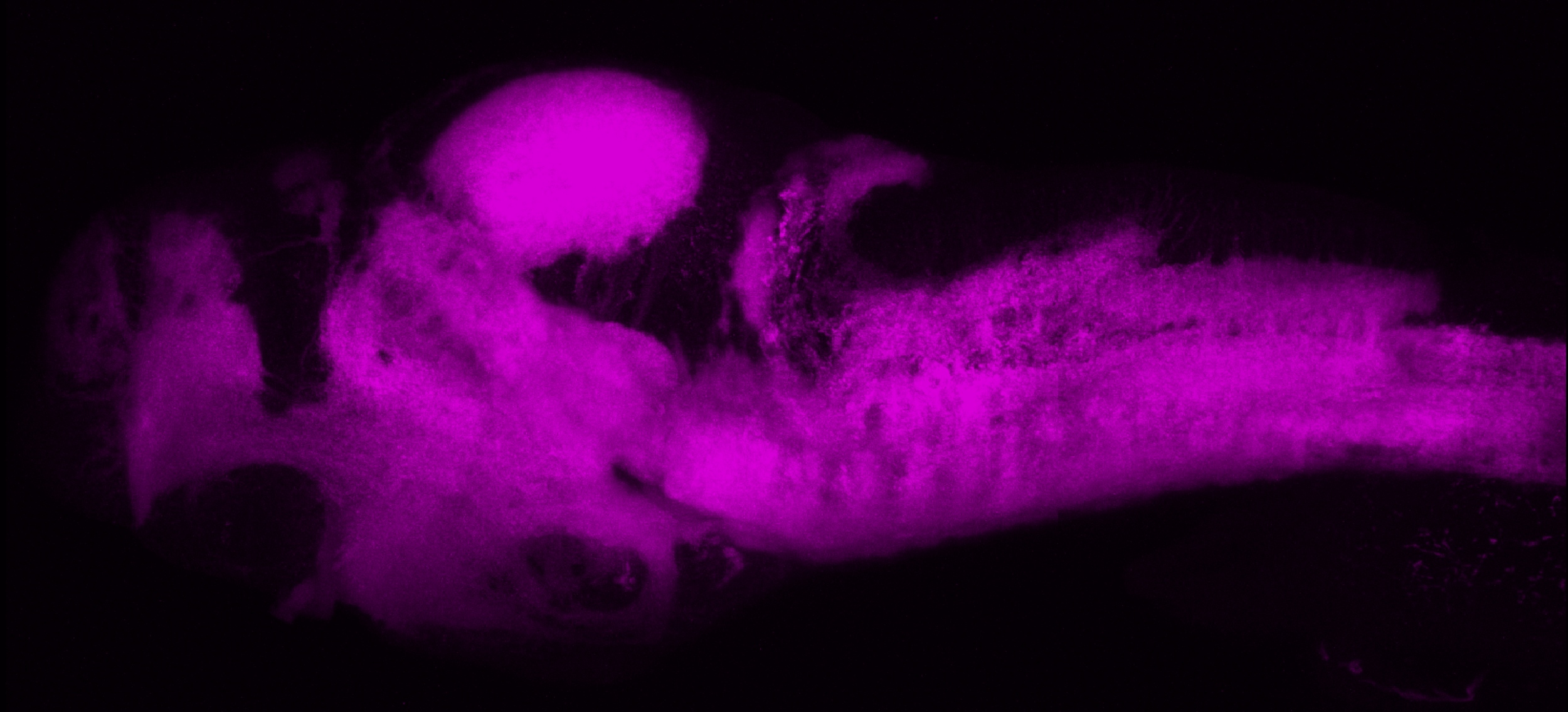
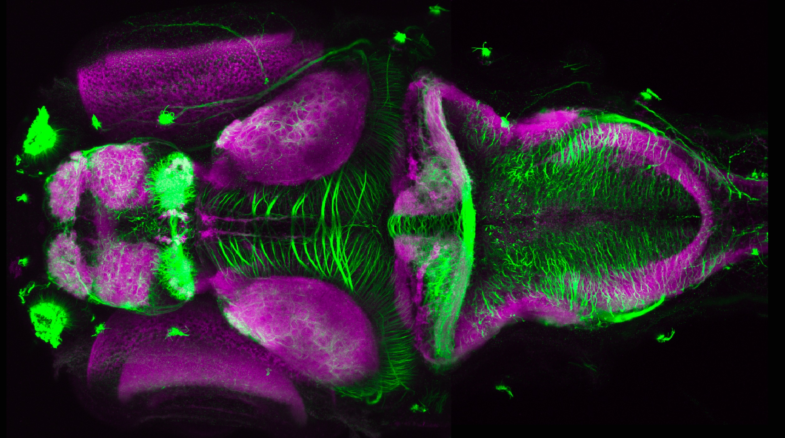
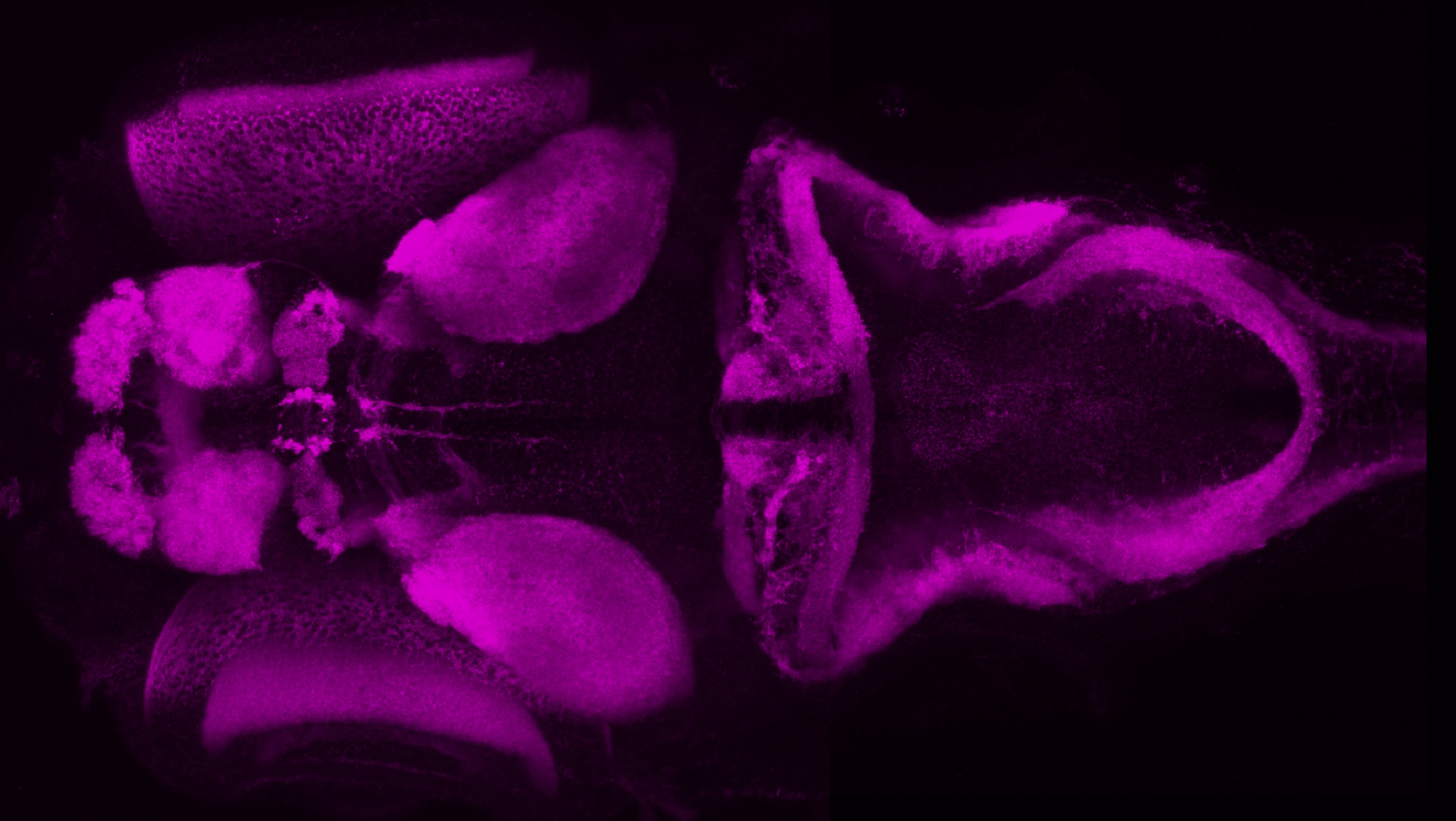
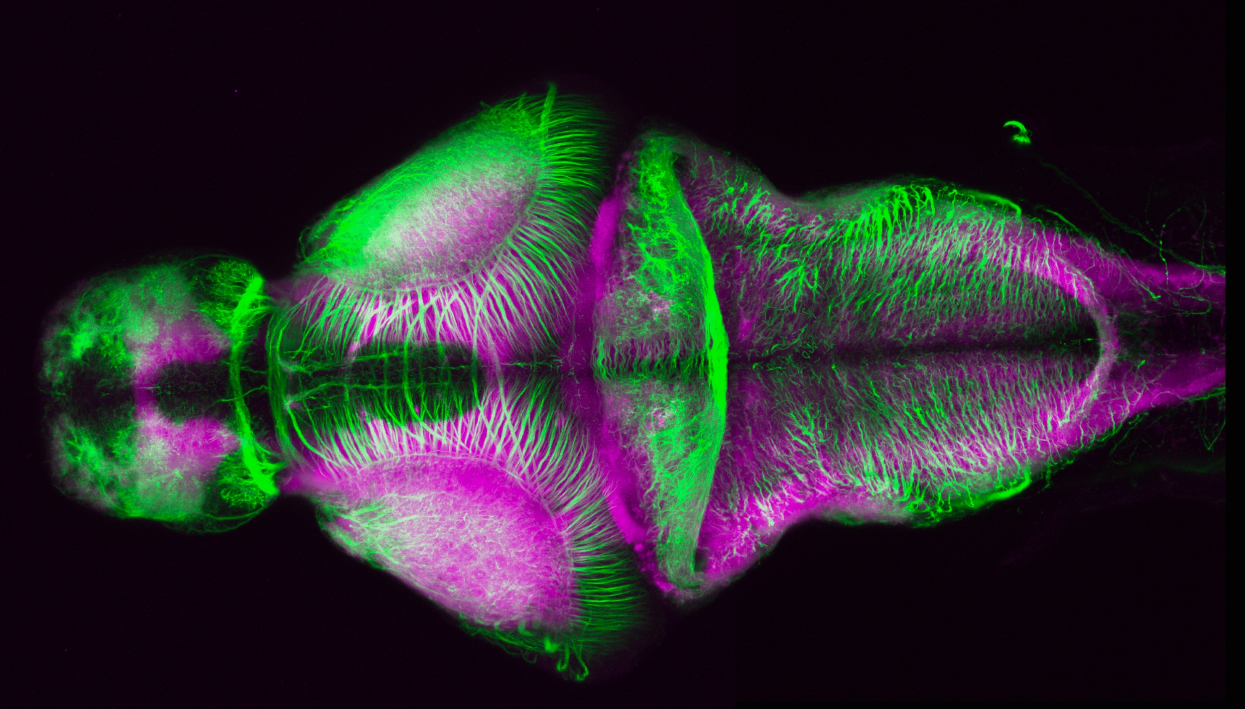
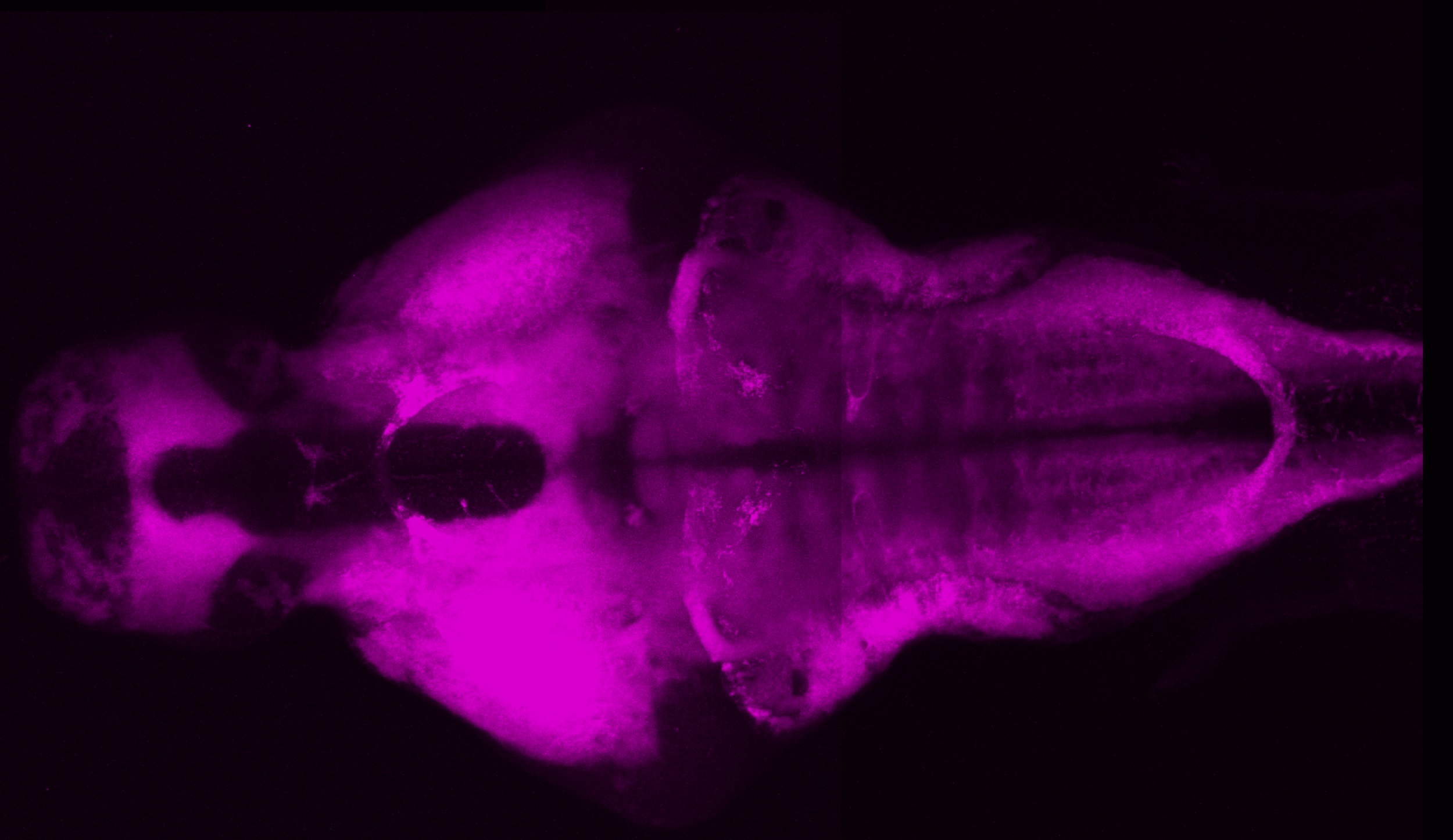
Synaptic Vesicle 2 labels synaptic neuropil.
We frequently use this antibody in conjunction with anti-acetylated tubulin (IgG2b, Sigma), which labels beautifully the axonal connections, to aid anatomical orientation in transgenic specimens. In addition to these antibodies being informative from a neuroanatomical perspective, they are also invaluable as a tool for anatomical localisation. These antibodies can be used as a framework to easily compare the expression patterns of different transgenic lines and locate GFP-positive structure in the context of the brain. Both antibodies are mouse monoclonals but can be detected in the same specimen using subtype-specific secondary antibodies.
Mouse monoclonal anti-SV2 (IgG1) (DSHB, Cat#AB 2315387, dilution 1:500)
Select images
External Links:
LABELS THESE BRAIN STRUCTURES:
Synaptic neuropil
KEY PUBLICATIONS
Buckley K, Kelly RB (1985)
Identification of a transmembrane glycoprotein specific for secretory vesicles of neural and endocrine cells.
J Cell Biol 100:1284–1294
Hendricks M, Jesuthasan S (2007)
Asymmetric innervation of the habenula in zebrafish.
J Comp Neurol 502:611–619
Turner KJ1, Bracewell TG, Hawkins TA.
Anatomical dissection of zebrafish brain development.
Methods Mol Biol. 2014;1082:197-214. doi: 10.1007/978-1-62703-655-9_14.