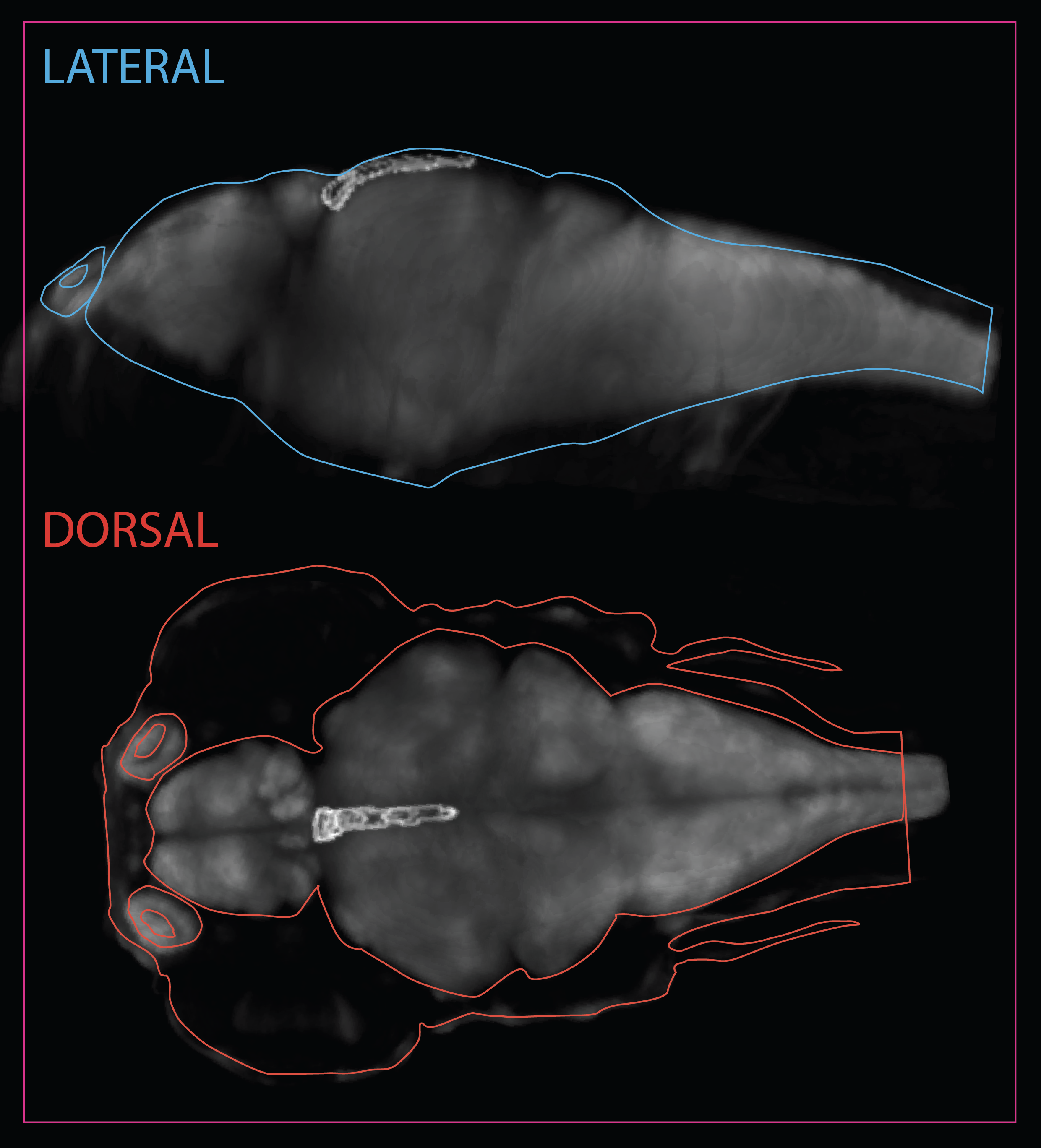
Midbrain > Torus Longitudinalis
Schematic showing the position of torus longitudinalis at 6dpf based on the 3D anatomical segmentation used by the Zebrafish Brain Browser by Gupta et al., 2018.
Description
The torus longitudinalis (TL) is a structure exclusive to ray-finned fish. It is a paired elongated neural structure lying along the medial margins of the optic tectum, suspended from the intertectal commissure and protruding into the tectal ventricle (Ito, 1971; Wullimann, 1994; Northmore, 2011). (Folgueira et al., 2020)
The adult TL can be roughly subdivided into three regions based on morphology and location along the rostro-caudal axis: rostral, intermediate and caudal. The rostral TL sits on top of the posterior commissure with its two halves fused forming a slightly flattened structure . The intermediate TL is longer than the other two, with both halves still fused at the midline. Rostrally, intermediate TL hangs into the tectal ventricle, while a bit more caudally it is flanked by the cerebellar valvula laterally and/or ventrally." (Folgueira et al., 2020)
Connectivity of TL
TL is reciprocally connected to the optic tectum. Toral cell axons exit the TL, then reach the stratum album centrale (SAC) of the optic tectum and finally ascend in separate bundles to the marginal layer, where they run parallel to the brain surface and form terminals. Other efferents project to the thalamic nucleus rostrolateralis.
Afferents to the TL come from visual and cerebellum-related nuclei in the pretectum, namely the central, intercalated and the paracommissural pretectal nuclei, as well as from the subvalvular nucleus in the isthmus. (Folgueira et al., 2020). A single type of tectal neuron projects to the TL in zebrafish that morphologically corresponds to the type X cell described in goldfish (Meek and Schellart, 1978) and in zebrafish (De-Marco et al., 2019).
Ontology
is part of: midbrain
has parts:
OLS Tree Diagram
Transgenic Lines that label this brain region
Antibodies that label this brain region
Key Publications
Folgueira M, Riva-Mendoza S, Ferreño-Galmán N, Castro A, Bianco IH, Anadón R and Yáñez J. Anatomy and Connectivity of the Torus Longitudinalis of the Adult Zebrafish.
Front. Neural Circuits (2020) 14:8. doi:10.3389/fncir.2020.00008
Back to Midbrain


