

Proposed identity of AF-8 in the adult:
Central pretectal nucleus or Nucleus pretectalis centralis (Baier & Wullimann 2021).

Schematic showing the approximate location of AF-8 in a 6dpf zebrafish. The optic tract is labelled in the Tg(atoh7:RFP) transgenic line and position of AFs are based on the data from Robles (2014).
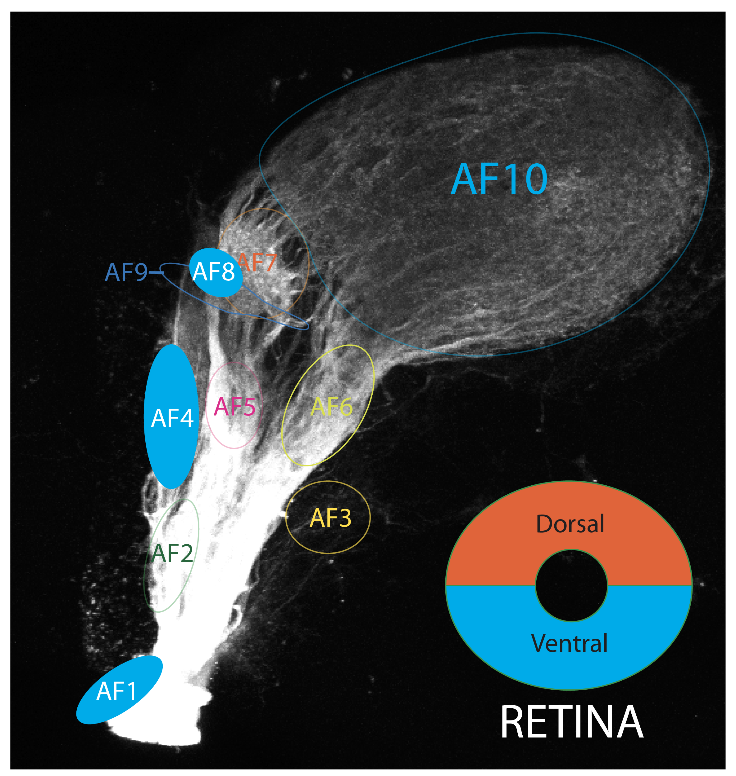
AF-8 is preferentially innervated by Retinal Ganglion Cells (RGCs) located in the ventral retina (Robles et al., 2014).
visual behaviours associated with AF-8
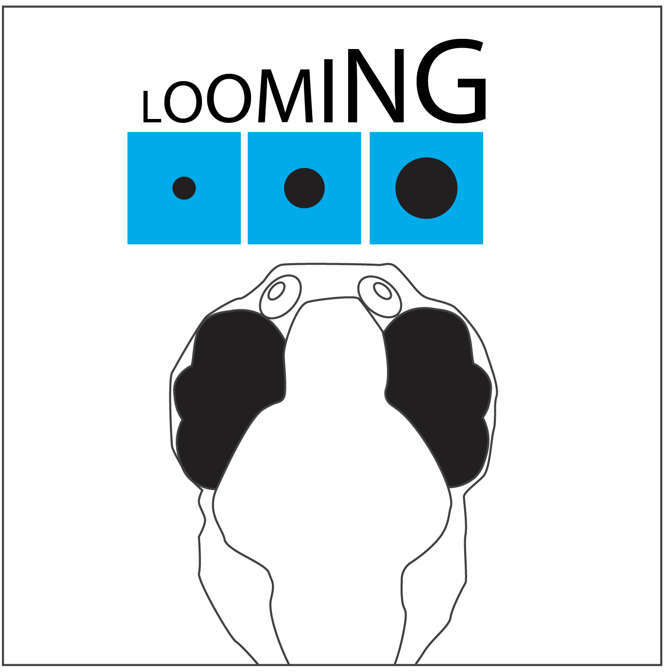
RGCs axons innervating AF-8 are robustly activated by dark looming and dimming stimuli. This AF receives input predominantly from RGCs with their dendrites embedded in the OFF layer of the inner plexiform layer. The role of AF-8 is still unclear, it may alert the tectum to the presence of a shadow facilitating an escape response. It may also play a role in phototaxis (Temizer et al., 2015).
Publications
Baier, H., Wullimann, M.F. (2021)
Anatomy and function of retinorecipient arborization fields in zebrafish.
The Journal of comparative neurology. 529(15):3454-3476.
Robles, E., Laurell, E., Baier, H. (2014)
The Retinal Projectome Reveals Brain-Area-Specific Visual Representations Generated by Ganglion Cell Diversity.
Current biology : CB. 24(18):2085-96.
Burrill JD & Easter Jr SS
Development of the Retinofugal projections.
J Comp Neurology, 2004 pp.1-18.
Temizer, I. et al (2015)
A Visual Pathway for Looming-Evoked Escape in Larval Zebrafish.
Current Biology, 25(14), pp.1823–1834.
Yáñez, J., Suárez, T., Quelle, A., Folgueira, M., Anadón, R. (2018)
Neural connections of the pretectum in zebrafish (Danio rerio).
The Journal of comparative neurology. 526(6):1017-1040.