

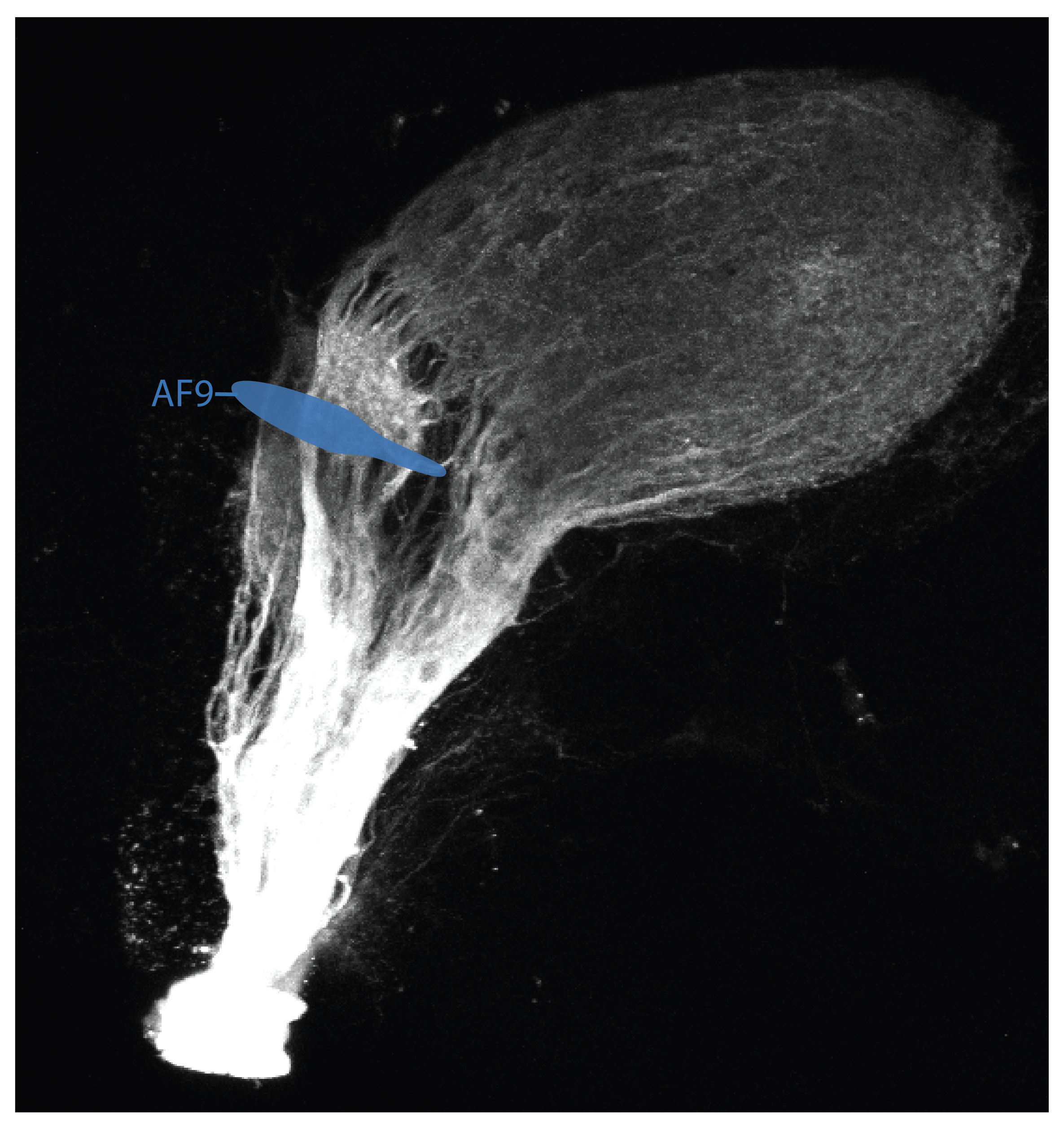
Burrill & Easter proposed identity of AF-9 in the adult:
n. pretectalis periventricularis pars dorsalis (nPPd)/ n.pretectalis periventricularis pars ventralis (nPPv)
synonym: pretectal periventricular nuclei.
“AF-9 was identified as the presumptive nPPd because its position relative to the posterior commissure was similar to that of the nPPd, and because some of the fibers that innervate both the nPPd and AF-9 continue dorsally and innervate the deep layers of the optic tectum (AF-10). In some adult cypriniformes (Braford and Northcutt, 1983) optic axons also project to the nPPv which is located caudal to the nPPd at the same dorsoventral level. We have not seen a separate AF in this region, and suggest that AF-9 represents a projection to both parts, dorsal and ventral, of the same nucleus.”(Burrill & Easter., 2004).
Schematic showing the approximate location of AF-9 in a 6dpf zebrafish. The optic tract is labelled in the Tg(atoh7:RFP) transgenic line and position of AFs are based on the data from Robles (2014).
visual behaviours associated with AF-9

Retinal Ganglion Cells projecting to AF-9 respond vigorously to only looming bright and receding bright (ON) stimuli.For the receding stimulus AF-9 axons were only activated by the appearance of the bright stimulus, not the receding motion. (Temizer et al., 2015)
Publications
Baier, H., Wullimann, M.F. (2021)
Anatomy and function of retinorecipient arborization fields in zebrafish.
The Journal of comparative neurology. 529(15):3454-3476
Temizer, I. et al (2015)
A Visual Pathway for Looming-Evoked Escape in Larval Zebrafish.
Current Biology, 25(14), pp.1823–1834.
Burrill JD & Easter Jr SS
Development of the Retinofugal projections.
J Comp Neurology, 2004 pp.1-18.
Robles, E., Laurell, E., Baier, H. (2014)
The Retinal Projectome Reveals Brain-Area-Specific Visual Representations Generated by Ganglion Cell Diversity.
Current biology : CB. 24(18):2085-96.
Yáñez, J., Suárez, T., Quelle, A., Folgueira, M., Anadón, R. (2018)
Neural connections of the pretectum in zebrafish (Danio rerio).
The Journal of comparative neurology. 526(6):1017-1040.