


HuC/HuD Monoclonal Antibody (16A11)
ABOUT THIS ANTIBODY
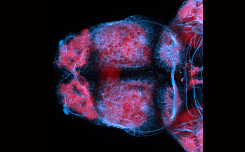
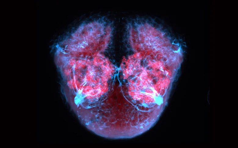
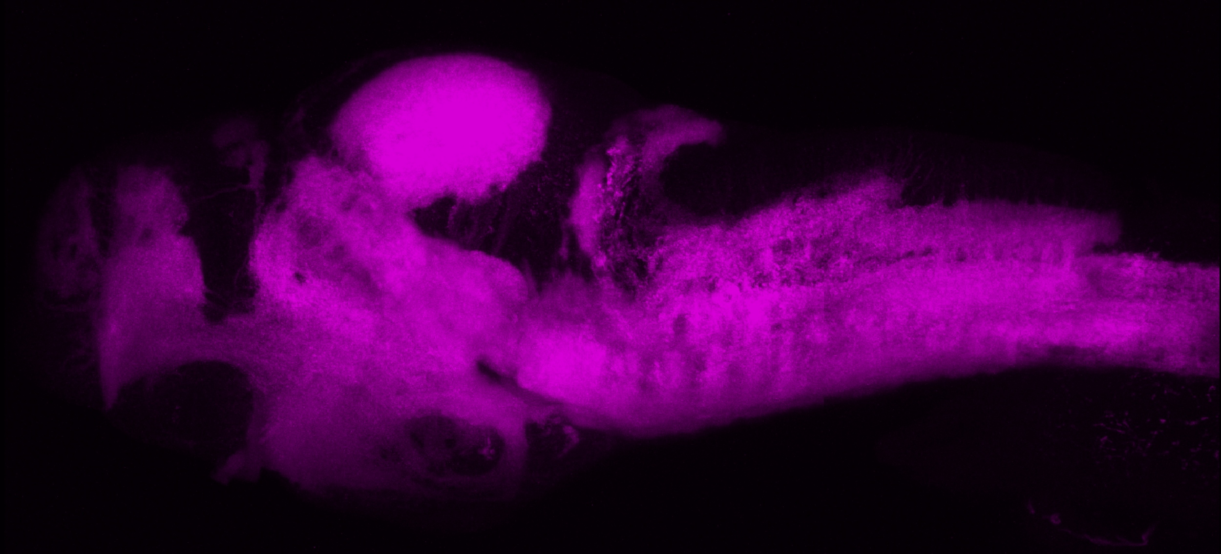
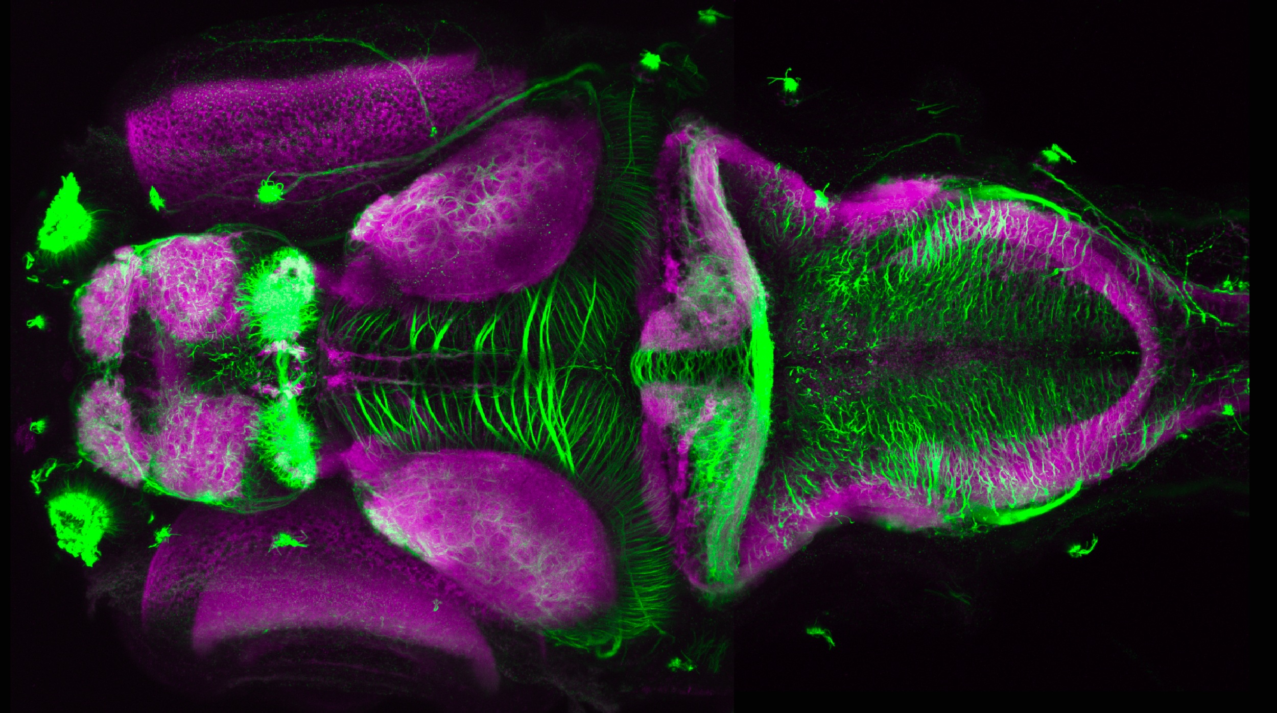
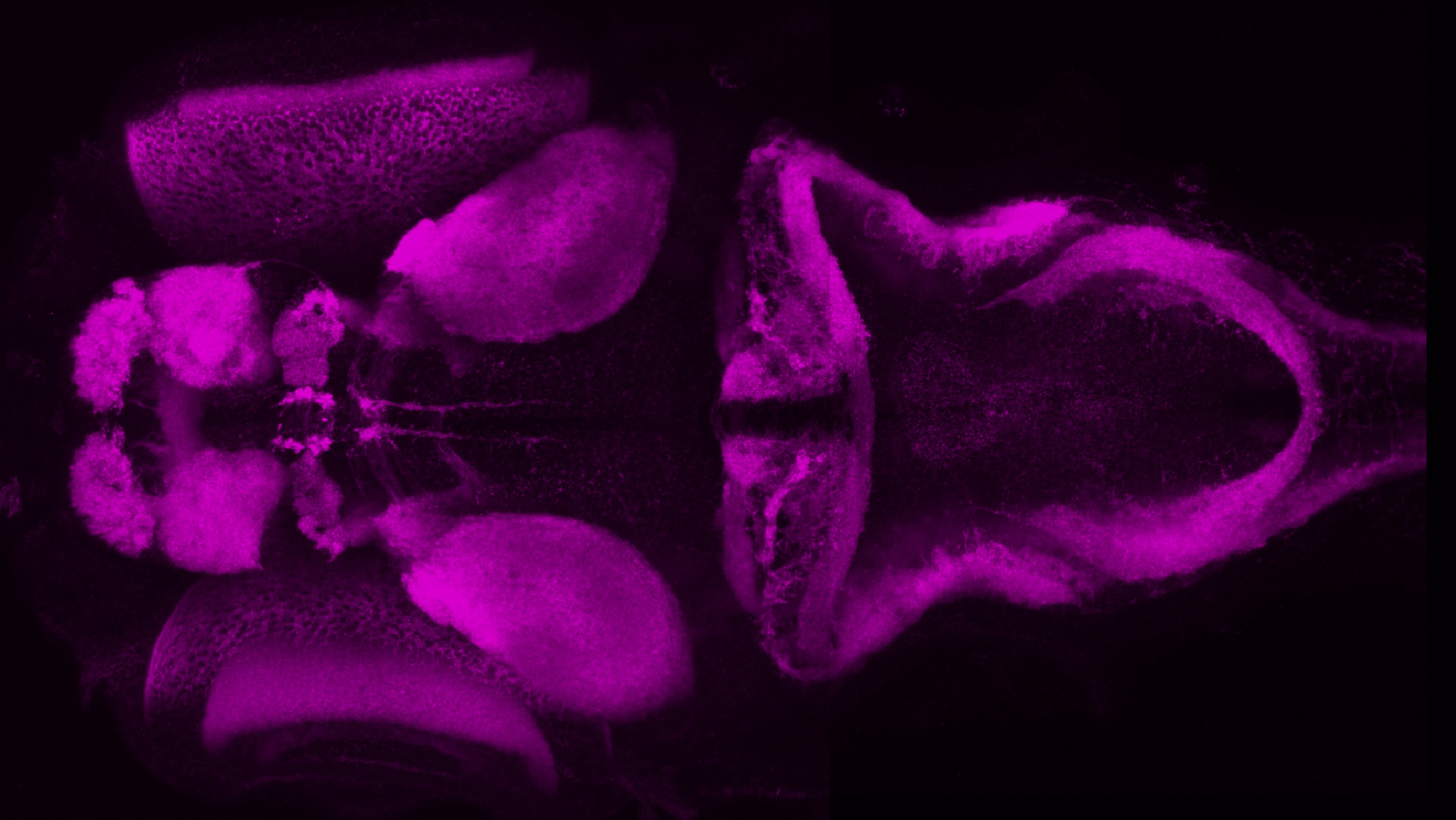
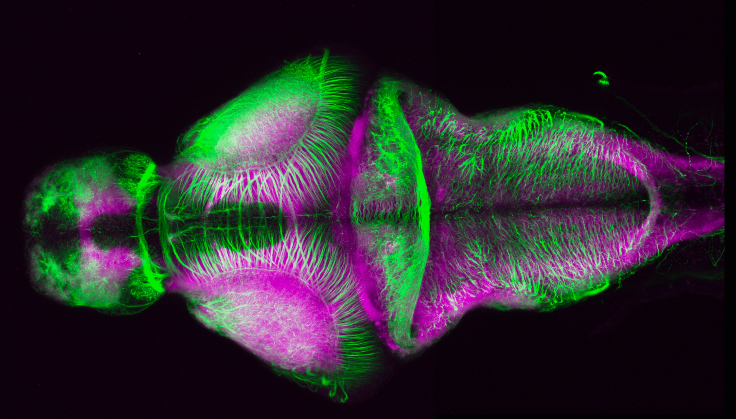
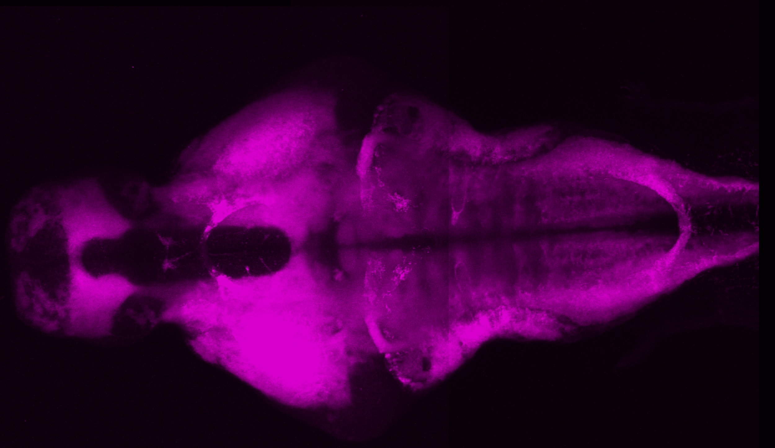
HuC/HuD is a pan-neuronal nuclear label and as such a very good counterstain for showing the structure of the brain.
Mouse anti-HuC/HuD (16A11)(IgG2B, Thermoscientific, cat#A-21271, dilution 1:200)
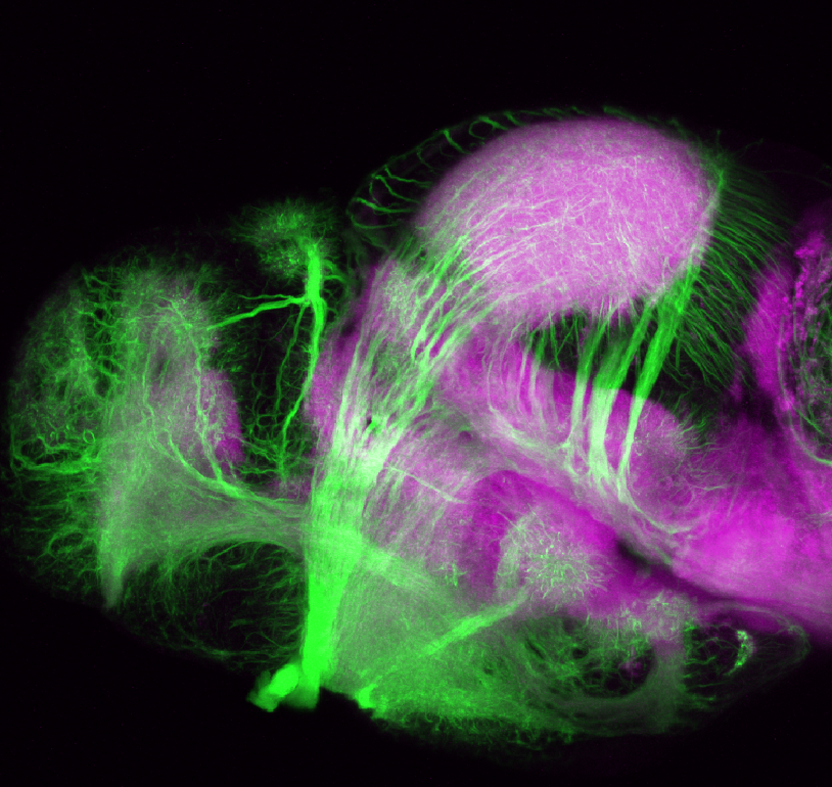
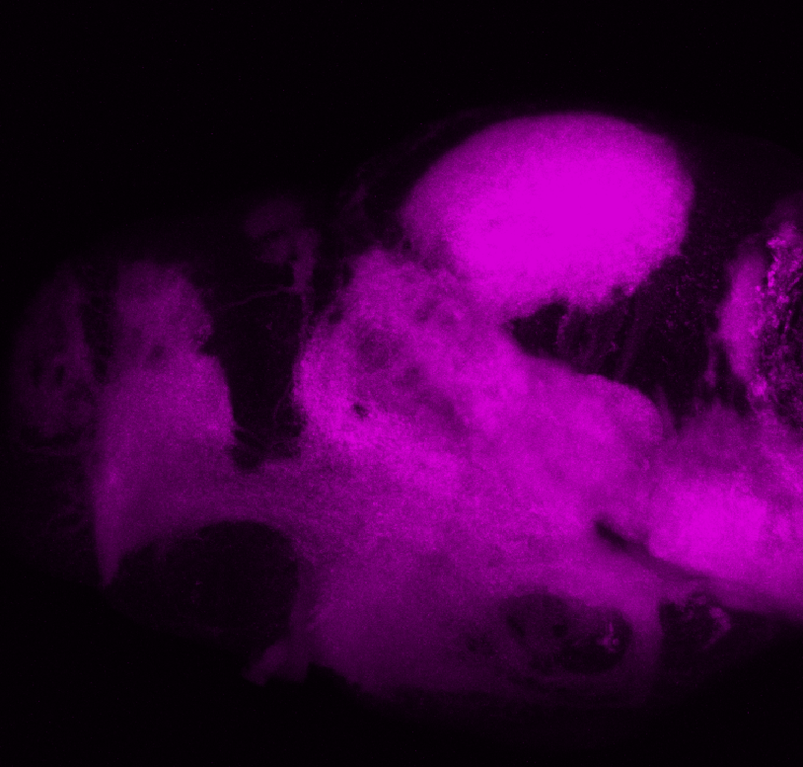
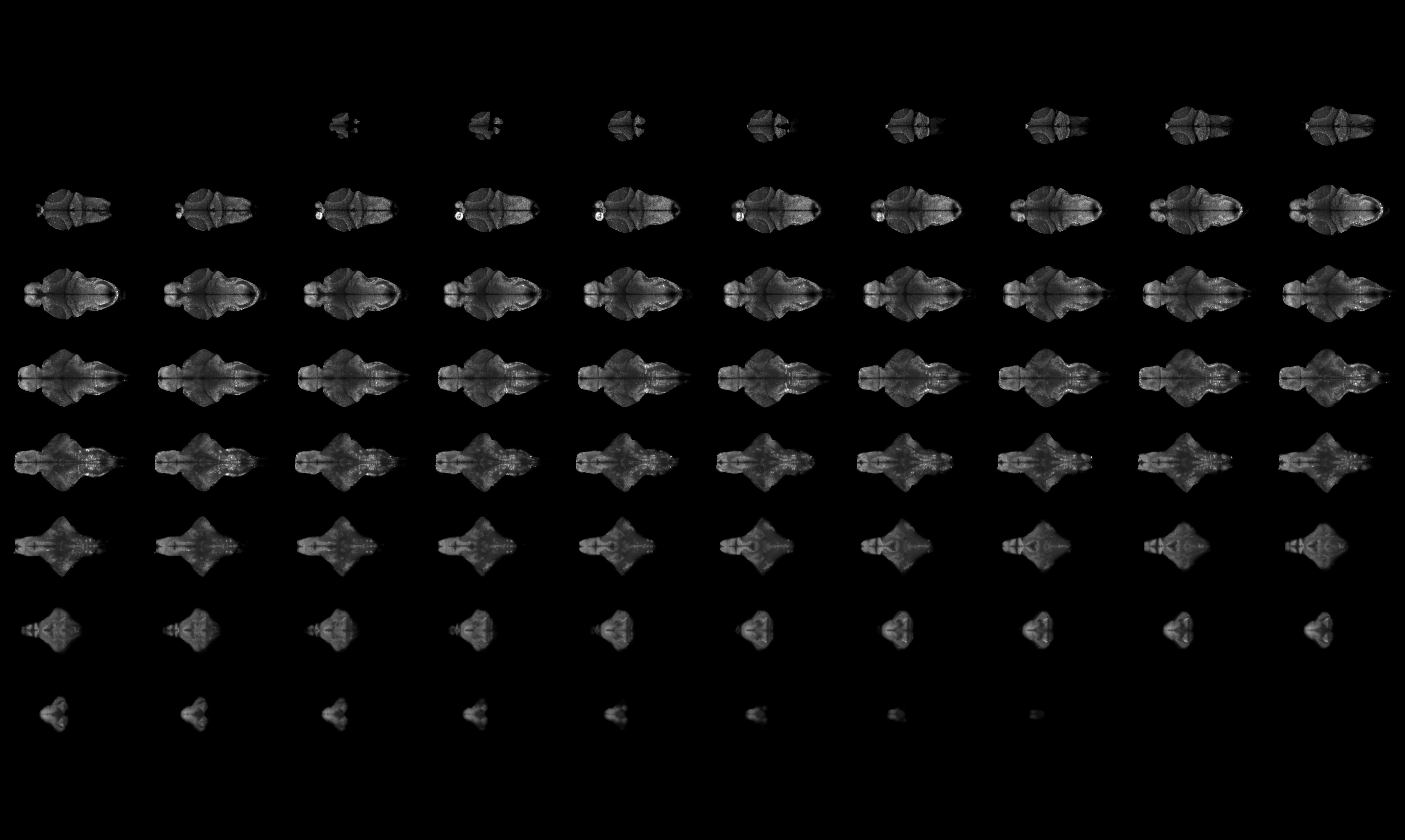
Select images by Chintan Trivedi imaged using a light-sheet microscope.

Flythrough movie of a 6dpf larval zebrafish brain labelled with anti-HuC and anti-HuD antibodies. Anti-HuC/D is a pan-neuronal nuclear label that labels all neurons in the nervous system.
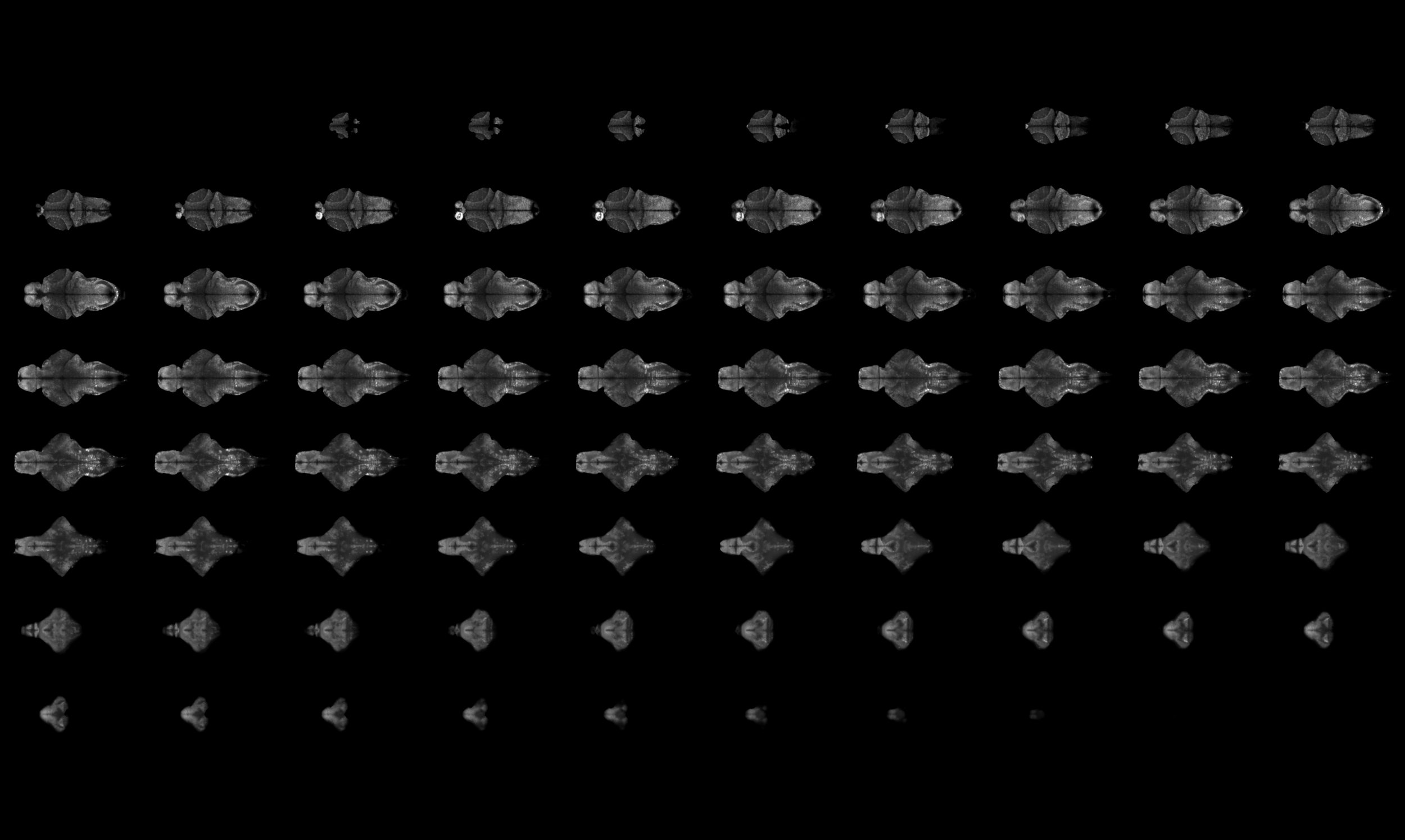
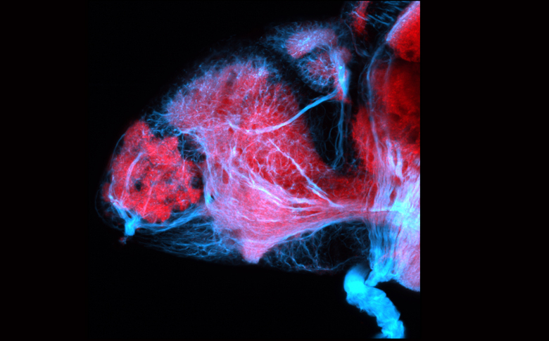
Montage of slices through the brain of a 6dpf zebrafish larvae labelled using anti-HuC/D antibody.

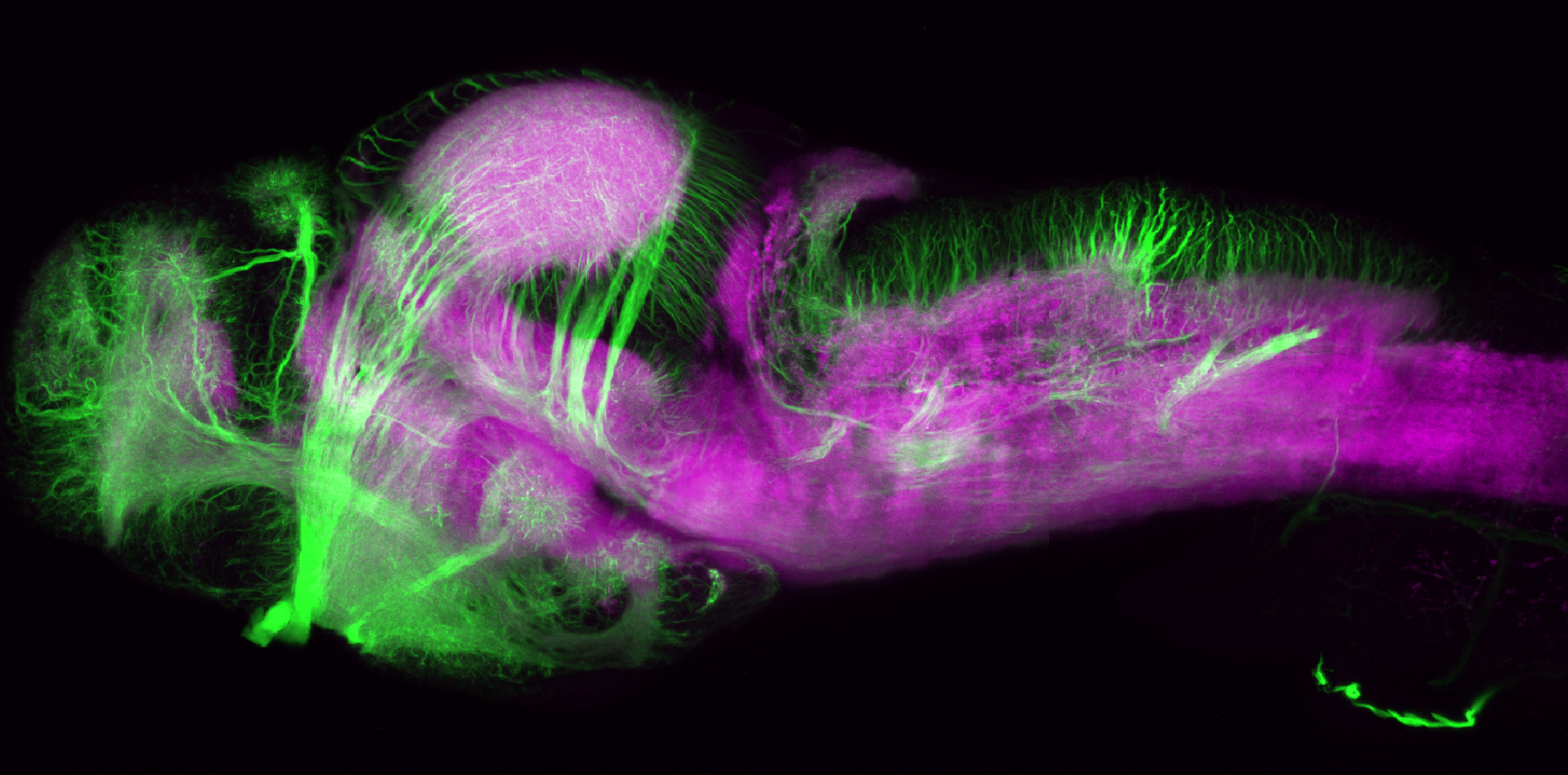
6dpf brain of a larval zebrafish labeled with anti-HuC/D. This stack was morphed to ZBB. This screenshot shows the HuC/D antibody labelling uploaded to ZBB and viewed using their online browser. If you would like to look at this data using ZBB please download the .png file using the link below and upload it to ZBB using the Lines> Custom>Load option.
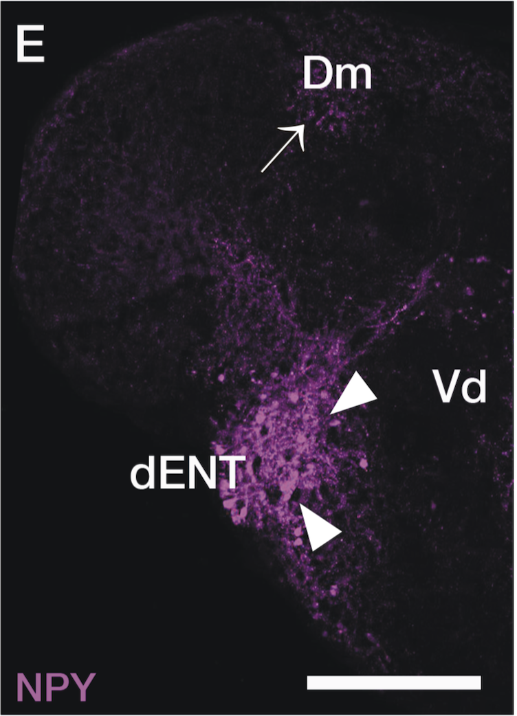
LABELS THESE BRAIN STRUCTURES:
pan-neuronal marker.
KEY PUBLICATIONS

