


MacDonald Lab
Mechanisms of eye development and disease
MacDonald Lab
Mechanisms of eye development and disease
Research
The overarching research aim of my laboratory is to understand how a healthy eye is built and maintained throughout life. More specifically, we are interested in how glial cells, the major support cells in the nervous system, are patterned and shaped during development to support neurons. We are also interested in what happens when the intricate glia-neuronal relationship breaks down due to increasing age or disease.
The retina is the light sensitive part of the eye that allows you to see. We use the zebrafish retina as a model as it contains the same neuron types and glial cells as the human eye. The zebrafish embryo is an incredible system to study development – it is transparent, we can label each cell with specific fluorescent markers and use time-lapse confocal microscopy to watch eye development happen in real time in a living fish! In addition to imaging we use CRISPR/Cas9 mutagenesis, RNA-sequencing and molecular biology to uncover and explore fundamental mechanisms of glial biology.
We are now interested in determining the cellular and molecular mechanisms regulating glial morphogenesis, with a particular focus on the consequences of disrupted glial contacts on neuronal function.
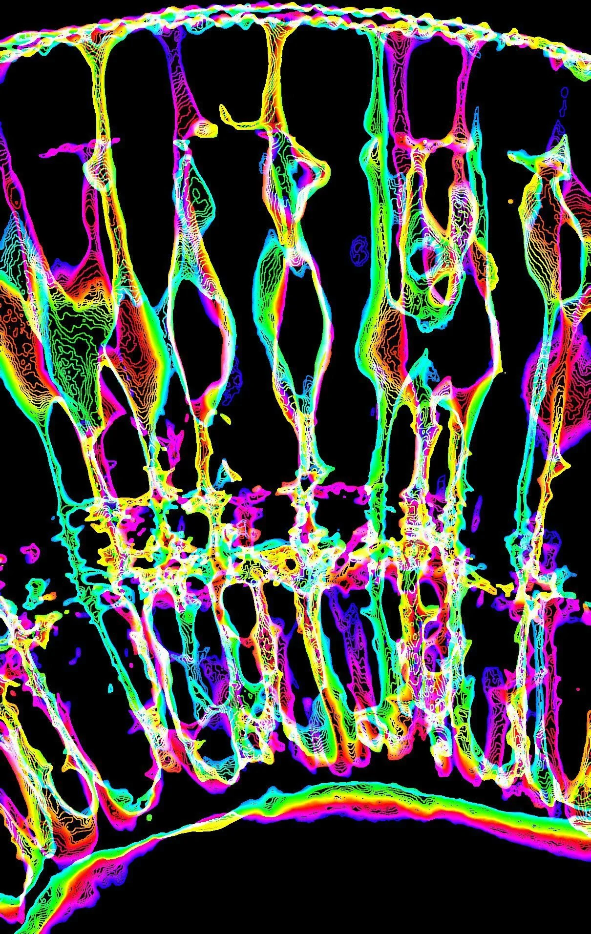
Retinal Structure
Credit to the undergraduate student Hiba Noor
Retinal Layers
Credit to the undergraduate student Hiba Noor
Click the buttons below to learn more about our different research topics. Visit our Resources page to access tools, protocols and view our antibody and transgenic line data.


Images
Images
To view our images and movies use the buttons below.


People
People
Current members


News
News
MacDonald Lab News


Publications
Publications
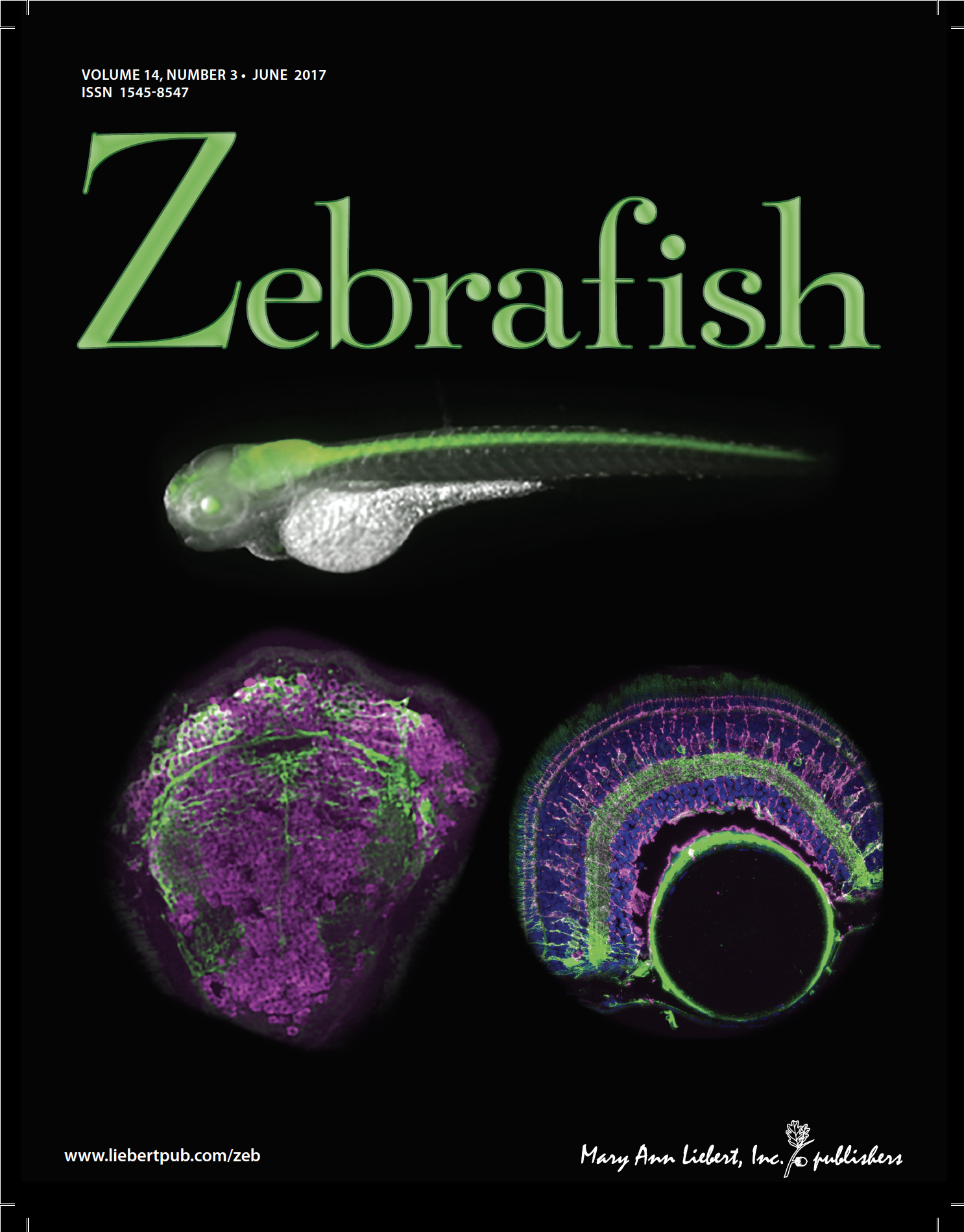
Our Covers


SELECTED Publications (Full list)
2026
Lahne M, Lungu R, Snorton M, Yoshimatsu T*, MacDonald R.B. 2026. Glia regulate local retinoic acid levels to specify neuronal specialisation for high-acuity vision. Submitted. bioRxiv: https://www.biorxiv.org/content/10.64898/2026.02.27.708512v1.
Reyes-Pinto, R., Arancibia-Altamirano, D., MacDonald, R.B., Kawakami, K., Valvida, L.E. 2026. A novel cyp26c1-driven reporter line to study boundaries of retinoic acid signalling during zebrafish development. Front. Cell. Dev. Bio. https://www.frontiersin.org/journals/cell-and-developmental-biology/articles/10.3389/fcell.2026.1753548/abstract
2025
Jaroszynska, N., Kothurkar, A.A., Jankovich, D., Cameron-Pack, M., MacDonald, R.B. 2025. Gelatinase activity is required for Muller glia morphogenesis and neuronal maintenance in the retina. Submitted. bioRxiv: https://www.biorxiv.org/cgi/content/short/2025.12.17.694830v1
Noel, C.L.N., Breitenbach, E.M., MacDonald, R.B.* 2025. Turquoise killifish naturally develop hallmarks of age-related macular degeneration with advancing age. Submitted. bioRxiv. https://doi.org/10.1101/2025.10.23.683644
Yaniv Z et al. 2025. The IBEX Imaging Knowledge-Base: A Community Resource Enabling Adoption and Development of Immunofluorescence Imaging Methods. eLife. 14:RP105737. https://doi.org/10.7554/eLife.105737.1
Radtke AJ et al. 2025. The IBEX Knowledge-Base: A central resource for multiplexed imaging techniques. PLoS Biol. 23(3): e3003070. https://doi.org/10.1371/journal.pbio.3003070
Farger E, Keatinge M, Pearce O, Piepponen P, Paula P, van Eden F, MacDonald RB, Bandmann O. 2025. A zebrafish model of acmsd deficiency does not support a prominent role for ACMSD in Parkinson’s disease. npj Parkinsons Dis. 11:118. https://doi.org/10.1038/s41531-025-00940-1
2024
Bergmans S§, Noel NCL§, Masin L, Harding E, Krzywanska AM, Rajagopal A, Hu CK, Arckens L, Ruzycki PA*, MacDonald RB*, Clark BS*, Moons L*. 2024. Age-related dysregulation of the retinal transcriptome in the African turquoise killifish. Aging Cell:e14192.
https://doi.org/10.1111/acel.14192 *Co-senior corresponding authors.
Kothurkar A§, Patient GS, Noel NCL, Krzywanska AM, Carr BJ, Chu CJ*, MacDonald RB*. 2024. Iterative Bleaching Extends Multiplexity (IBEX) Imaging facilitates simultaneous identification of all cell types in the vertebrate retina. Journal of Cell Science. 137(23):jcs263407. https://doi.org/10.1242/jcs.263407 *Corresponding authors. Cover Article. Research highlight: https://doi.org/10.1242/jcs.263407. Tutorial: Link YouTube: https://www.youtube.com/watch?v=yXuVVDuk18k.
Jaroszynska N§, Salzinger A§, Tsarouchas T, Becker CG, Becker T, Lyons D, MacDonald RB*, Keatinge M*. 2024. C9ORF72 deficiency results in degeneration of the zebrafish retina in vivo. J. Neurosci, e2128232024; https://www.jneurosci.org/content/early/2024/04/22/JNEUROSCI.2128-23.2024 *Co- corresponding authors.
2023
Uttley K, Papanastasiou AS, Lahne M, Brisbane J, MacDonald RB, Bickmore WA, Bhatia S. 2023. Unique functions of two overlapping PAX6 retinal enhancers. Life Science Alliance. 6(11):e20230212. https://doi.org/10.26508/lsa.202302126 *Co- corresponding author.
Keatinge M§, Gegg ME§, Watson L, Mortiboys H, Bui H, Lefeber DJ, van Rens A, MacDonald RB*, Bandmann O*. 2023. Unexpected phenotypic and molecular changes of combined glucocerebrosidase and acid sphingomyelinase deficiency. Disease models and mechanisms. https://journals.biologists.com/dmm/article/16/6/dmm049954/308903/ *Co- corresponding author.
Kugler E, Bravo I, Durmishi X, Marcotti S, Beqiri S, Carrington A, Stramer BM, Mattar P, MacDonald RB. 2023. GliaMorph: A modular image analysis toolkit to quantify Müller glial cell morphology. Development: 150(3):dev201008. https://doi.org/10.1242/dev.201008
Kugler E*, Breitenbach E-M, MacDonald RB*. 2023. Glia cell morphology analysis using the Fiji GliaMorph toolkit. Current Protocols. *Co- corresponding author. https://doi.org/10.1002/cpz1.654.
2022
Martins RR, Zamzam M, Moosajee M, Thummel R, Henriques CM*, MacDonald RB*. 2022. Müller Glia maintain their regenerative potential despite degeneration in the aged zebrafish retina. Aging Cell. 21:e13597. *Co- corresponding author. https://doi.org/10.1111/acel.13597
Larbalestier H, Keatinge M, Watson L, White E, Gowda S, Wei W, Koler K, Semenova SA, Rimmer N, Sweeney ST, Mazzolini J, Sieger D, McDearmid J, Panula P, MacDonald RB*, Bandmann O*. 2022. GCH1 Deficiency Activates Brain Innate Immune Response and Impairs Tyrosine Hydroxylase Homeostasis. J Neurosci. 42(4):702-716. *Co-corresponding authors. https://doi.org/10.1523/JNEUROSCI.0653-21.2021
Schlaeppi A, Adams W, Haase R, Huisken J, MacDonald RB, Eliceiri K and Kugler E. 2022. Meeting in the middle: towards successful multidisciplinary bioimage analysis collaborations. Frontiers in Bioinformatics. https://doi.org/10.3389/fbinf.2022.889755
Jurisch-Yaksi N, Kugler EC, MacDonald RB, Mosimann C, and Yaksi E. 2022. Microscopy techniques in zebrafish. RMS inFocus. https://www.rms.org.uk/resource/elisabeth-kugler-pdf.html
Join us/Contact
Join us/Contact
Join us
We’re amazed by glia development and hope you are too. If you would like to join us please get in touch. We enthusiastically encourage applications from prospective students or postdocs. To see examples of our previous work please see the images and publications sections on the website or look us up on Instagram!
Our lab is part of several excellent graduate programmes:
• UCL-Birkbeck MRC Doctoral Training Programme
• London Interdisciplinary Doctoral Programme
Please contact Ryan MacDonald more information.
Contact
Lab Address
MacDonald Lab
Institute of Ophthalmology
11-43 Bath St,
London EC1V 9EL
UK


Funding
Funding
Our work is generously supported by the following funders: