

About glyt2b (synonyms: slc6a5)
glycine transporter 2/ Solute Carrier Family 6 (Neurotransmitter Transporter), Member 5
glyt2b is orthologous to human GLYT2/ slc6a5 . This gene encodes a sodium- and chloride-dependent glycine neurotransmitter transporter. This protein is found in glycinergic axons and glycinergic synapses. Glycine is a major inhibitory neurotransmitter in the central nervous system of vertebrates.
Expression pattern
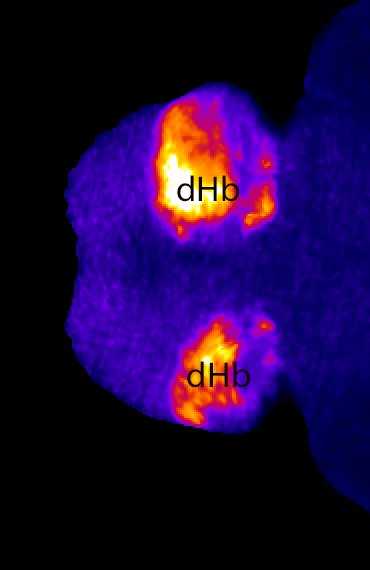
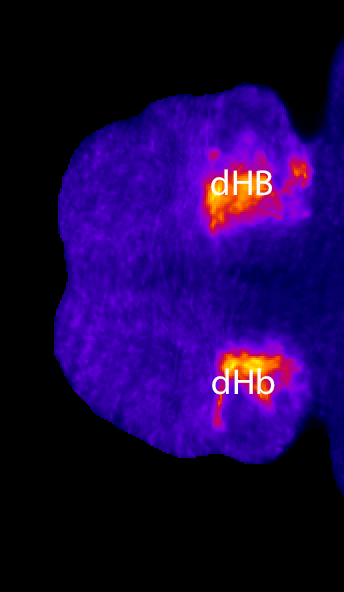
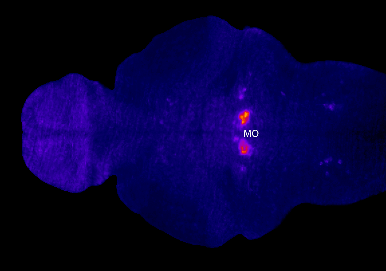
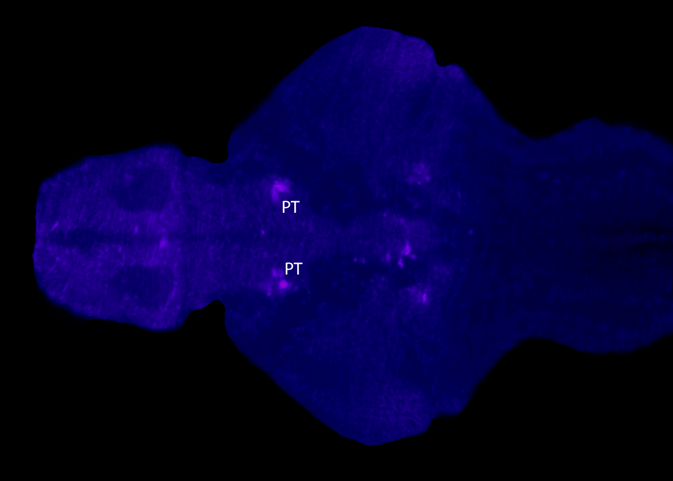
glyt2b is expressed in glycinergic neurons in the hindbrain.

Flythrough movie of a 6dpf larvae labelled using fluorescent in-situ hybridisation using a probe against glyt2b. This probe labels glycinergic neurons.

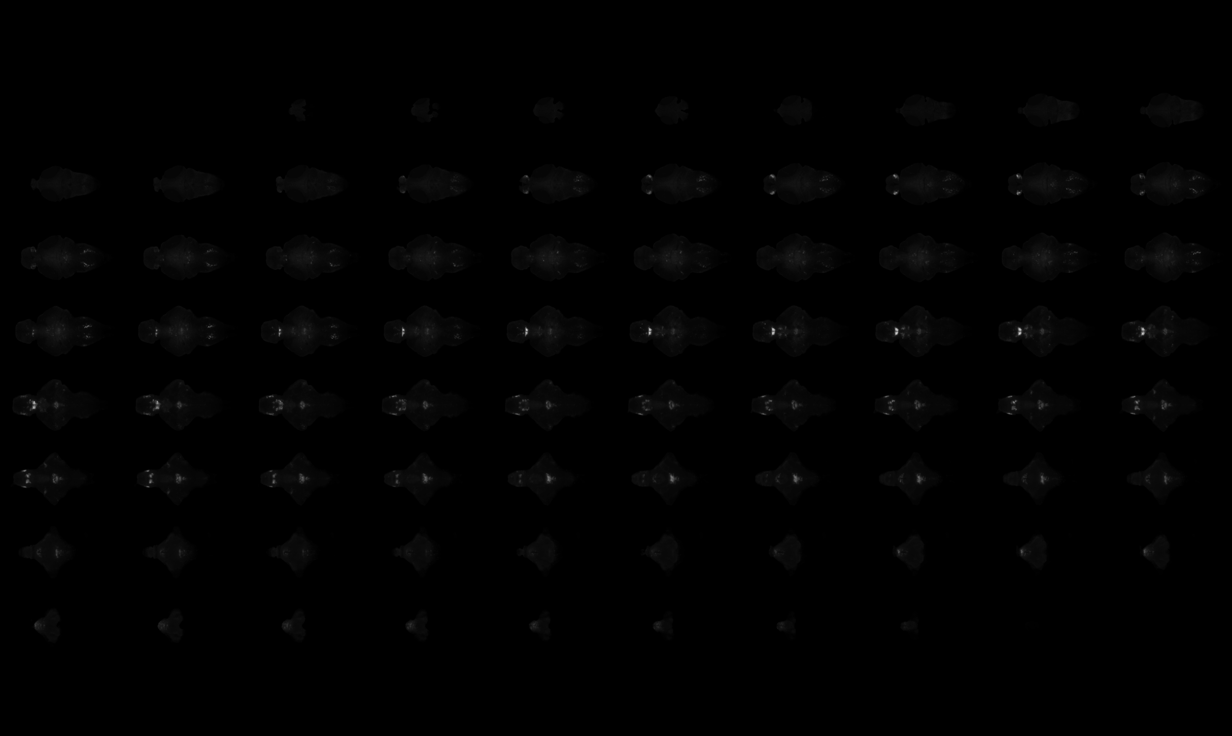
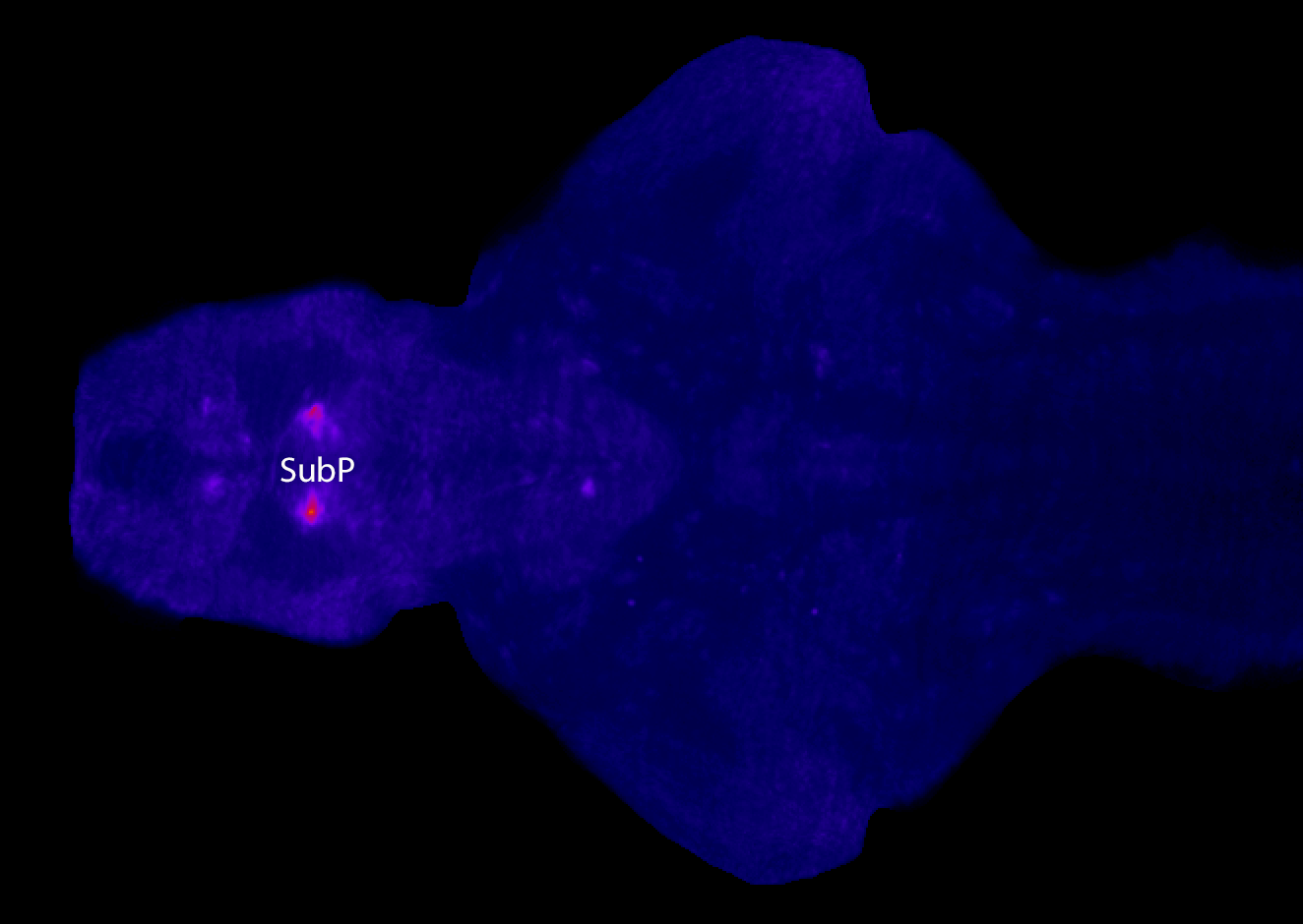
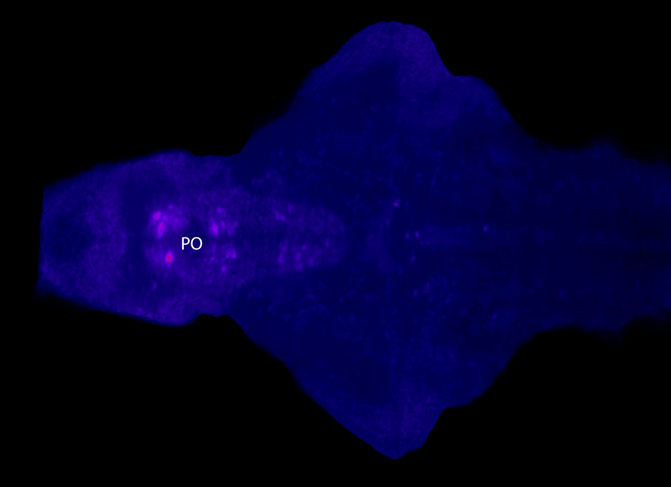
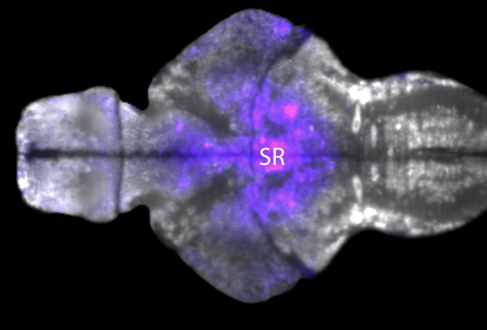
Montage of slices through the brain of a 6dpf zebrafish larvae labelled using fluorescent in-situ hybridisation using a probe against glyt2b.

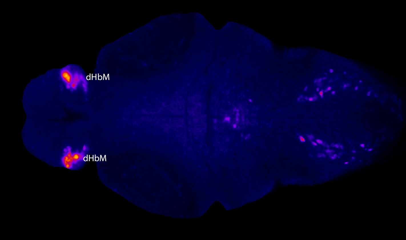
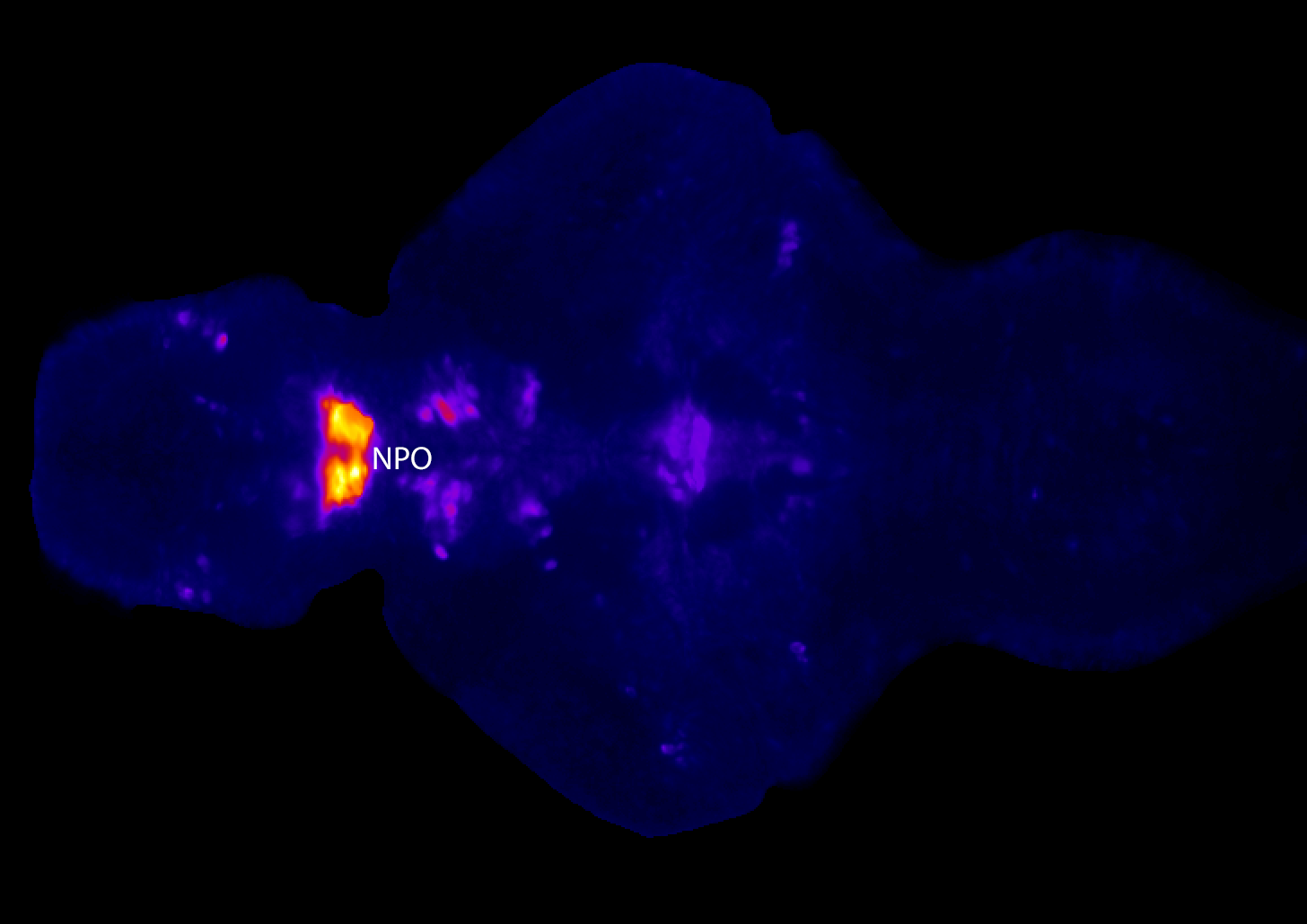
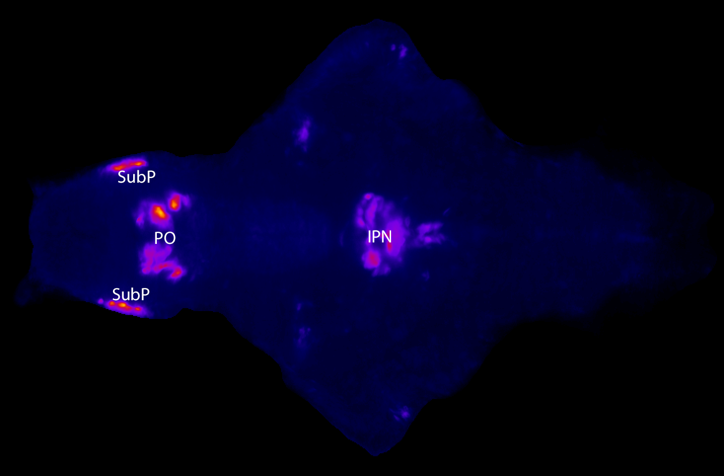
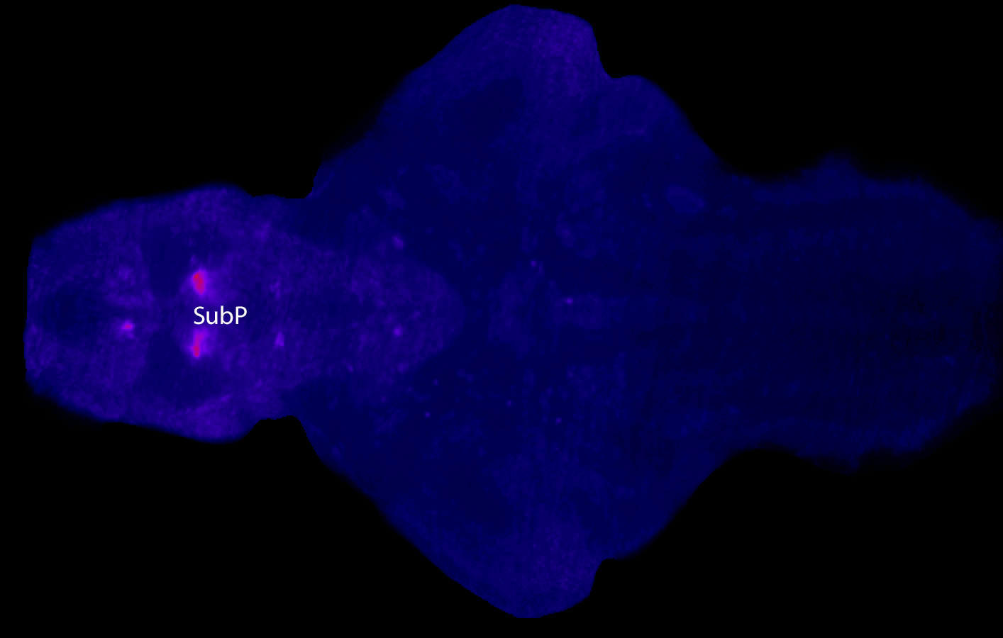
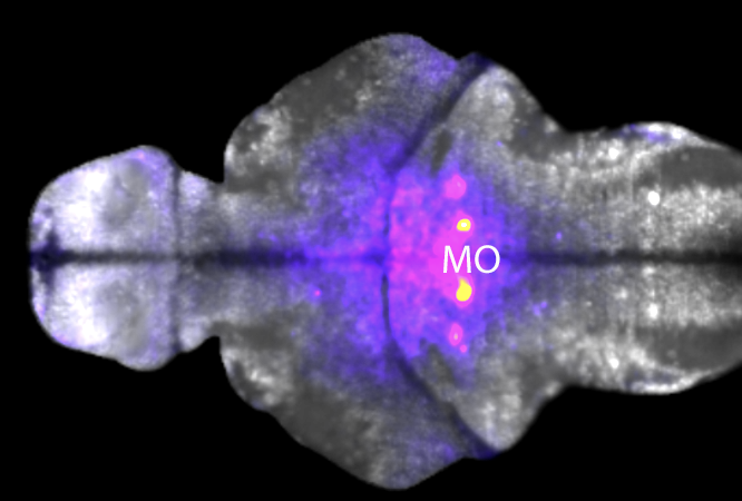
Z projection of a 6dpf larvae labelled using fluorescent in-situ hybridisation using a probe against glyt2b. This probe labels glycinergic neurons.

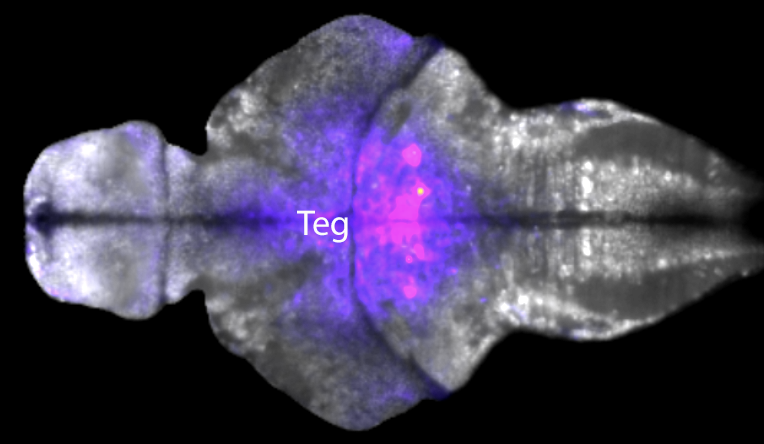
6dpf brain of a larval zebrafish labeled with a probe against glyt2b FISH. this stack was morphed to ZBB. This screenshot shows the glyt2b stack and a HuC/D antibody labelling uploaded to ZBB and viewed using their online browser. If you would like to look at this glyt2b data using ZBB please download the .png file using the link below and upload it to ZBB using the Lines> Custom>Load option.
Selected images
All imaging by Chintan Trivedi on a lightsheet microscope.
External links
Addgene probe link
Download the glyt2b .png file to view the data using Zebrafish Brain Browser
FISH Protocol
Key Publications
Higashijima, S.I., Mandel, G., and Fetcho, J.R. (2004)
Distribution of prospective glutamatergic, glycinergic, and GABAergic neurons in embryonic and larval zebrafish.
The Journal of comparative neurology. 480(1):1-18.
Moly, P.K., Ikenaga, T., Kamihagi, C., Tariqul Islam, A.F., and Hatta, K. (2014)
Identification of initially appearing glycine-immunoreactive neurons in the embryonic zebrafish brain. Developmental Neurobiology. 74(6):616-32.
Higashijima, S.I., Schaefer, M., and Fetcho, J.R. (2004)
Neurotransmitter properties of spinal interneurons in embryonic and larval zebrafish.
The Journal of comparative neurology. 480(1):19-37.


