
Tranverse section of a zebrafish eye at 3dpf

Zebrafish brain segmented
by Tom Hawkins & Kate Turner
Forebrain (purple), mid-brain (blue) and hindbrain (green).

Blood cover
by Gaia Gestri

Transverse section of a wild-type eye at 3dpf

Sarcomere
by Tom Hawkins
Sloth mutant and wildtype embryos arranged so to resemble a muscle sarcomere

Brn3a:GFP at 48hpf
by Kate Turner
Lateral view of a Brn3a:GFP embryo labelled wth anti-GFP and anti-tubulin antibodies. This transgenic line labels the sensory neuromasts, retina , optic tectum and the habenula.

2dpf-TOPdGFP
by Mate Varga

Optic chiasm
by Kate Turner
Ventral view of a Brn3a:GFP embryo labelled wth anti-GFP and anti-tubulin antibodies. This transgenic line labels the sensory neuromasts, the habenula, retina and optic nerves decussating at the optic chiasm on their way to the optic tectum.

Calretinin dorsal view
by Kate Turner
Calretinin positive nuclei (green) and their processes in the olfactory bulbs, epithalamus, optic tectum and cerebellum. Dorsal 3dpf embryo.

5HT
by Kate Turner
Ventral view of a 4dpf larvae labelled with anti-5HT antibody to label serotinergic neurons in the preoptic, posterior tuberculum, hypothalamus and raphe.

Calretinin neurons in the pretectum and optic tectum
by Kate Turner
Calretinin positive nuclei and their processes in the optic tectum and optic nerve (green). Frontal view of 3dpf dissected embryo.

Tg(Dlx4/6:GFP)
by Kate Turner
GFP expressed in many dopaminergic and GABAergic cells within the brain, shown here in the optic tectum, cerebellum, epithalamus and telencephalon of Tg(Dlx4/6:GFP) embryo. Dorsal view, 3dpf.

Foxd3:GFP
by Kate Turner
Pineal organ and the pineal processes that form the dorso-ventral diencephalic tract (green) descending ventrally in 60hpf Tg(FoxD3:GFP) embryo.

tela
by Kate Turner
Enhancer-trap transgenic expressing GFP in the tela choroidia, pineal organ and olfactory bulbs. Dorsal view, 60 hpf.

Axons and neuropil
by Jay Patel
Dorsal view of a 4dpf larva labelled with anti-tubulin(green) and anti-SV2(red) antibodies.

Habenula in Brn3a:GFP
by Kate Turner
Dorsal view of a 4dpf Tg(Brn3a:GFP)embryo.

Brn3a optic tract
by Kate Turner
Lateral view of a 4dpf Tg(Brn3a:GFP) embryo.

Neurons in the hindbrain
by Dave Lyons

Olfactory glomeruli in a Tg(DLx4/6:GFP) embryo
by Monica Folgueira

ET11
by Kate Turner
Dorsal view of a 4dpf Tg(ET11:GFP) embryo.
Kate Turner
The DDC
by Kate Turner
3D rendering of a 4dpf Tg(ET16:GFP) embryo.

Tg(vmat2:GFP)
by Kate Turner
Ventral view of a 4dpf Tg(vmat2:GFP) embryo. This enhancer-trap transgenic labels a subset of neurons in the olfactory bulbs and telencephalon, hypothalamus and posterior tuberculum and the raphe.

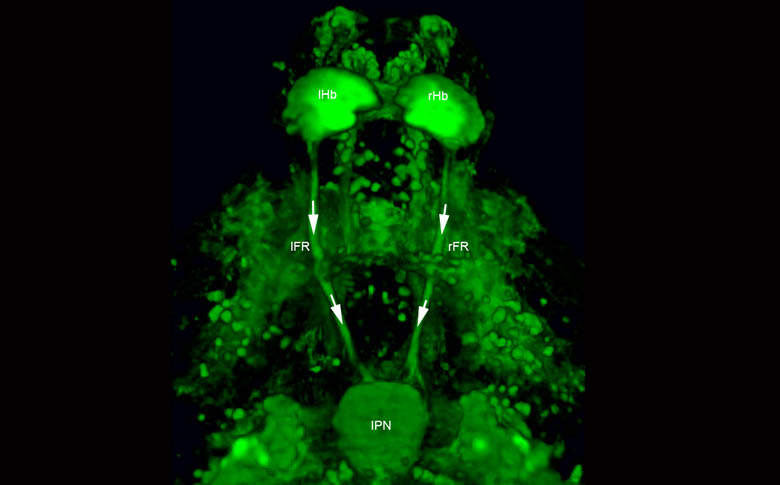
Tg(ET16:GFP) expresssion in the habenulae.
by Isaac Bianco

The left habenula of a Tg(ET16:GFP) embryo.
by Kate Turner
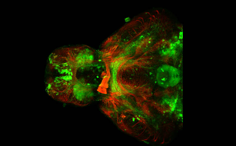
Ventral view of a 4dpf Tg(ET16:GFP) embryo.
by Kate Turner
This transgenic shows habenula terminals spiralling in the IPN.

Focal electroporation of an habenular neuron.
by Isaac Bianco

HuC:GFP in the retina
by Florencia Cavodeassi
Section through an eye of a 3 days old embryo immunostained to highlight a subset of neurons in the retina (green, Tg{HuC:GFP}) and their axonal projections (red, Zn5).

Coronal section through the head of a 2.5 days old embryo
by Florencia Cavodeassi
Coronal section through the head of a 2.5 days old embryo at the level of the eyes, stained with toloudine blue to reveal the highly organised architecture of the differentiating retina

Dorsal view of the anterior neural plate
by Florencia Cavodeassi
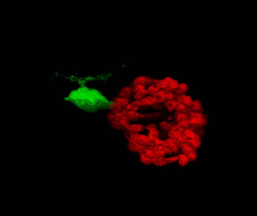
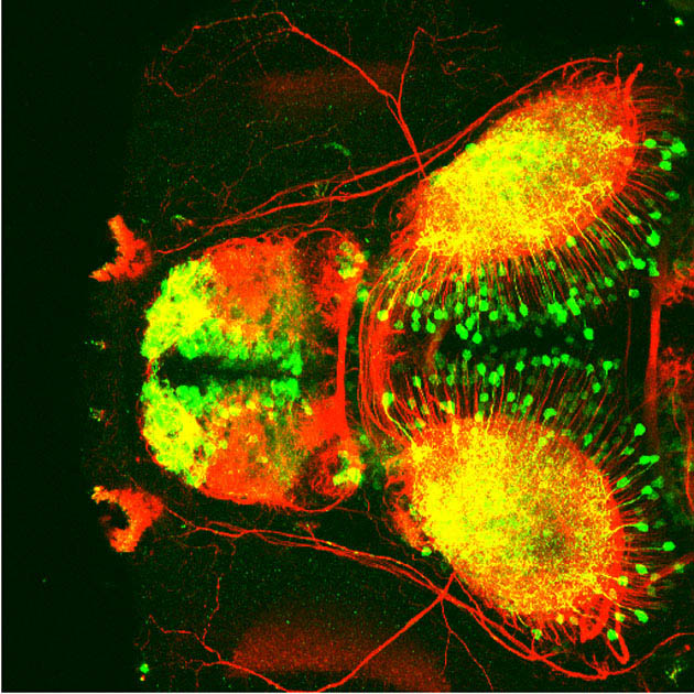
Dorsal view of the anterior neural plate, immunostained to reveal the eye field, the primordium of the eyes (red, Tg{rx3:GFP}) and the distribution of filamentus actin (green, phalloidin).
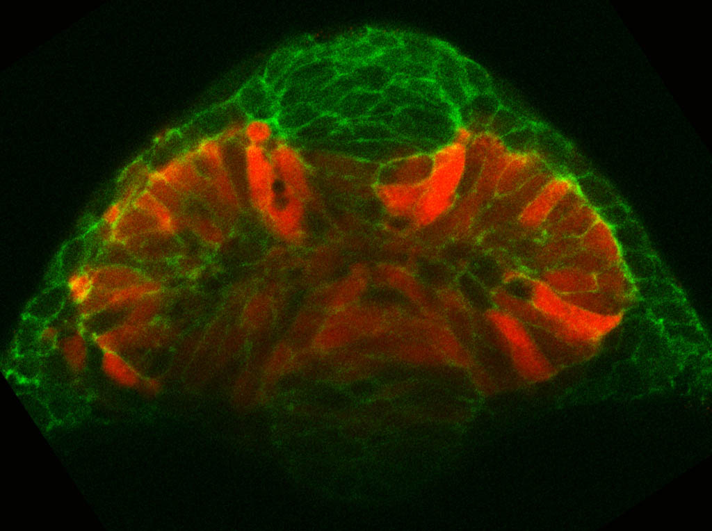
Frontal view of the anterior neural plate at the onset of evagination
by Florencia Cavodeassi
Frontal view of the anterior neural plate at the onset of evagination of the optic vesicles (red, Tg{rx3:GFP}; green, F-actin).

Innervation of the Interpeduncular nucleus.
by Isaac Bianco
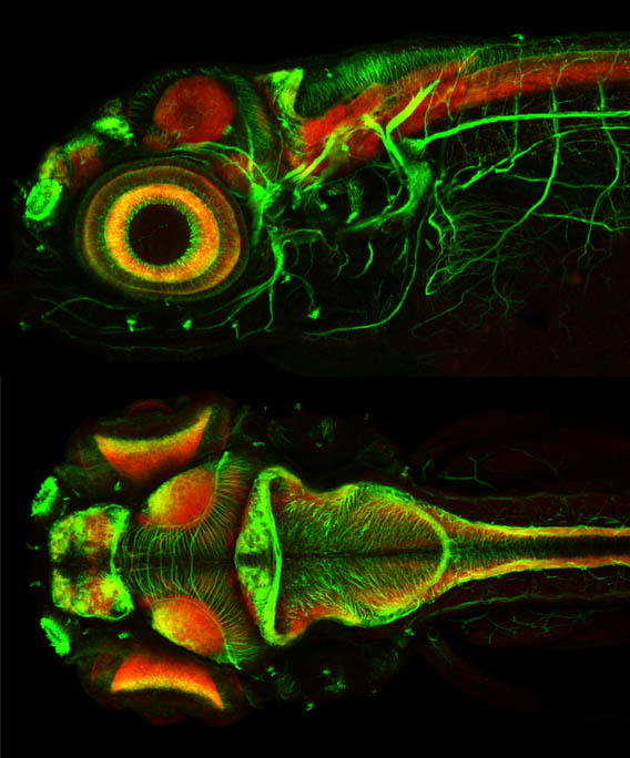


Sunrise in the eye: zebrafish retina
By Kara Cerveny
This photomicrograph shows the retina from the eye of a three-day-old zebrafish (Danio rerio).
The retina is viewed here from the front, as if the viewer is looking directly into the eye of the fish. This image is of the whole eye, created by reflecting half the image to represent the naturally occurring symmetry. It was created using a double in situ hybridisation - a staining technique that identifies spatial expression of gene products. Using this technique, different structures can be identified by staining for genes known to be found in specific tissues.
Retinal stem cells start to differentiate to become functional retinal neurons that are responsible for sending visual signals to the brain. Undifferentiated stem cells are shown in red, whereas the cells that have started to differentiate are shown in purple and are located at the periphery of the retina. The central yellow region is the lens.

GFP-expressing cranial motor neurons
by Kate Turner

Beautiful eye
by Kate Turner

Tg(tbr:GFP)
by Kate Turner
Lateral view of a Tg(tbr:GFP) and tubulin. GFP labelling cells in the telencephalon, midbrain and hindbrain, tubulin (red) labelling the axonal tracts in the brain.

Zebrafish
by Lukas Roth

Zebrafish BW
by Lukas Roth

Adult zebrafish eye
by Lukas Roth

diI and diO labelled retinal projections in a wingnut mutant
by Lukas Roth
Dorsal views of diI and diO labelled retinal projections in a wingnut mutant

the pineal organ and left-sided parapineal nucleus in a fish embryo
by Miguel Concha
Dorsal view of the pineal organ and left-sided parapineal nucleus in a fish embryo1

The pineal organ labelled in red and the left-sided parapineal nucleus in green
by Miguel Concha
The pineal organ labelled in red and the left-sided parapineal nucleus in green 2
by Miguel Concha

Pineal and parapineal
by Miguel Concha
The larger pineal organ in green (centre) with the left sided smaller parapineal nucleus. The bilateral asymetric (larger in the left) habenulae.
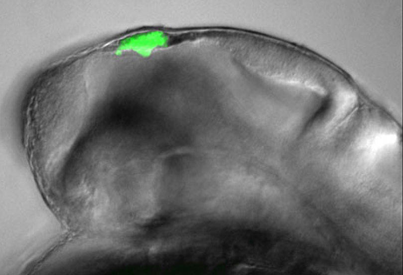
Floating head GFP in the epithalamus
by Miguel Concha

Dorsal view of an early zebrafish embryo
By Monica Folgueira
Confocal micrograph of a transgenic zebrafish embryo at 24 hours post-fertilization, showing expression of the fusion protein PARD3_GFP. This means that whenever the pard3 gene is expressed green fluorescent protein (GFP) will be produced, allowing the researcher to visualise the location of this gene in development.
In this image the embryo is viewed from the back (dorsally) sitting on top of a big yolk sac that provides nutrients for its development. GFP expression is seen in the surface of the neuroepithelium and thus highlights the ventricle of the brain, as well as in epithelial cells of the skin recovering the yolk.

Reticulospinal neuron
By Monica Folgueira
Confocal micrograph of a transverse section of the brain of a zebrafish embryo, at the level of the hindbrain. The brain section was stained using an antibody that marks a calcium binding protein (calretinin). The image shows a big reticulospinal neuron and its processes, surrounded by other smaller neurons. Reticulospinal neurons are specific neurons found in the brains of fish that are thought to be involved in coordinating swimming.

Tg(1.4dlx5a-6a:GFP) embryo stained against GFP and acetylated tubulin
by Monica Folgueira

Tg(1.4dlx5a-6a:GFP) embryo stained against GFP, SV2 and acetylated tubulin
by Monica Folgueira

GFP labelling areas of the forebrain and midbrain of a young zebrafish embryo
by Marina Mione

Dorsal view of a young zebrafish embryo; GFP labels cells in the forebrain and midbrain of the embryo
by Marina Mione
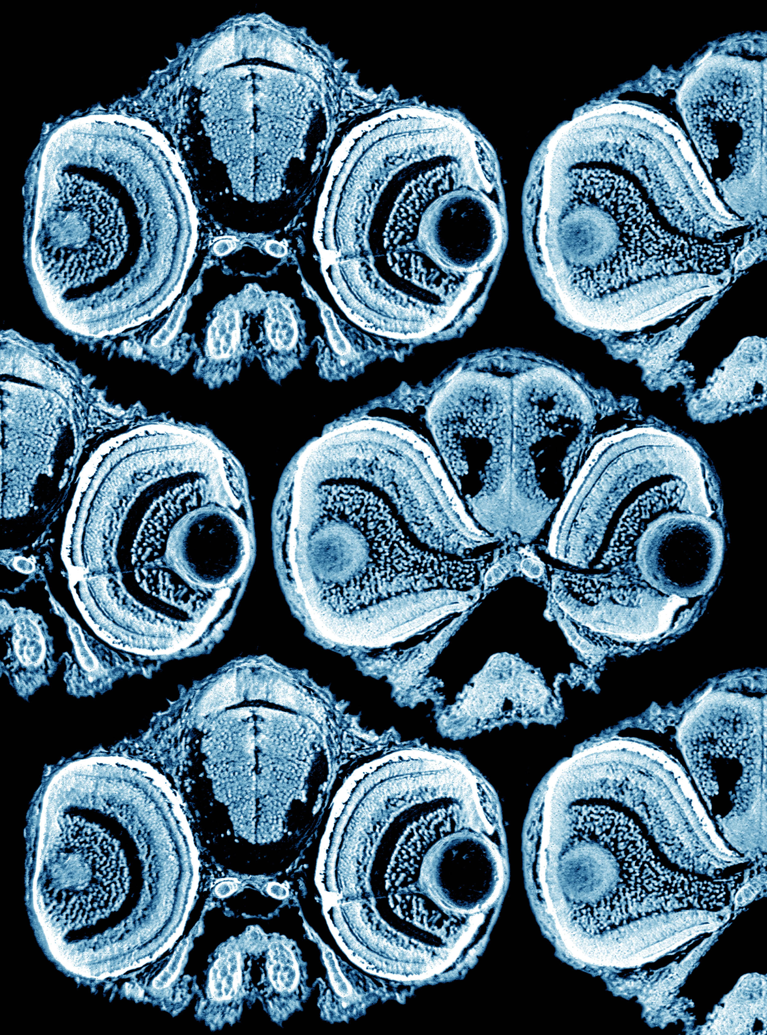
Transverse sections of zebrafish heads frontal view (blue) (cover)
by Masaya Take-Uchi

Transverse sections of zebrafish heads frontal view (orange) (cover)
by Masaya Take-Uchi

A few weird looking fish from an old print
by Anon

Pineal and parapineal in Tg(flh:GFP) embryo.
by Jenny Regan

Zebrafish eye development
By Steve Wilson and Rachel MacDonald
This differential interference contrast image
shows a section through the eye of a three-day old
zebrafish embryo. At this stage of development,
the nerve cells in the retina have begun to
separate into different layers. The outer layer of
cells are stained black with a marker for
photoreceptor cells, the neurons that respond to
light. (credit: R. MacDonald & S. Wilson)
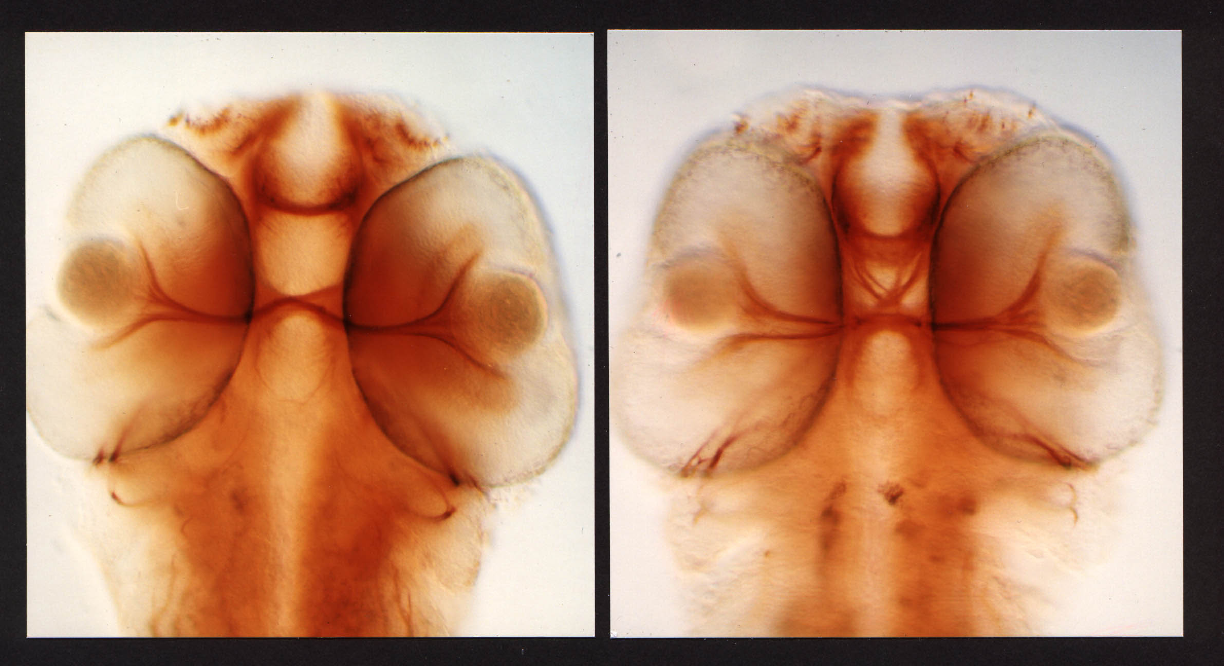
Zebrafish optic chiasm
By Rachel MacDonald and Steve Wilson
Light microscope image of a WT(left) and mutant(right) two-day-old zebrafish embryo which has been stained to show
all of the nerve cell processes in the eyes and
brain. This embryo has a mutation in the Noi gene
causing many of the processes (axons) to get lost
as they cross the midline from the eye to their
target brain region.
Zebrafish eye development
By Rachel MacDonald and Steve Wilson
This differential interference contrast image
shows a section through the eye of a three-day old
zebrafish embryo. At this stage of development,
the nerve cells in the retina have begun to
separate into different layers. The outer layer of
cells are stained black with a marker for
photoreceptor cells, the neurons that respond to
light.
Green labelled axons projecting to the optic tectum
by Silvia Castro

Fungal fissure
by Gaia Gestri

Scanning electron micrograph of a 4dpf larvae
by Leonardo Valdivia

Olfactory bulb(Tg(sox11a:GFP) and nerve(DiI)
by Kate Turner

Dorsal view of the forebrain of a zebrafish larva showing axons (blue) and neuropil (pink).
by Jen Cook

Frontal view of the forebrain of a zebrafish larva showing axons (blue) and neuropil (pink).
by Jen Cook

Lateral view of the forebrain of a zebrafish larva showing axons (blue) and neuropil (pink).
by Jen Cook

Pinhead Sculpture
by Keith Wilson
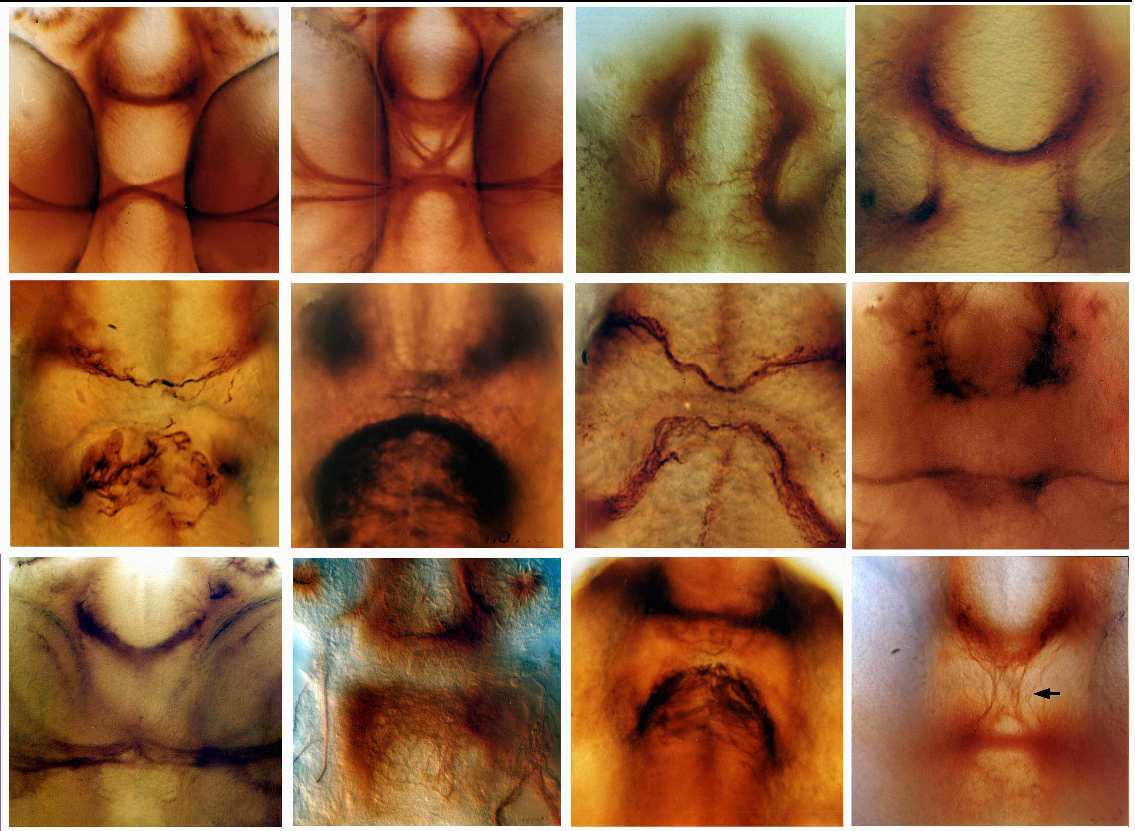
Various commissure mutants
by Steve Wilson

Epiphysis
By Steve Wilson
Lateral view of the head region in a zebrafish
embryo which shows neuronal structure in the
epiphysial region of the diencephalon, part of
the developing forebrain. This is the region where
where neurons first develop in the forebrain.
Here, axons labelled with diI can be seen
extending from epiphysial projection neurons.

Zebrafish growth cone
By Steve Wilson
This zebrafish embryo neuron is extending a long
process or axon through the brain, whose leading
edge is called the growth cone. The growth cone
explores the environment in order to navigate
towards the correct target nerve cells where it
will eventually form a synapse, a connection
through which nerve impulses pass from one neuron
to the next.

Axon extension
By Steve Wilson
Neurons extending numerous axons in the developing
forebrain of a zebrafish embryo.

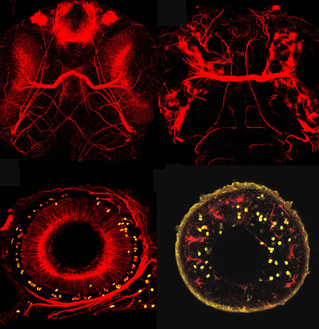
Commissural axons and dividing cells
by Zsolt Lele

Galanin mRNA
by Aisling O'Sullivan & Marcus Ghosh
The expression pattern of Galanin mRNA (by fluorescent in-situ hybridisation), at 6dpf, in 7 different WT fish. To make the samples comparable they have been registered using tERK.

Outer photoreceptor segments in an 8dpf zebrafish
by Gaia Gestri
Sagittal section across an 8 days post-fertilization zebrafish eye with the outer photoreceptor segments stained by anti-zpr1 (blue), UV cones expressing GFP (Tg(opn1sw1:GFP), magenta) and nuclei counterstained with DAPI (grey).

Outer photoreceptor segments in an 8dpf zebrafish
by Gaia Gestri
Sagittal section across an 8 days post-fertilization zebrafish eye with the outer photoreceptor segments stained by anti-zpr1 (blue), UV cones expressing GFP (Tg(opn1sw1:GFP), green) and nuclei counterstained with DAPI (magenta).

Pseudocoloured composite image of a zebrafish eye
by Gaia Gestri
Pseudocoloured composite image of a zebrafish eye expressing the UV cones transgenic line Tg(opn1sw1:GFP) and counterstained with anti-zpr1 (outer photoreceptor segments) and DAPI (cell nuclei).
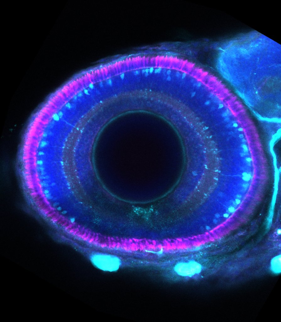
Zebrafish eye
by Gaia Gestri
Sagittal section across a zebrafish eye with the outer photoreceptor segments stained by anti-zpr1 (magenta), amacrine and retinal ganglion cells expressing GFP (Tg(islt1:GFP)), and counterstained with DAPI (blue).


