• Home
  • Groups
    • Alexandre Lab
    • Bianco Lab
    • Dreosti Lab
    • Frankel Lab
    • Tada Lab
    • RIhel Lab
    • MacDonald Lab
    • Mongera Lab
    • Payne Lab
    • Tuschl Lab
    • Wilson Lab
    • Wong Lab
    • Young Lab
    • Zebrafish Facility
  • ZebrafishBrain
  • UCL

Zebrafish UCL

  • Home
  • Groups
    • Alexandre Lab
    • Bianco Lab
    • Dreosti Lab
    • Frankel Lab
    • Tada Lab
    • RIhel Lab
    • MacDonald Lab
    • Mongera Lab
    • Payne Lab
    • Tuschl Lab
    • Wilson Lab
    • Wong Lab
    • Young Lab
    • Zebrafish Facility
  • ZebrafishBrain
  • UCL
1 2 3 4 5 6 7 8 9 10 11 12 13 14 15 16 17 18 19 20 21
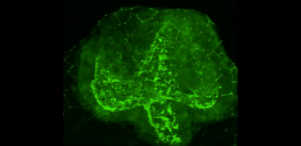
lef1 movie2
Lef1 movie1
Islet 1 Cranial Nerve Nuclei
An Introduction to the Zebrafish Brain
Habenula
Parapineal Organ
Brain
Shoaling Larvae 8x new
juvenile zebrafish
Zebrafish blood flow
Shoaling Juvenile Zebrafish
Optic vesicle evagination in a zebrafish embryo (Dorsal view)
Tissue macrophages RED interact with endotelial cells GREEN at site of vessel fusion
Optic vesicle evagination as shown by the apical marker Pard3 GFP
Timelapse Movie S1 from Gestri et al., 2018
Timelapse Movie S2 from Gestri et al., 2018
Timelapse Movie S3 from Gestri et al., 2018
Timelapse Movie S4 from Gestri et al., 2018
Timelapse Movie S5 from Gestri et al., 2018
Zrp1 labelled UV cones
Zpr1 labelled UV cones in the zebrafish retina by Gaia Gestri
Top
  • Alexandre Lab
  • Bianco Lab
  • Dreosti Lab
  • Frankel Lab
  • MacDonald Lab
  • Mongera Lab
  • Payne Lab
  • Rihel Lab
  • Tada lab
  • Tuschl Lab
  • Wilson Lab
  • Wong Lab
  • Young Lab
  • Zebrafish Facility


Contact us

1st Floor, Anatomy Building, University College London
Gower St, London, WC1E 6BT
tel 020 3549 5642

Copyright © 2017 UCL Disclaimer