Check out this paper describing the neuroanatomy of a cerebellum-like structure, the torus longitudinalis, in zebrafish.
The beautiful tectal pyramidal cells have spiny dendrites (c.f. Purkinje neurons).
Check out this paper describing the neuroanatomy of a cerebellum-like structure, the torus longitudinalis, in zebrafish.
The beautiful tectal pyramidal cells have spiny dendrites (c.f. Purkinje neurons).

Check out our paper in Nature Communications in collaboration with Filippo del Bene’s lab.
Zebrafish lack ipsilateral retinal projections yet they are likely to us binocular cue for judging distnace to their prey (Bianco et al 2011). Now we describe a population of ‘intertectal neurons’ that appear to transfer visual information across the midline and are required for zebrafish to strike at prey. Congratulations to everyone involved in the project and especially Christoph Gebhardt.

Ryan is an editor for the Frontiers in Cell and Developmental Biology Research Topic “"Molecular Mechanisms of Glia in Development and Disease" with Drs. Nathan Smith, Stefanie Robels and Tim Czopka.
About this Research Topic
Glial cells are critical for almost all aspects of nervous system development and function. This includes synapse formation and activity, signal conduction, blood flow regulation and the response to pathology, just to name a few. Yet, the full extent and mechanistic underpinnings of glia-glia and glia-neuronal interactions are not fully resolved. Notably, cellular and molecular mechanisms governing glial cell development are often also engaged in nervous system plasticity, trauma and neuronal degeneration, as well as repair. Leveraging developmental biology to gain insights into pathology and vice-versa with a systems biology approach is critical for our understanding of brain function and necessary for treating, or even preventing, neurological diseases.
This Research Topic aims to highlight the mechanisms involved in nervous system development and pathological conditions with a focus on glial cells. The content will include cellular and molecular mechanisms governing glial cells in establishing and maintaining contacts with neurons or other glia; comparative studies of glial cells between different model systems or different glia types; and characterization of common or unifying glial mechanisms in healthy or diseased tissues.
As such, this Research Topic is accepting manuscripts that cover the following themes:
• Glial specification and tissue morphogenesis during development.
• Evolutionary studies exploring conserved mechanisms of glial development.
• Establishment of functional connections between neurons and glia.
• Role of glia in regulation of CNS homeostasis and plasticity.
• Glial signaling mechanisms in CNS injury responses and repair.
• Role of glia in ageing and neurodegenerative diseases
• Novel methods to study glia and their interactions with neurons
• New models to study glial development and disease
• Potential therapeutic strategies focused on developmental pathways in neurodegenerative disease
Keywords: glial cell, neurodegeneration, development, pathology, neuron
Important Note: All contributions to this Research Topic must be within the scope of the section and journal to which they are submitted, as defined in their mission statements. Frontiers reserves the right to guide an out-of-scope manuscript to a more suitable section or journal at any stage of peer review.
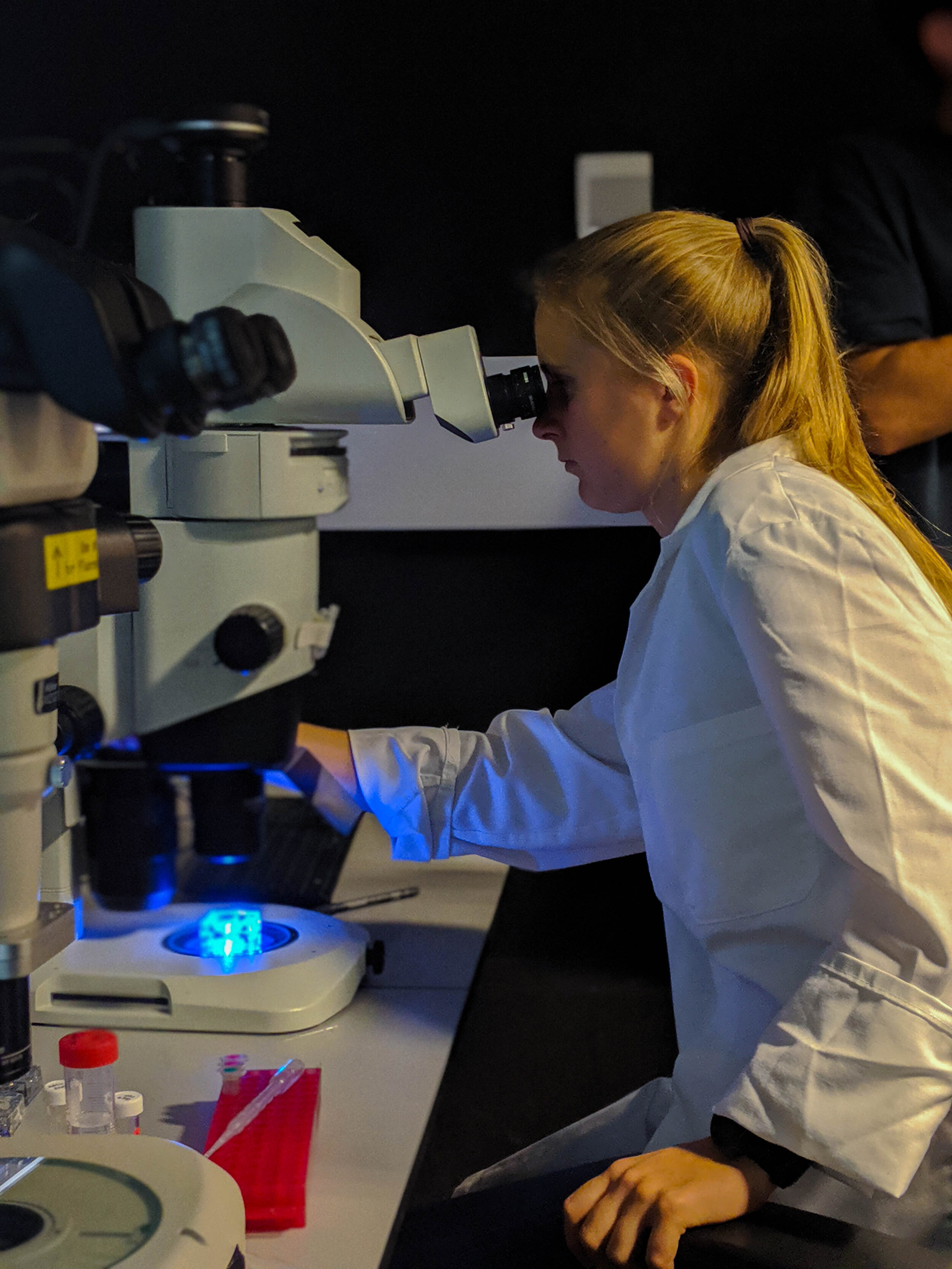
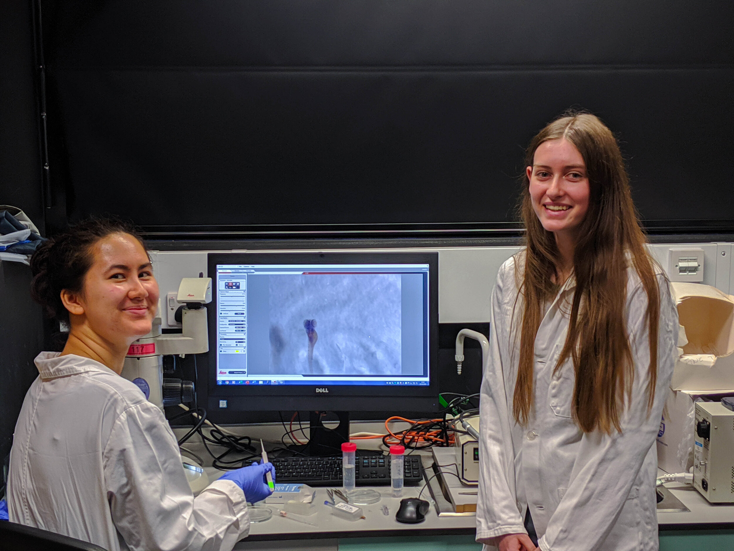
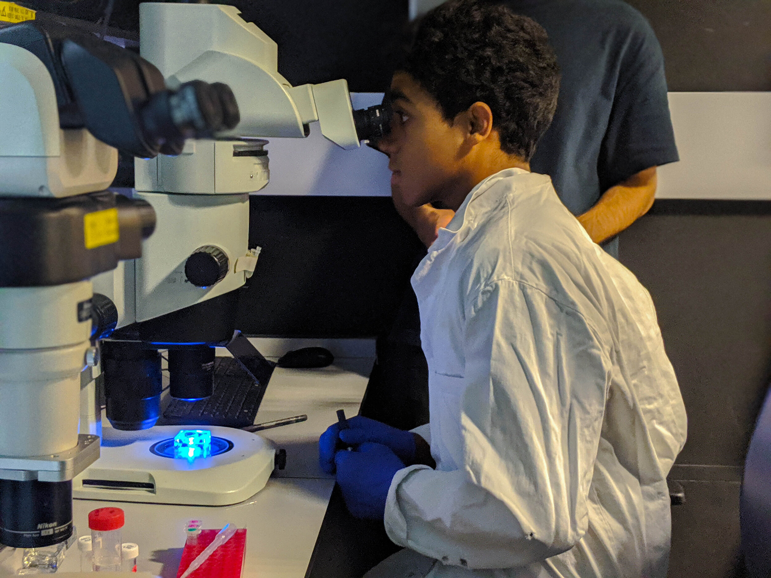
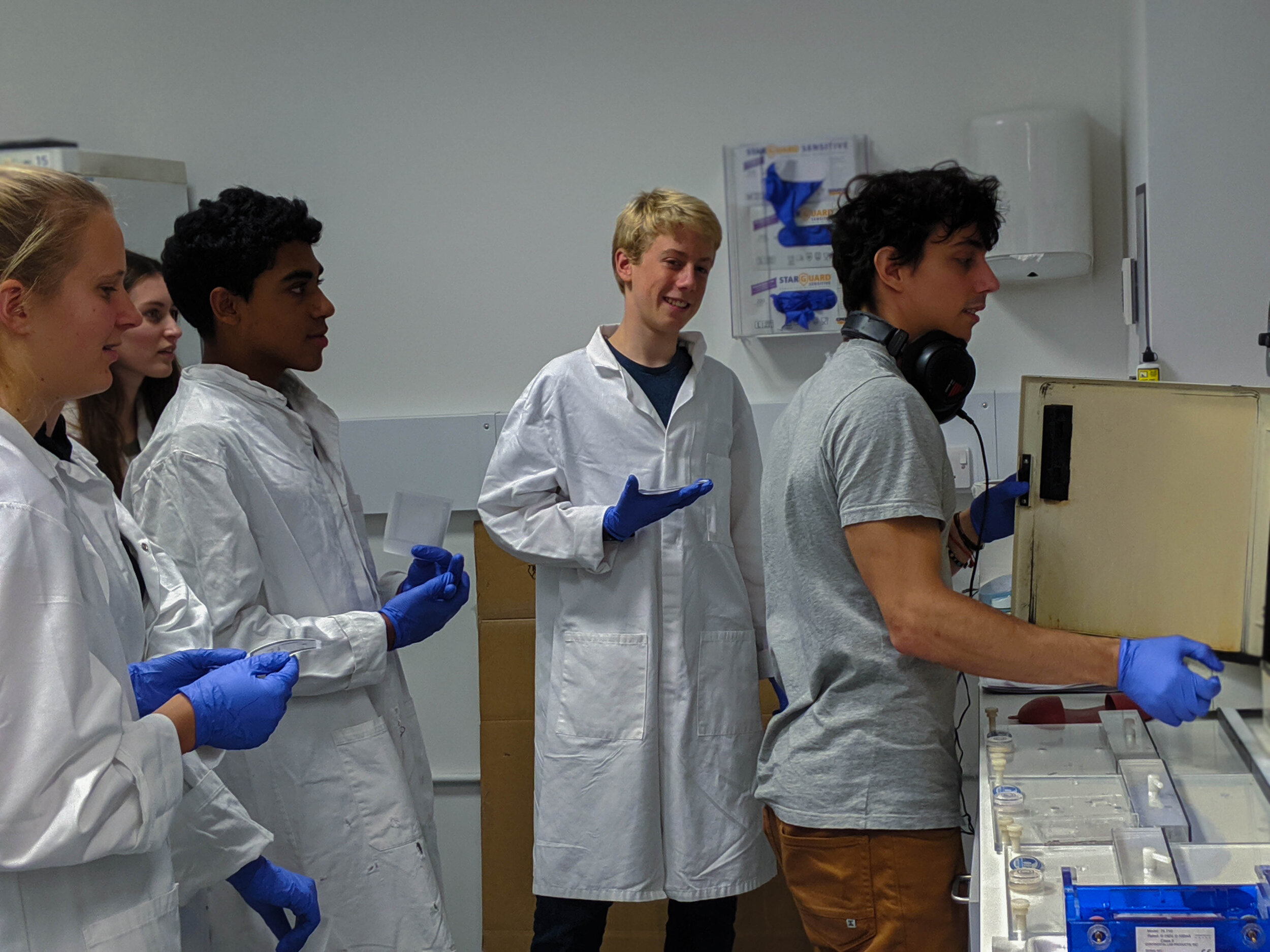
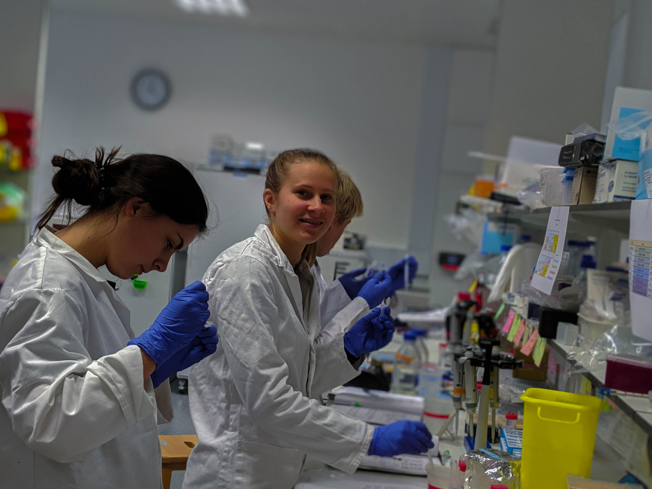
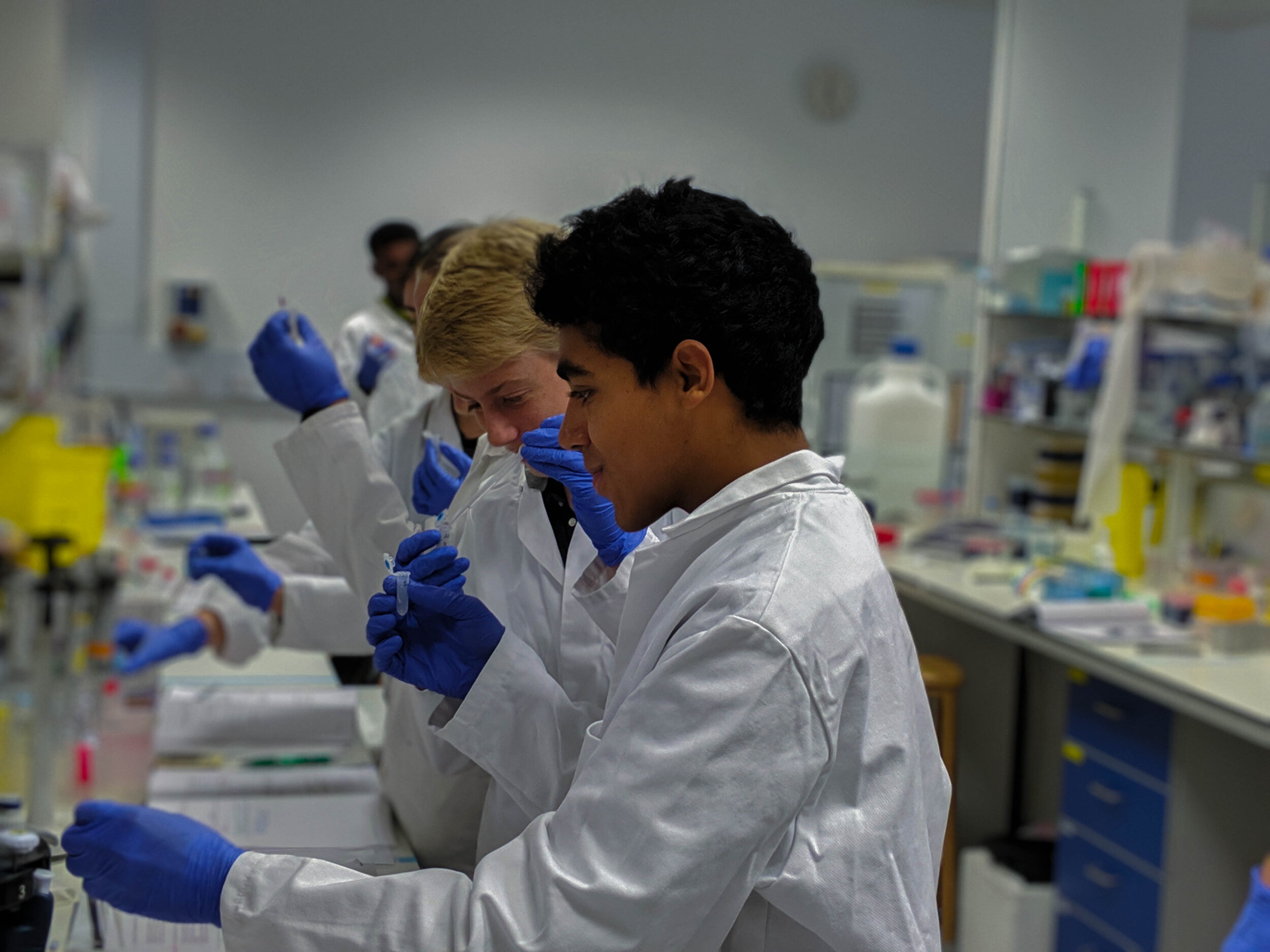
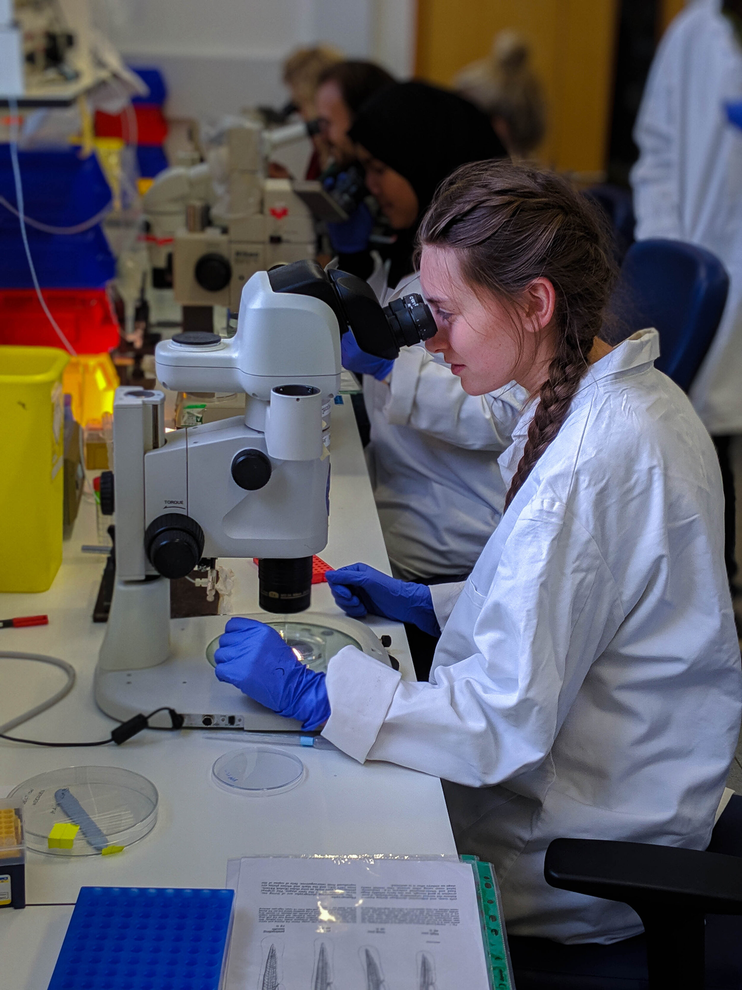
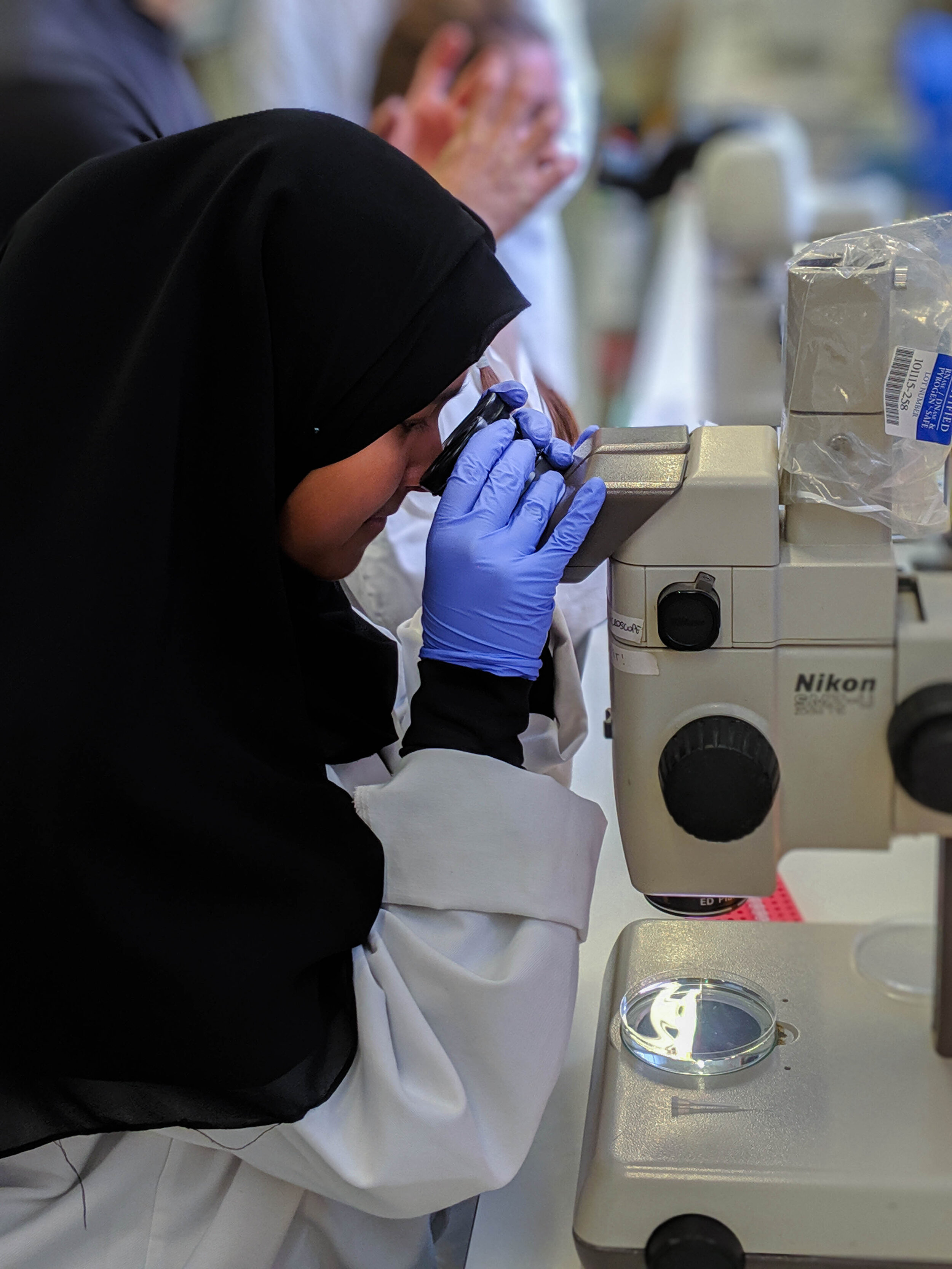
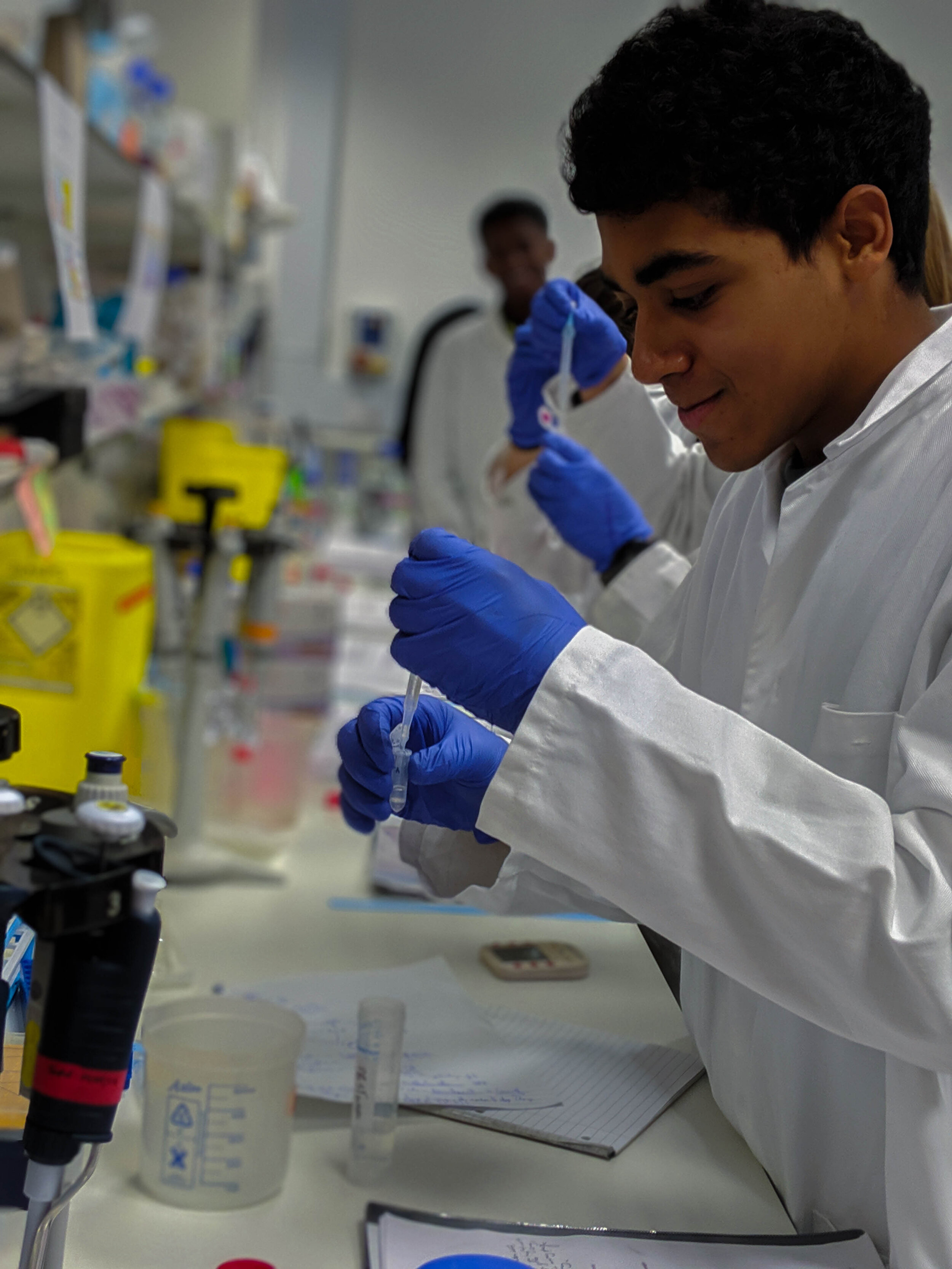
In 2019 the Zebrafish Lab had the pleasure to host 8 A-Level students for a period of one week, from 21st until 25th October. For many of our students, this was their first time in a research lab, so we made sure students received enough training and felt confident before they started with some hands-on experience. Students picked up the basic laboratory techniques very quickly. Once they were confident, we started delving deeper into the science behind our research and started the actual lab experiments. We were really happy to see how quickly the students picked up difficult concepts and how confident they grew in the lab during the course of the week. The students had the opportunity to learn and get hands-on research experience using many different techniques, such as traditional Molecular Biology methods, RNA in situ hybridisation and confocal microscopy. They also had the chance to see our cutting-edge behaviour assays and talk with our experts about their research. Despite being familiar with some of the theoretical concepts we were teaching, the majority of the students said they lacked in-depth knowledge on the subjects discussed. Nevertheless, on the final day, their presentations demonstrated their ability to grasp even complicated concepts and to place their work in the greater context of our research questions.
In 2019, we also launched a new mentorship programme, where we paired each student with a researcher in a mentoring relationship for the course of the week. This offered the opportunity for the students to engage one-to-one with a scientist, to discuss their careers goals and gather advise on academic paths. This initiative was extraordinarily well received, by students and mentors alike, and is one part of the programme we will continue in the following years.
All in all, our 2019 cohort was enthusiastic and a pleasure to interact with and supervise, and they really seemed to enjoy themselves, too, and worked extremely well together. We wish them all the luck in their future endeavours!

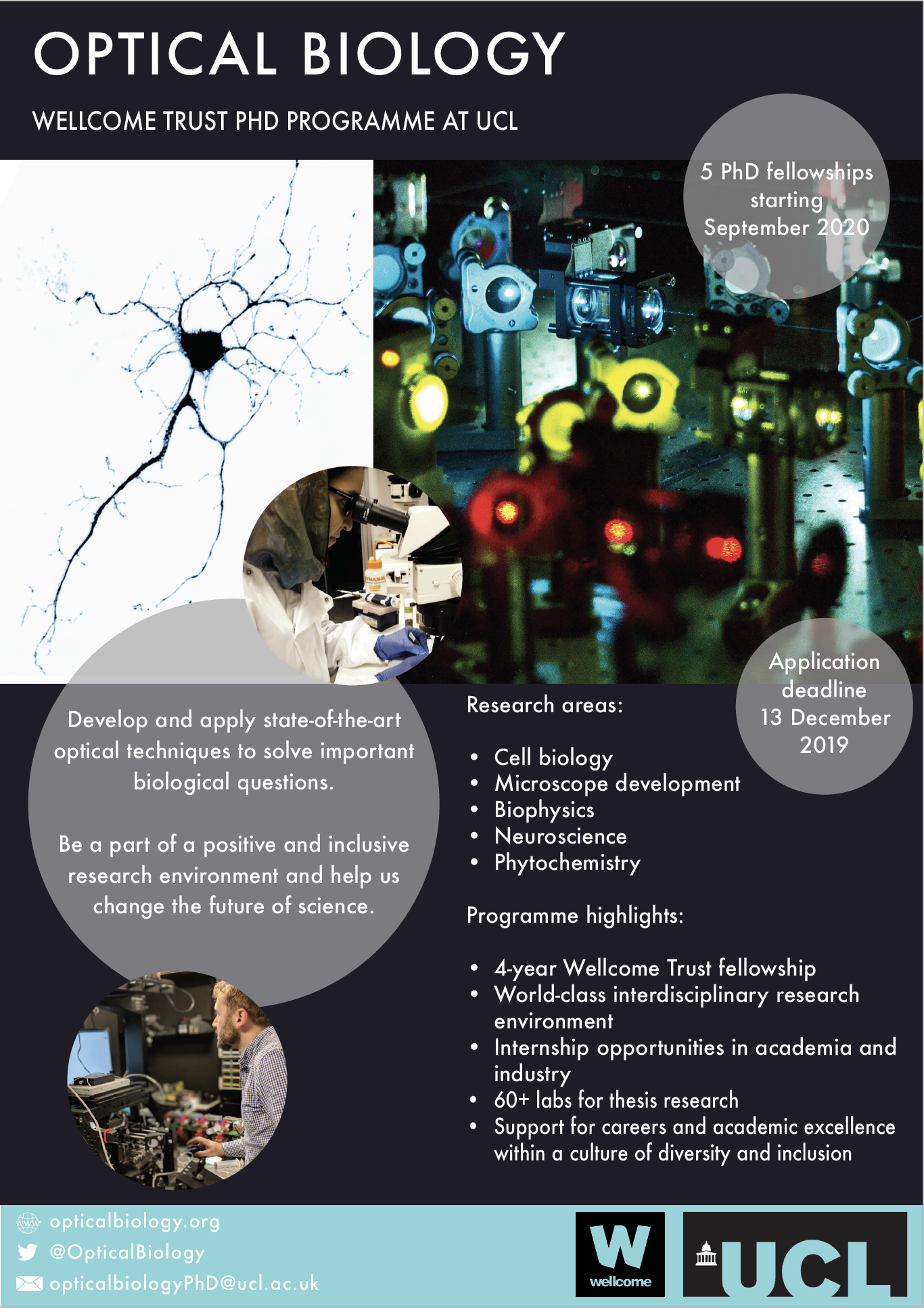
UCL is now accepting applications for the Wellcome funded Optical Biology PhD programme.
The first cohort of 5 students will start in September 2020. Please go to the
Optical Biology website at https://opticalbiology.org/ for information on how to apply.
Adverts will appear on findaphd.com and in Nature from 30th October.
The deadline for applications is 13 December 2019.
Follow the program on twitter for more updates.
To study with us as part of this exciting new program see https://opticalbiology.org/'
Our manuscript describing pretectal neurons that control hunting behaviour is now published in eLIFE! Congratulations to Paride! https://elifesciences.org/articles/48114
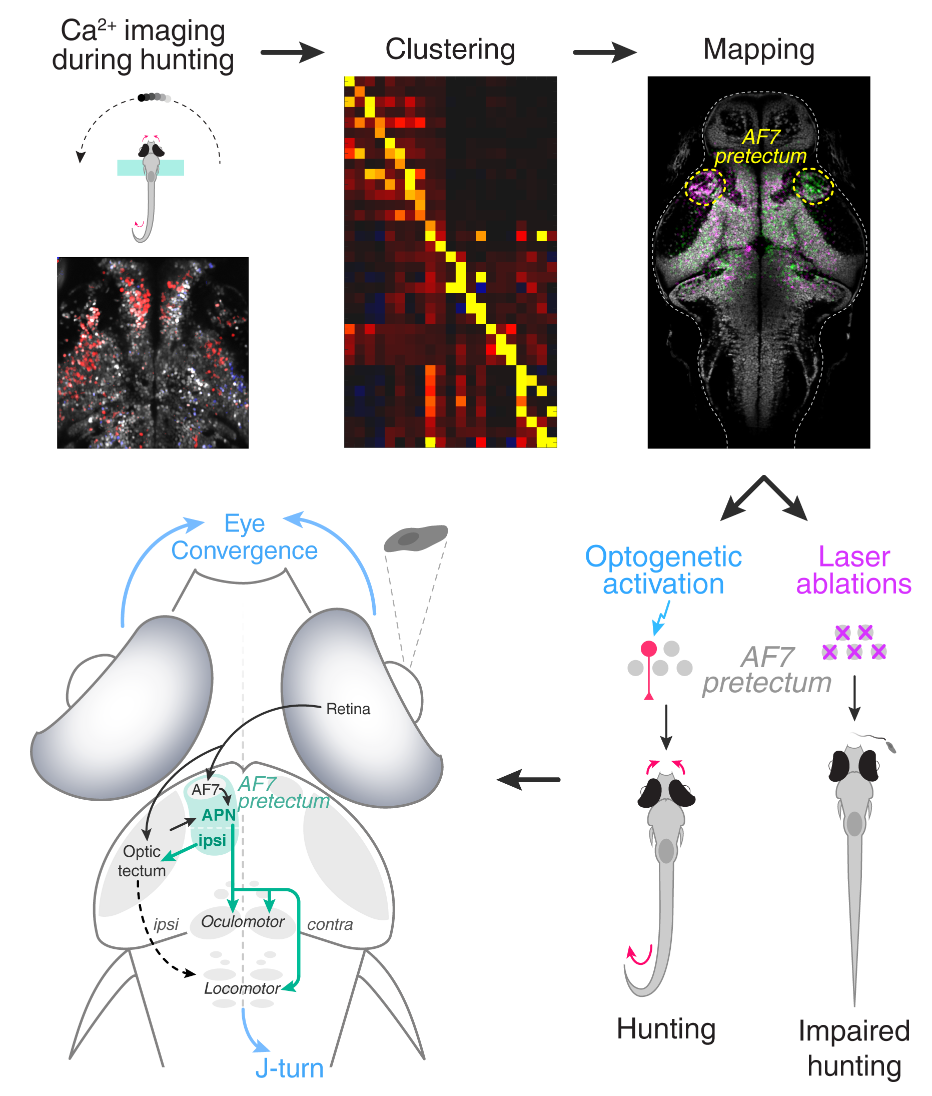
Hunting is an innate behaviour that relies on predators executing a precise set of actions to identify, approach, target, and strike prey. In vertebrates, the identity of the brain circuits that trigger hunting is still unclear. These networks are thought to link sensory perception (seeing prey) with a specialised action (starting an attack).
Larval zebrafish are a good model in which to study these circuits, because they have a tiny, transparent brain where neurons can be observed in real time. In addition, it is expected that the networks that control hunting in this species will be preserved across other vertebrates.
To discover these networks, Antinucci et al. genetically engineered zebrafish larvae so that their brain cells would ‘glow’ when they became active. This revealed which individual brain cells would turn on when zebrafish started to hunt. These neurons were in a part of the brain called the pretectum, which receives visual information about prey from the eye.
Next, Antinucci et al. harnessed a technique called optogenetics to artificially turn on these brain cells in the pretectum, which caused the fish to start hunting even when prey was absent. In fact, stimulating just one pretectal cell was enough to trigger the behaviour. Conversely, killing pretectal brain cells using precise laser surgery hindered hunting in zebrafish exposed to prey.
These experiments suggest that pretectal brain cells act like a command centre that controls hunting. They likely play a decision-making role, determining when animals do and do not respond to events in their surroundings. Similar neurons likely control other types of behaviour. Understanding how these circuits work at the cellular level in zebrafish may help scientist study them in other organisms, such as humans.

The Bianco and Dreosti labs are really excited to be part of the ZENITH (ZEbrafish Neuroscience Interdisciplinary Training Hub) programme.
ZENITH is now recruiting 15 graduate students to start their PhD’s in 2020. They will undertake interdisciplinary systems neuroscience projects investigating sensorimotor processing using larval zebrafish.
The Bianco and Dreosti labs at UCL will be recruiting three students.
Visit the official website to learn all about the scheme, the available projects and download the application pack. Applications close on Jan 5th 2020.
Read the press release here
“The intensity of brain activity during the day, notwithstanding how long we've been awake, appears to increase our need for sleep, according to a new UCL study in zebrafish.
The research, published in Neuron, found a gene that responds to brain activity in order to coordinate the need for sleep. It helps shed new light on how sleep is regulated in the brain.”
Full article below.
Sabine Reichert, Oriol Pavón Arocas, Jason Rihel
The Neuropeptide Galanin Is Required for Homeostatic Rebound Sleep following Increased Neuronal Activity
Neuron (2019) DOI:https://doi.org/10.1016/j.neuron.2019.08.010
Congratulations Sabine and Jason!

Are you a talented and enthusiastic post-doc looking to work on nervous system development and function in zebrafish? If so, we are currently recruiting to our exciting and collaborative research team.
Our group uses zebrafish to investigate brain and eye development, from early morphogenesis to circuit formation and behaviour (http://zebrafishucl.org/wilson-lab#wilson-lab/about-us ). One focus for our research is upon left-right asymmetry; the developmental and genetic mechanisms by which asymmetries are established and encoded in circuits and how such asymmetries impact behaviour.
The zebrafish community and UCL more widely offers a vibrant and very collaborative research environment, state-of-the art facilities and close proximity to many other research institutes of excellence.
Here is the link to the job description and application process:
We are also recruiting one or maybe two technical positions to our group.
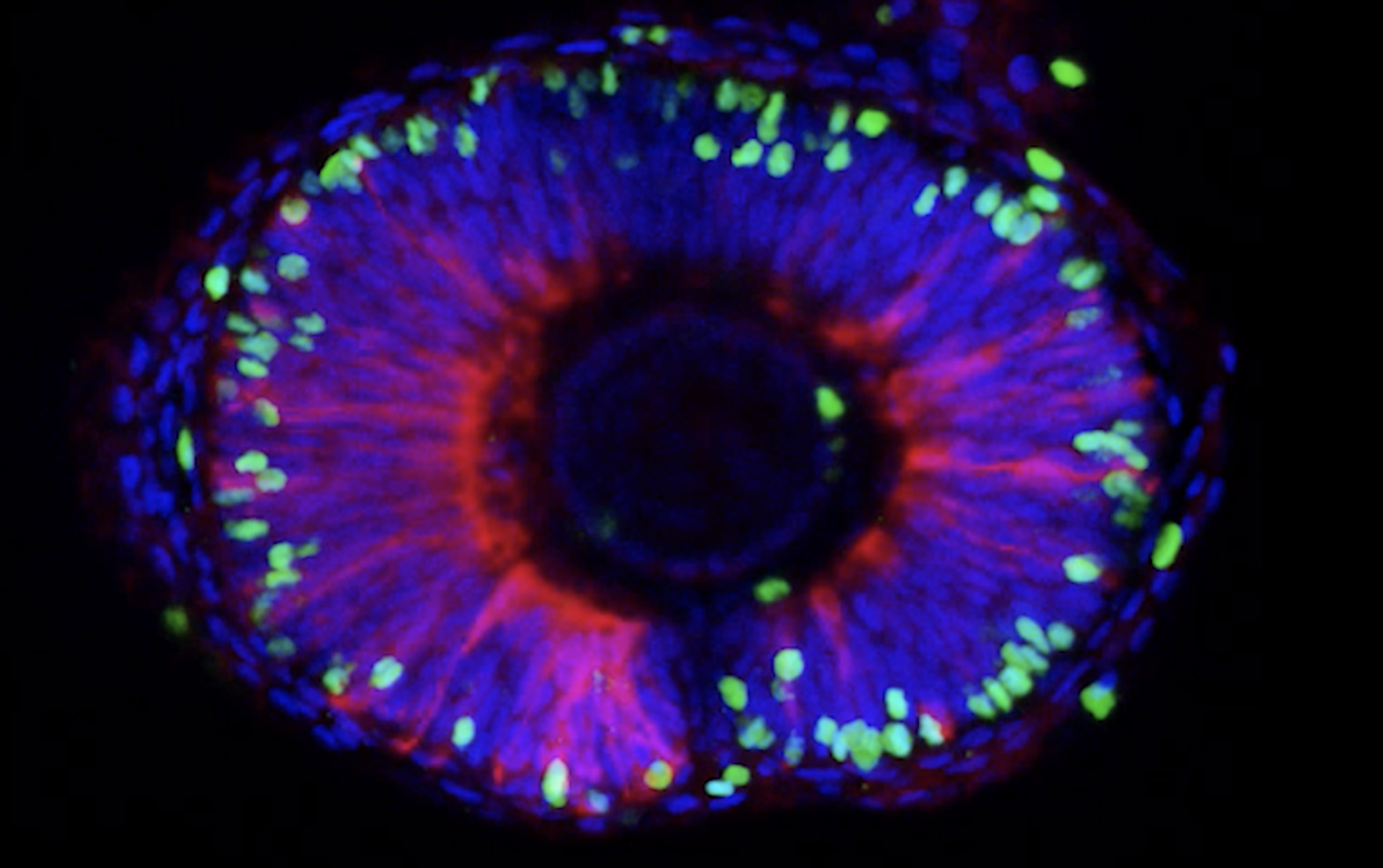
The Wilson labs latest paper on one of our favourite structures the parapineal has just been published in eLife. In this work , Ingrid shows that normal function of Sox1a is required for the parapineal to impart left-sided molecular character and efferent connectivity to habenula neurons, despite not being required for the specification and migration of the parapineal itself.
Read this open access paper in eLife:
Sox1a mediates the ability of the parapineal to impart habenular left-right asymmetry. eLife 2019;8:e47376 DOI: 10.7554/eLife.47376
or check out the publication summary.
Authors :Ingrid Lekk, Véronique Duboc, Ana Faro, Stephanos Nicolaou, Patrick Blader, Stephen W Wilson

The parapineal emerges from the pineal during zebrafish embryogenesis, as shown by confocal images of a transgenic line with GFP in the pineal complex (top pictures). The pineal complex lies at the midline of the epithalamus, flanked by left (green on the scheme) and right (magenta on the scheme) habenulae. The habenulae are innervated by the parapineal and themselves send neuronal projections to the midbrain interpeduncular nucleus (IPN). The side of the parapineal, on the left in wild-type (wt), determines which habenula acquires a left-type identity projecting to the dorsal part of the IPN (dIPN).
Congratulations to Ingrid!
Dr Ryan McDonald, a BBSRC David Phillips Fellow is joining the Institute of Ophthalmology, UCL from the University of Sheffield where he is JG Graves Medical Research Fellow in department of Infection, Immunity and Cardiovascular Disease.
Ryan’s lab will study the role of cell adhesion molecules in glial morphogenesis using the zebrafish retina as a model system. Click here to see the UCL press release and read more about the MacDonald Lab research.

What brain circuits control predatory behaviour?
Our paper identifying a pretectal command system that induces hunting behaviour is now on @biorxivpreprint. Congratulations to @anti_paride and Mónica Folgueira.

Congratulations to Renato on getting his doctorate.
Read the Elife Digest of Rodrigo’s paper, “Compensatory growth renders Tcf7l1a dispensable for eye formation despite its requirement in eye field specification” here:
https://elifesciences.org/digests/40093/eye-size-is-pre-programmed-in-zebrafish
or watch this great video for a simple explanation of Rod’s findings.
The review we wrote with Dr Kristen Severi about the cellular organisation of supraspinal circuits is already available online!
Share link providing free access until May 10, 2019: https://authors.elsevier.com/c/1Ym208nMlta80e
https://www.sciencedirect.com/science/article/pii/S2468867319300136

Congratulations to Asaph who was recently awarded a prestigious BBSRC Discovery Fellowship. It is all the more impressive because this is the first Discovery Fellowship ever awarded to any UCL researcher!
The award will allow Asaph to combine cellular resolution whole-brain functional imaging in larval zebrafish and modern machine learning techniques, to study how the brain transforms visual input to behaviour.

Follow the Wilson lab on twitter at @stevewilsonlab!

We’re all extremely proud of Pedro who by all accounts nailed his viva today [err, actually yesterday - ed]! First Bianco lab PhD… Celebrations much deserved!


The Bianco lab are pleased to welcome Kristie, Nicole and Adam (from left to right), who will be doing their research projects with us this year.