Zebrafish UCL A-Level Work Experience 2022

Zebrafish UCL A-Level Work Experience 2022

We are happy to announce after 2 years hiatus the return of the UCL Zebrafish Academy A-level work experience placement.

Wilson/Rihel/Bianco/Tada Lab Work Experience 2022

24th-28th October 10:00-16:00

***Applications for the 2022 work experience are now being accepted. ***

Please note you must be in Sixth Form at a UK school at the time of application and at the time of the work experience placement.

***SUBMISSION DEADLINE EXTENSION TILL 29th May 2022***

Please note applications will not be accepted beyond this date.

For more information about the programme click the button below.  

The Tuschl Lab secure further funding

The Tuschl Lab secure further funding

The Tuschl lab secure further funding from:

GOSH BRC Junior Faculty Consumable Support (£10,000): to develop antisense oligonucleotide treatment for manganese neurotoxicity.

&

UCL Therapeutic Innovation Networks (TINs) Pilot Data Scheme – Small Molecules (£10,000): to develop novel chelators for the treatment of manganese neurotoxicity in collaboration with the group of Prof John Spencer and Dr George Lostakis at the University of Sussex.

Elena Dragomir awarded a  Sir Henry Wellcome Fellowship

Elena Dragomir awarded a Sir Henry Wellcome Fellowship

Elena Dragomir has been awarded a four-year Sir Henry Wellcome Fellowship to work on the role of the asymmetric habenular nuclei in state-dependent modulation of behavioural preference. Elena’s research will span between Steve’s and Isaac Bianco’s groups with input on modelling approaches from James Fitzgerald at Janelia Research Campus.

Postdoc position in  Wong Lab

Postdoc position in Wong Lab

The Wong Lab is looking to recruit one Postdoctoral Research Fellow. The position is funded by the Wellcome Trust and Royal Society for 3 years. Please apply if you are interested in morphogenesis, cell migration, signalling and doing some great imaging and fun science. Contact Mie (mie.wong@ucl.ac.uk) if you are interested to find out more.

More details can be found here:

https://thenode.biologists.com/jobs/postdoctoral-position-wong-lab-ucl-departmental-of-cell-and-developmental-biology-uk/

https://www.wongmorphogenesislab.com/

UCL vacancy reference number: 1881595

The application deadline is 2 Feb 2022.

The zebrafish lateral line primordium.

The Wong Lab joins the UCL Fish Floor

The Wong Lab joins the UCL Fish Floor

Mie Wong has joined the Fish Floor and the Department of Cell and Developmental Biology, UCL, as a Wellcome Trust and Royal Society Sir Henry Dale Fellow. She was previously at the University of Zurich, Switzerland.
Mie's lab studies how moving cells shape the developing embryo. The lab is especially interested in understanding the interplay between extrinsic signals and collective cell behaviours in establishing and maintaining the directionality of cell migration during development. The primary model used in the lab is a system of sensory organs called the lateral line. The Wong Lab employs interdisciplinary approaches including genetic, chemical and optical manipulations combined with advanced quantitative imaging, mass spectrometry and mathematical modelling.

Click here (https://www.wongmorphogenesislab.com/) to find out more about the lab.

Gold award in the Laboratory Efficiency Assessment Framework (LEAF)

Gold award in the Laboratory Efficiency Assessment Framework (LEAF)

First Floor Zebrafish Labs were awarded GOLD in the Laboratory Efficiency Assessment Framework (LEAF) for our efforts to improve sustainability and efficiency in all areas of laboratory practice. Check out the Zebrafish Green Lab section of our homepage to see what practices we have put in place and get some ideas on how to make your lab greener. Keep up the good work!

Screenshot 2021-09-03 at 11.37.07.png

The Tuschl Lab are hiring a postdoc!

The Tuschl Lab are hiring a postdoc!

Karin_header banner2.jpg

Interested in whole brain activity and neurochemistry mapping, in vivo calcium imaging of neuronal activity, single-cell transcriptomics, behavioural analysis and genome editing? Then join us at ZEBRAFISH UCL to study the role of manganese in health and disease.

Details here: tinyurl.com/3yweh4cn

UCL vacancy reference number: 1876901

UCL Neuroscience Symposium 2021

UCL Neuroscience Symposium 2021

We all enjoyed another excellent UCL Neuroscience Symposium, which featured fantastic talks by our own Charlie and Asaph.

az.png
cd.png

Headloop PCR protocol

Headloop PCR protocol

Advances in CRISPR/Cas9 technologies allow researchers to rapidly test the function of genes by knocking them out in injected embryos, reducing the number of animals and time needed to generate stable mutant lines (Kroll et al. 2021 eLife 10: e59683; https://doi.org/10.7554/eLife.59683). However, determining the efficacy of CRISPR/Cas9 ribonucleoproteins (RNPs) often requires access to expensive and specialised equipment (e.g. deep sequencing or high resolution melting analysis) or restricts the potential target sequences (e.g. restriction fragment length polymorphism analysis).

Recently, we adapted and piloted a method of suppression PCR called headloop PCR (Rand et al. 2005 Nucleic Acids Research 33: e127, https://doi.org/10.1093/nar/gni120; Kroll et al. 2021 eLife: 10:e59683). This proved to be a sensitive and robust way to identify non-functioning and low efficiency guide RNP complexes. We’ve observed that headloop PCR is sensitive to a wide range of mutations, including small compound indels, and those at low copy number. Its performance correlates well with deep sequencing analysis. It is simple, cheap and easily adoptable, requiring only basic molecular biology reagents and equipment, and is flexible, with no constraints on guide RNA sequences.

Importantly, we believe it could also be used to support the aims of the 3Rs project – to help reduce the use of animals in retesting negative results and in the generation of stable mutant lines. Having identified genes of interest for further study, researchers can use the same validated RNPs to create inherited stable mutations. Unfortunately, most methods only indicate that mutations have been made in the respective targets; determining the nature of those mutations can be difficult from ambiguous sequencing data. Because headloop PCR is effectively allele-specific, the reaction products can be sequenced to determine the nature of the inherited lesions directly.

For further details about headloop PCR and its use in testing CRISPR/Cas9 guides please see our article at eLife: Kroll et al. 2021 . More detailed information and resources for headloop PCR are available at our Protocols page.

headloop-pcr.gif

Postdoc positions in Bianco lab - updated

Postdoc positions in Bianco lab - updated

The Bianco lab is looking to recruit one/two postdoctoral research fellows.

Positions are Wellcome funded for 4 years. Experience with custom design of optical systems or electrophysiology would be especially great, but most important is enthusiasm to understand the circuit basis of behaviour.

NOTE: We have extended the deadline for applications so you can now apply up until 30 NOVEMBER. Contact Isaac if you’re interested to find out more.

BIANCO_1_ESR7 image.png

Isaac receives Wellcome SRF

wellcome-logo-black.png

We’re delighted to announce that the Wellcome Trust are generously funding the Bianco lab for the next five years.

We will be using fast calcium imaging, electrophysiology and holographic optogenetic stimulation to figure out how zebrafish command and pattern their hunting sequences.

We’ll be advertising postdoc positions soon, so please contact Isaac if you’re interested.

Fast online motion correction

As anyone who does 2-photon imaging in behaving animals will know, motion artifacts are a pain the neck and can be difficult to correct. We were lucky enough to contribute to a fantastic project in Angus Silver’s lab, which led to technology enabling real-time motion correction in 3D. As Joanna showed, it works beautifully in larval zebrafish!

3dmc.png

Congratulations Dr. Lau!

Congratulations Dr. Lau!

Congratulations to Joanna Lau who was just awarded her PhD. Thanks also to her examiners, Jon Clarke and Andrew Murray.

main.jpeg
image1.jpeg
image4.jpeg
image3.jpeg

Bianco lab publishes an optogenetic toolbox for zebrafish

In collaboration with Claire Wyart’s lab and Herwig Baier’s lab we have generated nine transgenic lines expressing optogenetic actuators and calibrated their efficacy using behavioural assays and in vivo electrophysiology. This should be a great resource for the zebrafish neuroscience community. Congratulations to first authors Paride Antinucci and Adna Dumitresco!

opsinpaper.png

Understanding rare diseases affecting eye formation

Understanding rare diseases affecting eye formation

Our new MRC programme grant “Resolving the basis of phenotypically variable hereditary abnormalities of eye formation” has started!

Although abnormalities of eye formation are a major cause of blindness, the genetic bases of such phenotypes are poorly understood. The list of genes implicated in Microphthalmia (small eyes), Anophthalmia (lack of eyes) and Coloboma (a failure in optic fissure fusion) is growing but only accounts for a minority of cases. This proposal will identify new genes involved in eye formation and will elucidate why MAC phenotypes show variable penetrance. We have established (thanks to a previous MRC grant) novel zebrafish mutants lines that show MAC phenotypes with very low penetrance (eg. only in one eye and/or only in some of the mutants). We suggested that due to the robustness of eye formation, MAC phenotypes may only be consistently present when embryos carry more than one genetic mutation (eg. FIG 1).

Fig 1: Combining tcf7l1a and gdf6a mutations gives rise to variably penetrant microphthalmia (B,C) or anophthalmia (D). tcf7l1a mutants alone show no overt phenotype and gdf6a mutants have misshapen eyes.

For this grant, we will use Crispr/Cas9 F0 gene editing to screen for genetic interactions between hundreds of genes in different mutants sensitised to showing coloboma, anophthalmia and microphthalmia phenotypes. Once the world returns to more normal times and we can get experiments under way, we will create a database that will make all the results of the screen and some of the resources available, so stay tuned to our web pages!! We also aim to understand the molecular and tissue level functions of selected genes that are critical in the expression of MAC phenotypes. We will clarify the conserved roles for Yap and Mab21l1/2 proteins in eye morphogenesis using genome wide screens in worms to complement novel transgenic approaches in zebrafish. Among our collaborators on this project will be Rod Young, Florencia Cavodeassi, Leo Valdivia and Tatjana Sauka-Spengler.

This research will both inform and be informed by large-scale human genomics studies, will deliver a comprehensive analysis of genetic interactions that build the eyes and aims to provide a wealth of information to inform diagnosis and understanding of debilitating abnormalities of eye formation.

Gaia Gestri and Steve Wilson

31 March 2020

Anatomy of the Torus Longitudinalis

Anatomy of the Torus Longitudinalis

Check out this paper describing the neuroanatomy of a cerebellum-like structure, the torus longitudinalis, in zebrafish.

The beautiful tectal pyramidal cells have spiny dendrites (c.f. Purkinje neurons).

pyr.png

Bianco lab publishes collaborative paper on intertectal neurons

Check out our paper in Nature Communications in collaboration with Filippo del Bene’s lab.

Zebrafish lack ipsilateral retinal projections yet they are likely to us binocular cue for judging distnace to their prey (Bianco et al 2011).  Now we describe a population of ‘intertectal neurons’ that appear to transfer visual information across the midline and are required for zebrafish to strike at prey.  Congratulations to everyone involved in the project and especially Christoph Gebhardt.

geb.png

Contribute to "Molecular Mechanisms of Glia in Development and Disease"

Contribute to "Molecular Mechanisms of Glia in Development and Disease"

Ryan is an editor for the Frontiers in Cell and Developmental Biology Research Topic “"Molecular Mechanisms of Glia in Development and Disease" with Drs. Nathan Smith, Stefanie Robels and Tim Czopka.

About this Research Topic

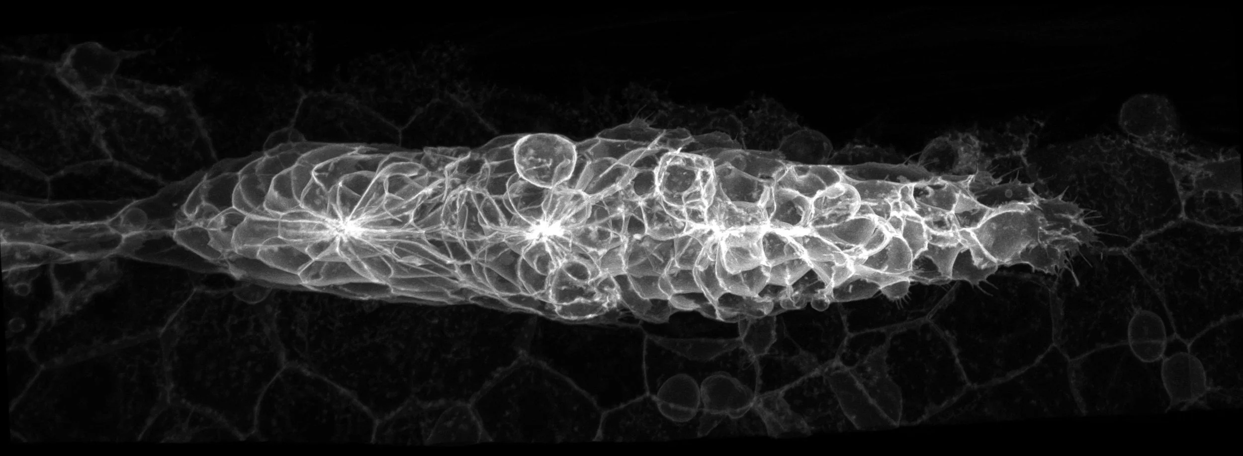
Glial cells are critical for almost all aspects of nervous system development and function. This includes synapse formation and activity, signal conduction, blood flow regulation and the response to pathology, just to name a few. Yet, the full extent and mechanistic underpinnings of glia-glia and glia-neuronal interactions are not fully resolved. Notably, cellular and molecular mechanisms governing glial cell development are often also engaged in nervous system plasticity, trauma and neuronal degeneration, as well as repair. Leveraging developmental biology to gain insights into pathology and vice-versa with a systems biology approach is critical for our understanding of brain function and necessary for treating, or even preventing, neurological diseases.

This Research Topic aims to highlight the mechanisms involved in nervous system development and pathological conditions with a focus on glial cells. The content will include cellular and molecular mechanisms governing glial cells in establishing and maintaining contacts with neurons or other glia; comparative studies of glial cells between different model systems or different glia types; and characterization of common or unifying glial mechanisms in healthy or diseased tissues.

As such, this Research Topic is accepting manuscripts that cover the following themes:

• Glial specification and tissue morphogenesis during development.

• Evolutionary studies exploring conserved mechanisms of glial development.

• Establishment of functional connections between neurons and glia.

• Role of glia in regulation of CNS homeostasis and plasticity.

• Glial signaling mechanisms in CNS injury responses and repair.

• Role of glia in ageing and neurodegenerative diseases

• Novel methods to study glia and their interactions with neurons

• New models to study glial development and disease

• Potential therapeutic strategies focused on developmental pathways in neurodegenerative disease

Keywords: glial cell, neurodegeneration, development, pathology, neuron

Important Note: All contributions to this Research Topic must be within the scope of the section and journal to which they are submitted, as defined in their mission statements. Frontiers reserves the right to guide an out-of-scope manuscript to a more suitable section or journal at any stage of peer review.