A visual guide to some of the main tracts and commissures in the larval zebrafish brain. Tracts and commissures have been grouped into forebrain, midbrain and hindbrain white matter.
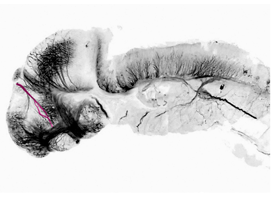
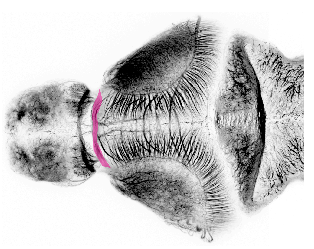
forebrain white matter
Featured


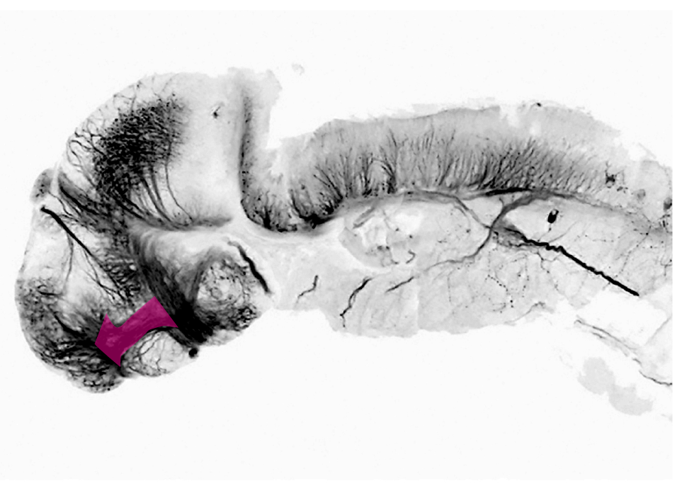


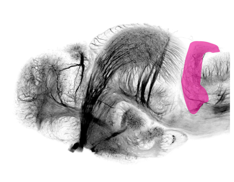
midbrain white matter
Featured


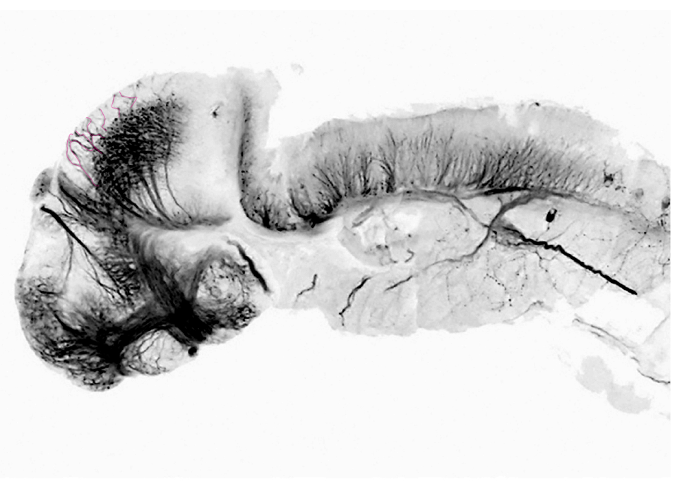
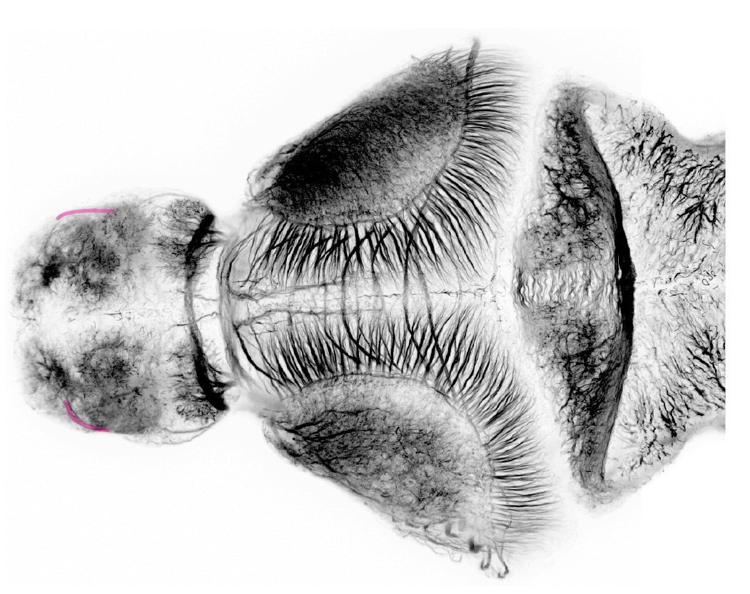
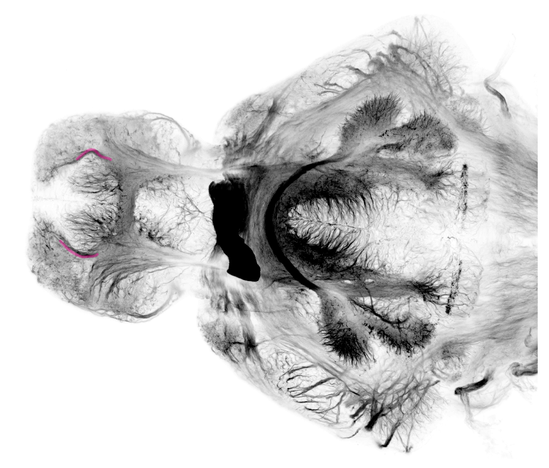
Hindbrain white matter
cerebellar commisure
Featured