Burrill & Easter proposed identity of AF-10 in the adult:
Optic Tectum/Tectum Opticum
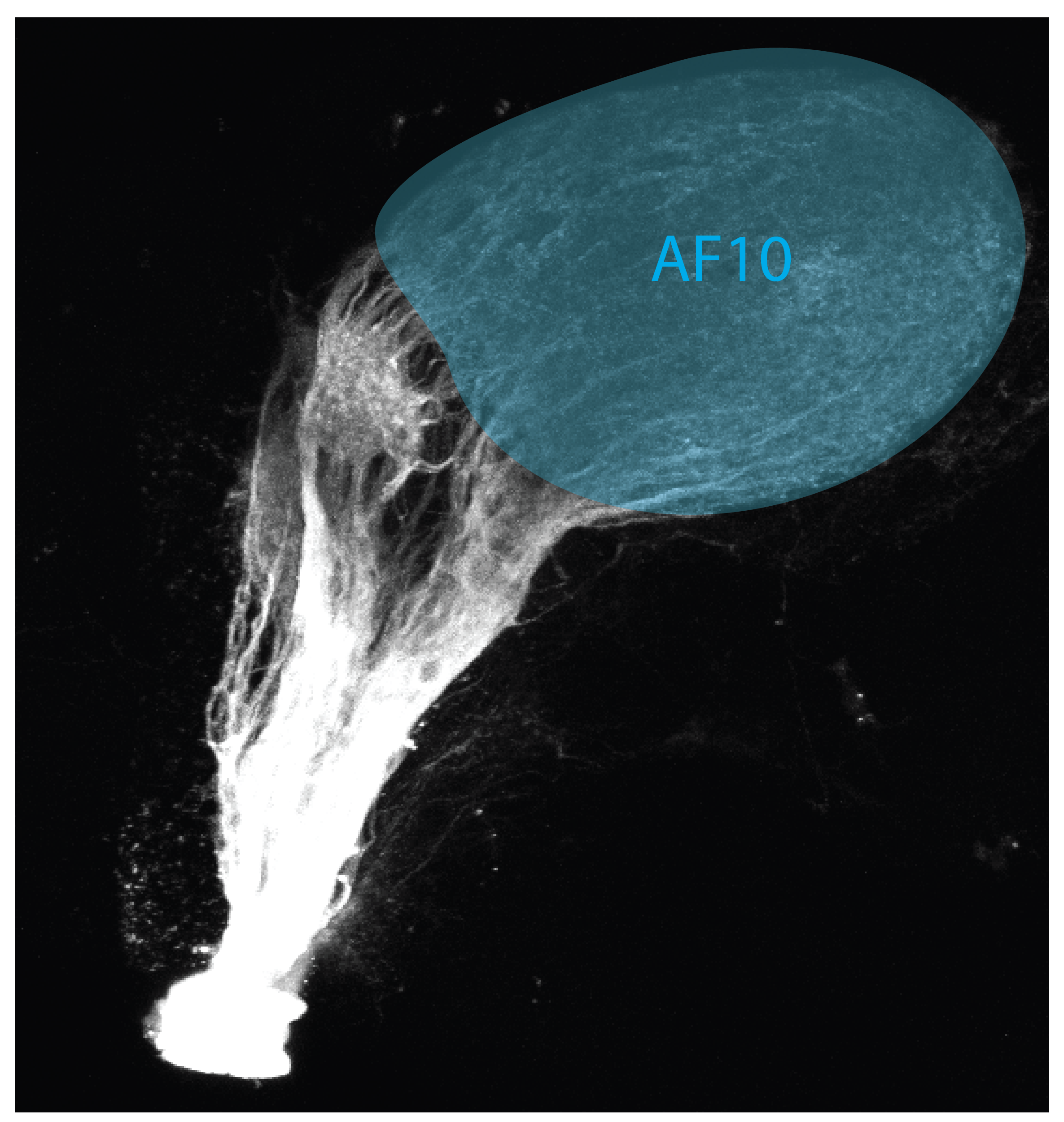
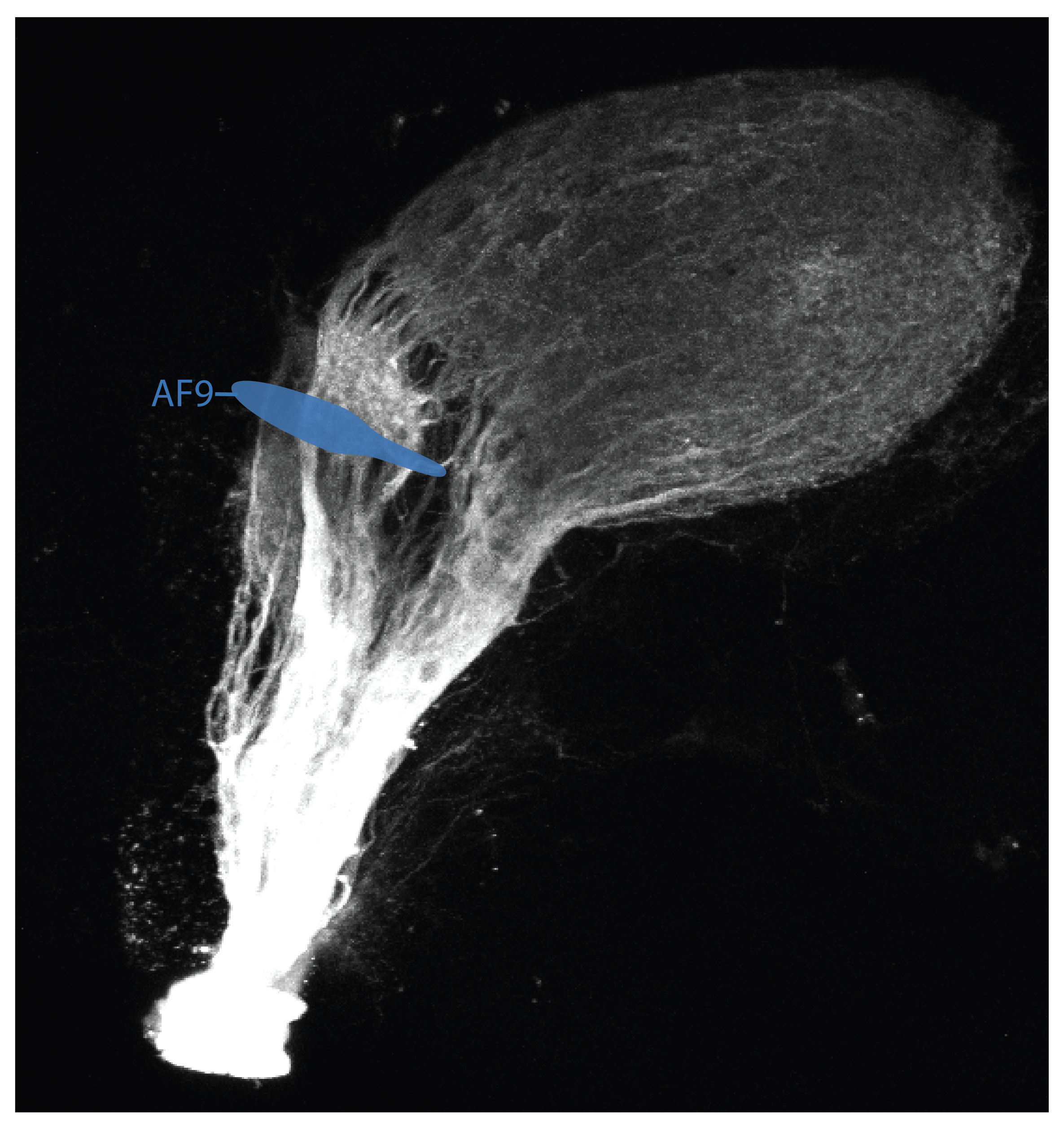
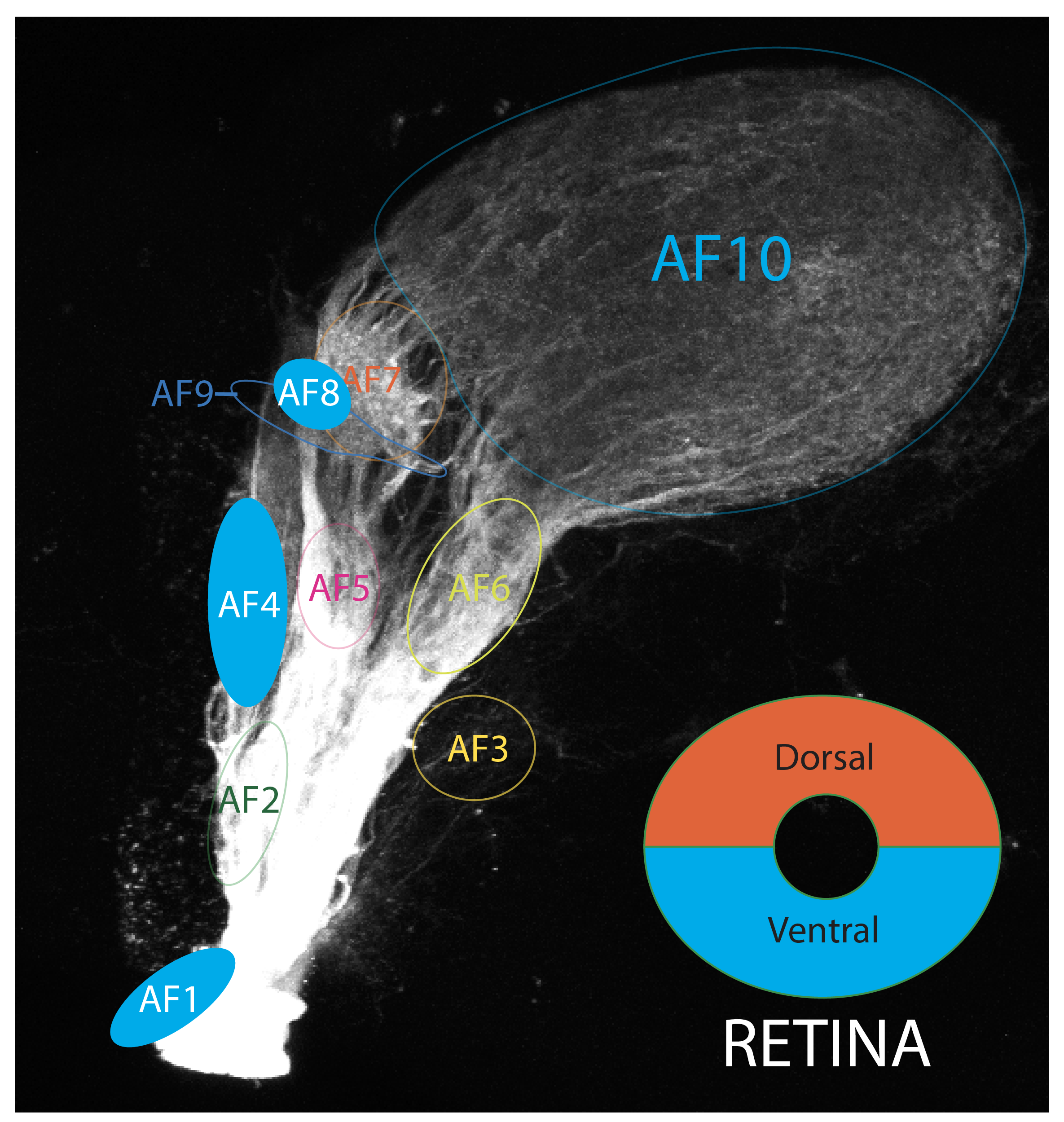
Schematic showing the approximate location of AF-10 in a 6dpf zebrafish. The optic tract is labelled in the Tg(atoh7:RFP) transgenic line and position of AFs are based on the data from Robles (2014).
VISUAL BEHAVIOURS ASSOCIATED WITH AF-10

The tectum contains a high-resolution map of visual space. The OT has been shown to be involved in localising objects and directing orienting movements toward or away from salient objects, such as prey or predators.

Intact tectal neuropil Is necessary for the looming-evoked escape response (Temizer et al., 2015).
Temizer et al show the direction of the escape behavior is dependent on the location of the stimulus within the visual field. Thus, the location of looming-responsive neurons within the tectum could be read out to generate a directional motor response.
Publications
Nevin, L.M., Robles, E., Baier, H., and Scott, E.K. (2010)
Focusing on optic tectum circuitry through the lens of genetics.
BMC Biology. 8:126.
Robles, E., Laurell, E., Baier, H. (2014)
The Retinal Projectome Reveals Brain-Area-Specific Visual Representations Generated by Ganglion Cell Diversity.
Current biology : CB. 24(18):2085-96.
Burrill JD & Easter Jr SS
Development of the Retinofugal projections.
J Comp Neurology, 2004 pp.1-18.
Temizer, I. et al (2015)
A Visual Pathway for Looming-Evoked Escape in Larval Zebrafish.
Current Biology, 25(14), pp.1823–1834.
Antinucci P, Folgueira M, Bianco IH.(2019)
Pretectal neurons control hunting behaviour.
eLife 8 doi.org/10.7554/eLife.48114
Bianco IH & Engert F(2015)
Visuomotor Transformations Underlying Hunting Behavior in Zebrafish
Current Biology 25, 831–846
Ethan Gahtan, Paul Tanger and Herwig Baier (2005)
Visual Prey Capture in Larval Zebrafish Is Controlled by Identified Reticulospinal Neurons Downstream of the Tectum.
Journal of Neuroscience 25 (40) 9294-9303 DOI: https://doi.org/10.1523/JNEUROSCI.2678-05.2005
Förster, D., Helmbrecht, T.O., Mearns, D.S., Jordan, L., Mokayes, N., Baier, H. (2020)
Retinotectal circuitry of larval zebrafish is adapted to detection and pursuit of prey.
eLife ;9:e58596. DOI: https://doi.org/10.7554/eLife.58596
Helmbrecht, T.O., Dal Maschio, M., Donovan, J.C., Koutsouli, S., Baier, H. (2018)
Topography of a Visuomotor Transformation.
Neuron. 100(6):1429-1445.e4.
Del Bene, F., Wyart, C., Robles, E., Tran, A., Looger, L., Scott, E.K., Isacoff, E.Y., and Baier, H. (2010)
Filtering of visual information in the tectum by an identified neural circuit.
Science. 330(6004):669-673.