Viewing entries tagged
Eyes

Sizing the eyes- a study of the mechanisms by which the developing eye copes with deleterious genetic mutations

Sizing the eyes- a study of the mechanisms by which the developing eye copes with deleterious genetic mutations

A summary of the article “Compensatory growth renders Tcf7l1a dispensable for eye formation despite its requirement in eye field specification” Elife. 2019 Feb 19;8. pii: e40093. doi: 10.7554/eLife.40093.


Although some genetic mutations lead to congenital abnormalities and/or disease, DNA sequencing projects have shown that compromised function of many genes has no obvious effect.  Indeed, people that carry such mutations live an apparently normal life that is not impacted by altered gene function.  This suggests that genetic pathways and developmental processes are robust and able to cope when challenged by altered gene function.  An important goal in biomedicine is to identify the compensatory mechanisms that enable such mutations to be carried without consequence  

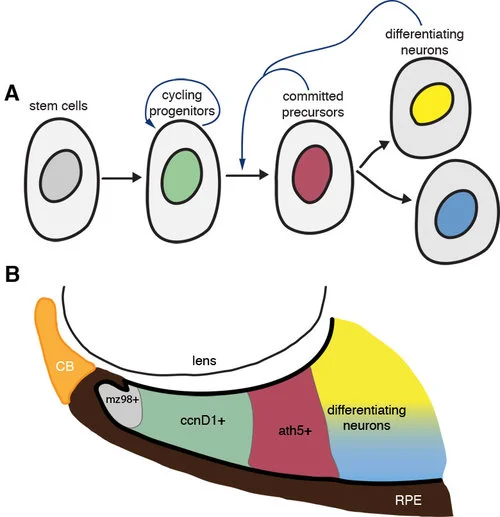
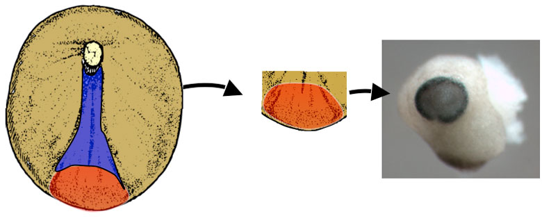
Zebrafish embryos are well suited to explore the link between genetic mutations (genotype) and developmental outcome (phenotype).  In a paper recently published in eLife (elifesciences.org/articles/40093), we studied zebrafish carrying a mutation in a gene called tcf7l1a that encodes a protein functioning in the Wnt pathway, one of the most critical pathways by which cells signal to each other during development.  The Wnt pathway is particularly important for development of the eyes.  Zebrafish embryos with the tcf7l1a mutation initially have tiny forming eyes, but within a day or two, these eyes undergo compensatory growth to reach a size comparable to that in unaffected individuals. We found that the smaller eyes delayed the onset of differentiation, enabling them to continue growing until they reached the right size. How does the eye know what the right size is? We propose that there is a mechanism by which eye size is intrinsically assessed and this informs retinal cells when to stop dividing and start differentiating into neurons. This mechanism may also explain how it is that left and right eyes develop independently and yet still grow to the same size.

 Although compensatory growth could restore eye size when tcf7l1a function was absent, we speculated that developing eyes in these mutants may be very sensitive to the effect of additional genetic mutations.  Indeed, we found additional interacting mutations that completely impaired eye development such that no eyes were formed, and others in which the developing eyes were unable to compensate their growth and remained small.

 These results contribute to a better understanding of the origin of developmental eye pathologies such as microphthalmia and anophthalmia, in which people are born with small eyes or no eyes at all.

Read the digest of this paper on Elife or treat yourself to the full text here.

Key Publications

Young RM, Hawkins TA, Cavodeassi F, Stickney HL, Schwarz Q, Lawrence LM, Wierzbicki C, Cheng BY, Luo J, Ambrosio EM, Klosner A, Sealy IM, Rowell J, Trivedi CA, Bianco IH, Allende ML, Busch-Nentwich EM, Gestri G, Wilson SW.
Compensatory growth renders Tcf7l1a dispensable for eye formation despite its requirement in eye field specification.
Elife. 2019 Feb 19;8. pii: e40093. doi: 10.7554/eLife.40093.

Sizing the eyes-a study of the mechanisms by which the developing eye copes with deleterious genetic mutations

A gene and a population of cells important for shaping the eye

Back to publication summaries


A GENE AND A POPULATION OF CELLS IMPORTANT FOR SHAPING THE EYE

Gaia Gestri and Steve Wilson
Lmx1b coordinates ocular FGF signaling to regulate patterning and morphogenesis of multiple eye tissues in zebrafish

LMX1B is a gene that when mutated in humans causes Nail-Patella Syndrome (NPS), a condition with many different features including limb, joint and renal (kidney) defects and disrupted function of the central nervous system (brain and spinal cord) and eyes. Approximately 50% of patients develop elevated intraocular pressure and glaucoma. LMX1B encodes a LIM-homeodomain transcription factor - a protein that functions of a kind of genetic switch to regulate the activity of many other genes. Individuals can exhibit the condition if they lack function of just of their two copies of the gene (one from mother and one from father). Although the identity of the causative gene is known, how defective function of the gene leads to the wide range of abnormalities observed in patients is poorly understood. In this study, a collaboration with Brian Link, we studied the function of the zebrafish version of the LMX1B gene to elucidate how it might function during embryonic development of the eye.

Figure 1: A. The lmx1b gene (in blue) is expressed in cells that surround the eye and in an open groove in the retina, the choroid fissure (indicated by the black line in B), that is present in the eye early in development. Eyes in embryos with comp…

Figure 1: A. The lmx1b gene (in blue) is expressed in cells that surround the eye and in an open groove in the retina, the choroid fissure (indicated by the black line in B), that is present in the eye early in development. Eyes in embryos with compromised Lmx1b function exhibit varying degrees of coloboma (the condition where the choroid fissure doesn't properly close).

The eyes form during embryogenesis as outgrowths from the developing brain - these outgrowths are called optic vesicles and they will give rise to the retina and the pigmented cells at the back of the eye (for more information, see our research pages on eye development www.ucl.ac.uk/zebrafish-group/research/eye.php). Other groups of cells contribute to other eye structures such as the lens, blood vessels and protective coat of the eyeball. How all of these different groups of cells interact in a highly coordinated way during eye formation is poorly understood. In this study, we show that the lmx1b gene is expressed in highly motile cells that migrate around the optic vesicles as they are forming (Figure 1A and Movies 1 and 2). To test the function of the gene in these cells, we removed its function using anti-sense reagents that prevent formation of Lmx1b protein. This resulted in the motile cells failing to properly migrate around the eye and indeed many of these cells subsequently died (Movie 2). This disruption to the migratory cells was correlated with a failure in the optic vesicle to under the tissue movements that enable the eye to take on its spherical shape. Indeed, we often observed that eye exhibited coloboma, a condition in which two lips of the forming eye fail to fuse together in the ventral (lower) part of the eye (Figure 1B-D). These results strongly suggest (but have not proven) that the migratory cells are required for the optic vesicle to form the spherical eye. If correct, how might this happen? We suspect that the migratory cells may secrete proteins that help the nascent eye cells to undergo tissue re-shaping but this will require further research to resolve.

We also found that retinal cells are incorrectly patterned when Lmx1b function is disrupted - this would lead to defects in visual map formation when connections are made between the eye and the brain. We do not know if this problem is a consequence of the death of the motile cells around the eye or to some other action of the Lmx1b gene.

Many of the deficits we describe in fish are more severe than observed in human patients - this is most likely due to the fact that our experiments deplete Lmx1b function more severely than in patients. If the function of the gene is severely compromised in humans, it would lead to early lethality as we observe in fish. Our studies in fish will continue to elucidate how genes function and cells and tissues interact during vertebrate eye formation and will help us to understand human diseases, conditions and syndromes in which these processes are disrupted.

Other links
UK Nail Patella Syndrome charity website
Wikepedia
NIH Genetics page

If you have any further questions, please contact Gaia Gestri or Steve Wilson

This study was a collaboration with Brian Link and our work received financial support from

The complex choreography of eye formation

Back to publication summaries
 

the complex choreography of eye development

Florencia Cavodeassi and Stephen Wilson, October 2009

Original paper reference
Dynamic Coupling of Pattern Formation and Morphogenesis in the Developing Vertebrate Retina

If we are to interpret correctly what we see, a precise representation of the visual input received the retina must be formed in our brain. This "visual map" forms during embryogenesis, when retinal neurons (nerve cells) project long processes (axons) to the brain to establish the wiring between the eye and those parts of the brain that process visual information. Each retinal axon targets a very specific area of the superior colliculus (or optic tectum), a region of the brain that interprets visual information, thus generating a map of connections that reproduces the external visual field in the brain. For the visual map to be accurate, the neurons in the retina must be able to interpret where in the retina they are located, and who are their neighbours. We know quite a lot about the genetic "codes" that give the retinal cells their identity but much less about how the retinal cells acquire this knowledge of who they are and where they must go.

Until now, it has been particularly challenging to resolve the mechanisms that instruct positional information in the retina, since this process happens in parallel to the extensive remodelling (or morphogenesis) of tissues that occur during formation of the eye. Using a combination of imaging techniques and genetic manipulations, in this study, we describe how the processes of morphogenesis and allocation of retinal identity are coordinated during maturation of the optic cup.

In a previous study, our collaborators Alexander Picker and Michael Brand proposed that a combination of secreted Fibroblast Growth Factor (Fgf) proteins are required by the eye cells to 'know' where they are. The study determined that retinal cells acquire their identity during a very early stage of eye development, when the nascent eye (optic vesicle/cup) is just beginning to emerge from the brain. This presented a puzzle - Fgfs work through being secreted from one group of cells and received and interpreted by nearby cells - however, during the stage when Fgfs appear to function in the eye, the nascent eye tissues are undergoing complex rearrangements. How then, could this process of morphogenesis be temporally and spatially coordinated with the localised action of Fgfs on retinal cells? This study found that as the prospective eye cells undergo various movements, it brings them transiently into proximity with other tissues that produce Fgfs which can act consequently act upon their temporarily neighbouring nascent retinal cells.

Figure 1: cartoon illustrating the reorganisation of the nasal-temporal axis during maturation of the optic cup. The source of Fgf signals is shown in blue, the future nasal region of the retina is shown in green and the future temporal region in re…

Figure 1: cartoon illustrating the reorganisation of the nasal-temporal axis during maturation of the optic cup. The source of Fgf signals is shown in blue, the future nasal region of the retina is shown in green and the future temporal region in red. The olfactory placode (the future nasal epithelium, dark blue) is one source of Fgfs and the other is the telencephalic region of the forebrain (pale blue).

The retina is divided into two axes - a nasal to temporal axis (nose to ear) and a dorsal to ventral axis (up to down) and Fgf promotes nasal identity. One surprising finding was that Fgfs are produced by tissues dorsal to retina at early stages (Figure 1) suggesting that nasal retina is initially located dorsally in the nascent eye and that the eye somehow rotates by 90 degrees during subsequent stages of development. But how is this shift of the axis effected?

To answer this question required labelling subsets of retinal cells with green fluorescent protein and tracking them in live zebrafish embryos. In this way, it was possible to follow the complex reorganisation movements that transform the initial dorsal-ventral axis of the optic vesicle into the nasal-temporal axis of the mature retina. During this process, the dorsal, future nasal cells of the retina become compacted, the future temporal cells move from ventrally to join the nasal cells in the forming retina and simultaneously the whole optic vesicle rotates to bring the dorsal domain to its final location anteriorly in the eye. Remarkably, this reorganisation is coordinated with a similar shift of the source of Fgfs, which act as choreographers of some aspects of the complex morphogenetic process. In this way, one set of signals (the Fgfs) instructs positional information in the retina and simultaneously ensures that the axis is maintained during the subsequent maturation of the eye.

The study also revealed that many aspects of morphogenesis proceed very well in the absence of Fgfs indicating that other pathways must be involved. We hope that future collaborations will help to identify these additionally choreographers of eye formation.

If you have any further questions, please contact Florencia Cavodeassi or Steve Wilson. Alexander Picker and Michael Brand would also be very happy to receive any questions on the study.

This study was a collaboration with Alexander Picker and Michael Brand and our contribution to the research received financial support from the MRC and Wellcome Trust.

Back to publication summaries


Big eyes/small eyes: identifying signals that regulate eye formation

Florencia Cavodeassi and Steve Wilson
Original paper reference:
Early stages of zebrafish eye formation require the coordinated activity of Wnt11, Fz5 and the Wnt/β-catenin pathway

You might not realise it but your eyes are in fact part of your brain. Early in embryonic development, the eyes arise as outgrowths from the forming brain. How does a subset of brain cells become destined to form the eyes and how do these cells then undergo the complex movements that must occur during eye formation? In this study, we addressed the role of genes functioning in the Wnt pathway, a cell-to-cell signalling pathway, in the regulation of early steps of eye formation.

The primordium of the mature eyes is specified as a single "eye-field" of cells at very early stages of embryonic development. At this stage, the nervous system is just a simple sheet of cells called the neural plate. As the neural plate folds up to form the brain, the eye-field splits in two and evaginates from the brain to form left and right eyes.

In zebrafish embryos, the eye field and other regions of the prospective brain can be readily visualised within the neural plate by the overlapping expression of various genes encoding transcription factors. The eye field is surrounded anteriorly by the prospective telencephalon, and posteriorly by the prospective diencephalon (Figure 1).

Figure 1: (A) Organisation of brain territories at neural plate stage. The groups of cells giving rise to each territory are shown in different colors. (B-D) The same colour code is used to show the relative positions of the different territories in…

Figure 1: (A) Organisation of brain territories at neural plate stage. The groups of cells giving rise to each territory are shown in different colors. (B-D) The same colour code is used to show the relative positions of the different territories in embryonic brains at a later stage of development, from lateral (B), dorsal (C) and ventral (D) views. Modified from Cavodeassi et al., (2009) Squire LR (ed.) Encyclopedia of Neuroscience, vol. 4, 321-325

The Wnt signalling pathway has been well studied and work done by our group and others, suggested a model whereby a gradient of Wnt activity specifies different regional fates within the anterior neural plate. The model suggested high levels of Wnt activity specify diencephalic fates and lower levels of Wnts specify gradually more anterior fates, such as the eye field and the telencephalon. In this study, we re-examined the role of the Wnt pathway during eye field specification, and found that it is more complex than expected. Wnt proteins can activate several different signalling cascades: simplistically, Wnt/β-catenin signalling leads to changes in gene expression and the identity of cells (like brain versus eye) whereas Wnt/PCP signalling leads to changes in cell shape and movements.

By doing a series of experiments where we activated or switched off the Wnt pathway locally within the anterior neural plate, we found that these two branches of the pathway have very different effects on eye formation (Figure 2). High levels of Wnt/β-catenin signalling tells cells to become diencephalic brain and blocks eye formation. In contrast, Wnt/PCP activity within the eye field promotes eye formation, and it does so, at least partially, by antagonising the Wnt/β-catenin pathway. Each branch of the Wnt pathway appears to be activated by a different combination of Wnts and their receptors (called Fzs) in the nascent forebrain. Thus, Wnt8b and Fz8a activate the Wnt/β-catenin pathway, while Wnt11 and Fz5 activate the non-canonical pathway.

Figure 2: Manipulation of two branches of the Wnt pathway has opposite effects on eye formation. Dorsal views of embryos with transplants of cells (labelled in brown) activating different branches of the Wnt pathway. Wnt8b/βcatenin activity blocks e…

Figure 2: Manipulation of two branches of the Wnt pathway has opposite effects on eye formation. Dorsal views of embryos with transplants of cells (labelled in brown) activating different branches of the Wnt pathway. Wnt8b/βcatenin activity blocks eye formation (asterisk in B), while Wnt11/PCP activity leads to the formation of bigger, misshapen eyes (asterisk in C). A control transplant expressing GFP does not have any effect on eye development (A). The optic vesicles are labelled by the expression of the rx2 gene (blue). Anterior is to the left.

Six ways to make a brain

Gaia Gestri and Steve Wilson
Original paper reference:
Six3 functions in anterior neural plate specification by promoting cell proliferation and inhibiting Bmp4 expression

During development of the embryo, cells make decisions about the tissues they will contribute to and how they will differentiate. The outer layer of the embryo is called ectoderm and cells in this layer make the nervous system (brain and spinal cord) and the skin as well as migratory cells called neural crest. How do the ectoderm cells know which of these tissues to form? Within cells there are genes that encode proteins called transcription factors that act as molecular switches turning on different developmental programmes that control cell identity. In this study, we explored how one such transcription factor, called Six3, promotes the ability of cells to form eyes and brain.

Six3 is sometimes called a 'master regulator' for eye and forebrain development in that it is necessary and sufficient for the eyes and brain to form. What this means is that in gain of function experiments (in which the embryos express more Six3 protein), embryos show brain enlargement and ectopic eye structures (see the arrow pointing to 'the third eye' in Fig.1) while in loss of function experiments (where the Six3 protein is absent or not functioning) embryos show reduction of the most anterior part of the brain and no eyes (Fig. 1, right).

Figure 1. Pictures of the heads of frog tadpoles the black blobs are the forming eyes. In the tadpole on the left there is a small extra eye on top of the head when there is too much Six3 protein and the tadpole on the right is missing an eye due to…

Figure 1. Pictures of the heads of frog tadpoles the black blobs are the forming eyes. In the tadpole on the left there is a small extra eye on top of the head when there is too much Six3 protein and the tadpole on the right is missing an eye due to lacking Six3 protein on one side of its brain.

So how might Six3 control brain and eye development? With this in mind, we asked when and how Six3 exerts it function taking advantage of both the frog, Xenopus laevis, and the zebrafish as model systems for our experiments.

The processes that regulate brain development start at the early neural plate stage, this is much earlier than the first morphological appearance of the eye and the brain. The neural plate is a sheet of cells that will form both the brain and the eyes and at first, it is continuous with a sheet of cells called the epidermis that will form the skin. It is the anterior part of the neural plate, ANP, (Fig. 2 red) that gives rise to the brain and eyes (Fig. 2).

Figure 2. The schematic shows a very young frog embryo where the cells that are going to form the nervous system are coloured in red and blue. The red cells are those that will develop into the brain and eyes (right photo)

Figure 2. The schematic shows a very young frog embryo where the cells that are going to form the nervous system are coloured in red and blue. The red cells are those that will develop into the brain and eyes (right photo)

Given that the ANP has the capacity to form the brain and eyes, then the key genes that control eye and brain development must already be active in the ANP. These ANP genes must positively confer brain/eye identity as well making sure that the cells don't acquire the identity of the surrounding tissues such as the posterior neural plate (blue in Fig. 2), that will give rise to the spinal cord, and the surrounding epidermis that will give rise to the skin (brown in Fig 2). In our study we found that Six3 plays key roles in both specification and maintenance of the brain/eye promoting properties of the ANP.

Figure 3. Schematic and two photos of the anterior neural plate (blue in the schematic and white in the two photos. The neural plate is surrounded by epidermis (yellow in the schematic and blue in the photos). The little brown dots in the photos are…

Figure 3. Schematic and two photos of the anterior neural plate (blue in the schematic and white in the two photos. The neural plate is surrounded by epidermis (yellow in the schematic and blue in the photos). The little brown dots in the photos are individual cells that either have a lot of Six3 (middle), or lack Six3 (right)

One of the properties of the ANP is that it is more highly proliferative compared with the posterior neural plate and we found that Six3, expressed in this territory, promotes proliferation and represses the production of neurons.

The ANP is surrounded by the epidermis, (Fig. 3) that will later form the skin. The epidermis produces a signal, called Bmp, that has the ability to inhibit expression of ANP genes. We found that Six3 switches off Bmp expression (Fig. 3 middle) ensuring the maintenance of the ANP as a Bmp-free domain. This is really important given that if Six3 function is lost, Bmp signalling encroaches into the ANP (Fig. 3 right) and doing so, leads to embryos with severely reduced anterior brain and eyes and expanded epidermis (Fig. 1 right panel).

In humans, mutations in Six3 can lead to severe congenital abnormalities of the brain and eyes, and this study has helped to elucidate the function of this gene, one of the most important genes for brain and eye development.

This study was done as a collaboration between Guiseppina Barsacchi's lab in Pisa and Steve Wilson's lab here at UCL.