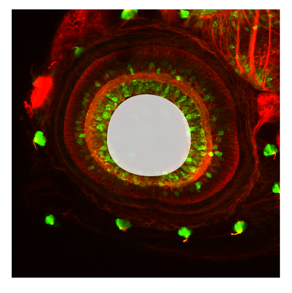
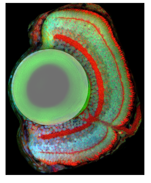
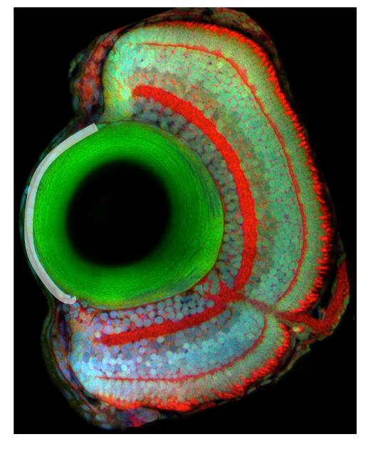
Viewing entries tagged
INL T

Cornea

Cornea

cornea-03.png

Description

The cornea develops from the ectoderm overlying the lens. Corneal epithelial layers proliferate and generate multiple layers. the corneas main function is to refract or bend light. It also acts as a protective outer layer to the eye.


Ontology
 

is part of: eye

has parts:


Transgenic Lines/Antibodies that label this brain region


Key Publications

lens

lens

Schematic showing the position of the lens in sagittal, horizontal and coronal sections through the zebrafish brain.  Based on the anatomical segmentation of 3 day old zebrafish larval brain by Thomas Müller, Olaf Ronneberger, Wolfgang Driever and c…

Schematic showing the position of the lens in sagittal, horizontal and coronal sections through the zebrafish brain.
Based on the anatomical segmentation of 3 day old zebrafish larval brain by Thomas Müller, Olaf Ronneberger, Wolfgang Driever and colleagues. For details see Ronneberger et al., Nat. Meth. 2012 and http://vibez.informatik.uni-freiburg.de

Abbreviations: GCL, ganglion cell layer; INL, inner nuclear layer ; ipl, inner plexiform layer ; ONL, outer nuclear layer ; opl, outer plexiform layer; RPE, retinal pigmented epithelium.

Description

Upon neural retina specification, signals from the neural retina induce lens fate in the overlying surface ectoderm. The lens placode then invaginates to give rise to the lens vesicle, which buds out at around 24hpf. The lens vesicle keeps growing throughout adulthood and fibre cells differentiation is maintained from a pool of undifferentiated proliferating epithelial cells located at the most anterior region of the vesicle.


Ontology
 

is part of: eye

has parts:


Transgenic Lines/Antibodies that label this brain region


Key Publications

retinal inner plexiform layer

retinal inner plexiform layer

ipl-03.png

Description

Within the inner plexiform layer are localized synapses between the amacrine and bipolar interneurons and the retinal ganglion cells. Connections between cells that are depolarized upon an increase in illumination (ON) occupy approximately the inner half of the IPL, whereas synapses of cells hyperpolarized (OFF) by light are confined to the outer half of the IPL. 


Ontology
 

is part of: neural retina

has parts:


Transgenic Lines/Antibodies that label this brain region


Key Publications