Viewing entries tagged
preoptic area

Tg(1.4dlx5a-dlx6a:GFP)ot1

Tg(1.4dlx5a-dlx6a:GFP)ot1

About

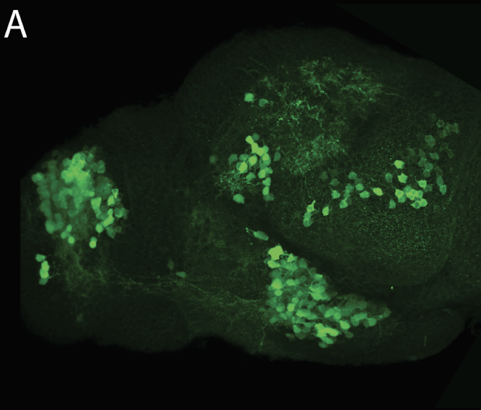
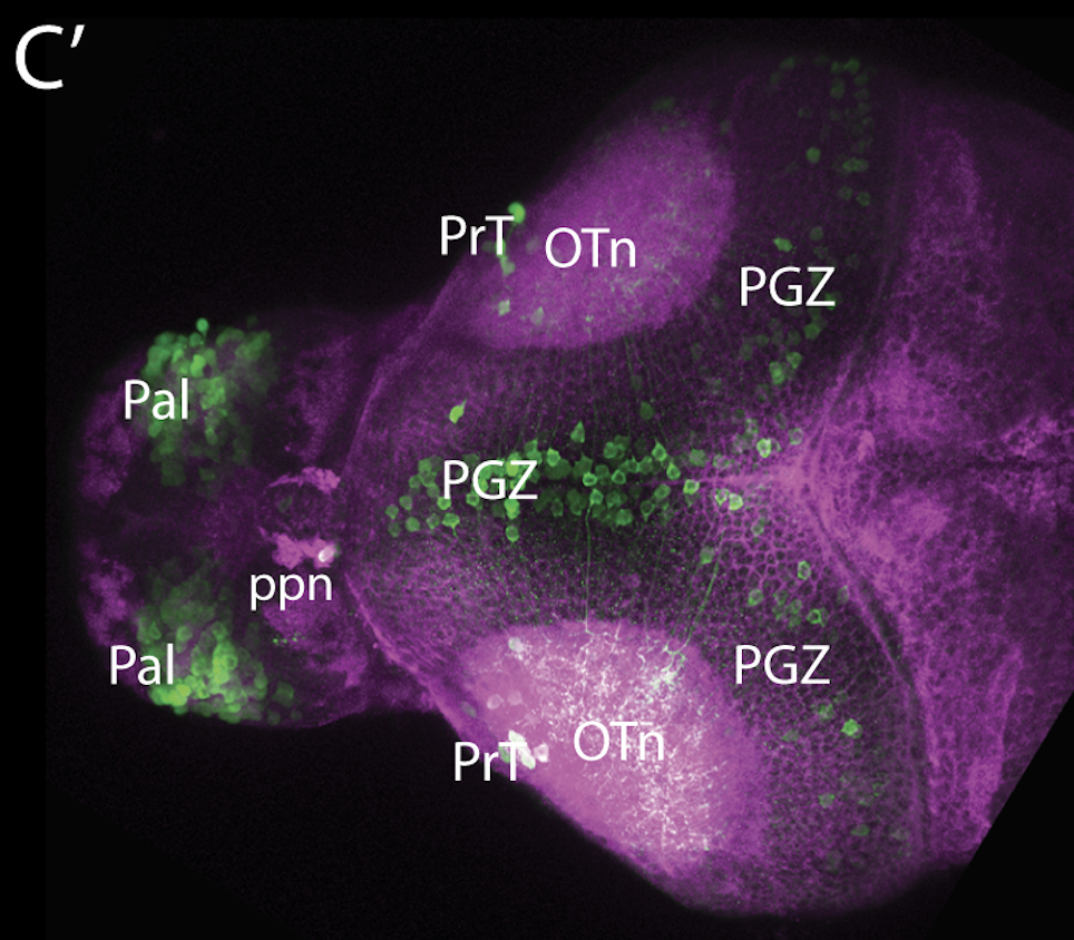
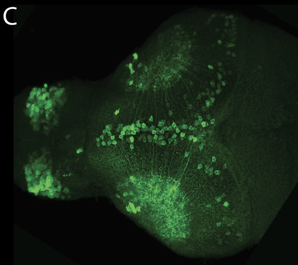
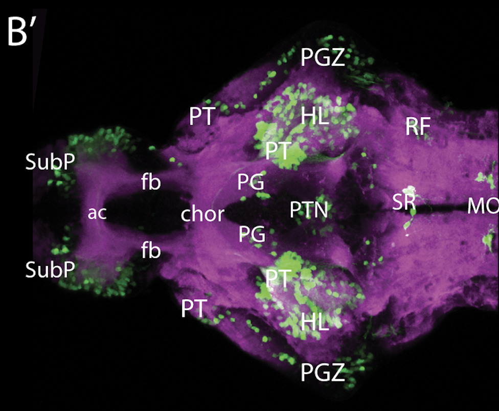
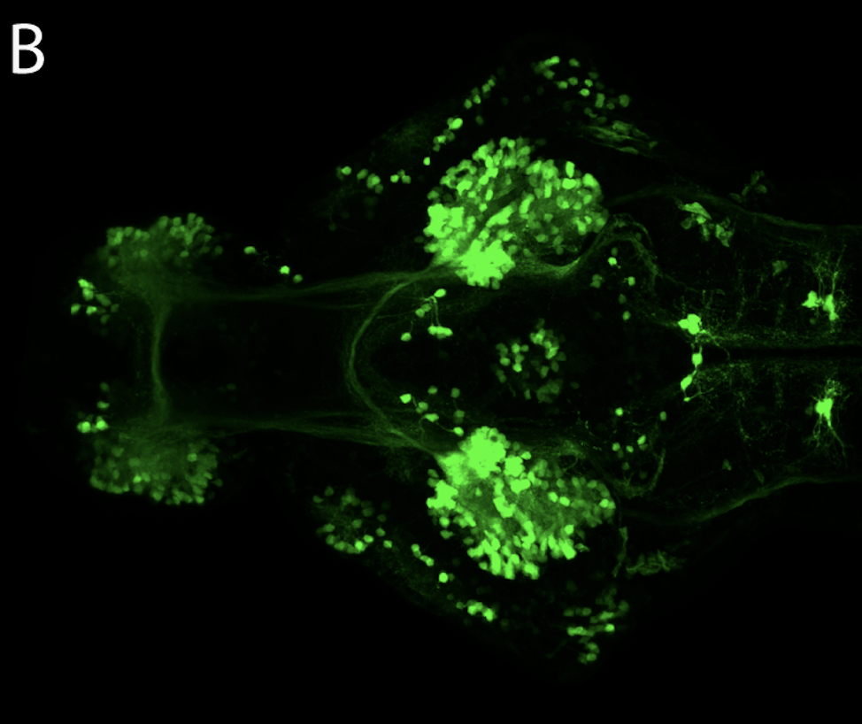
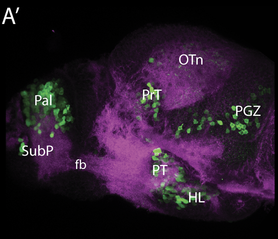
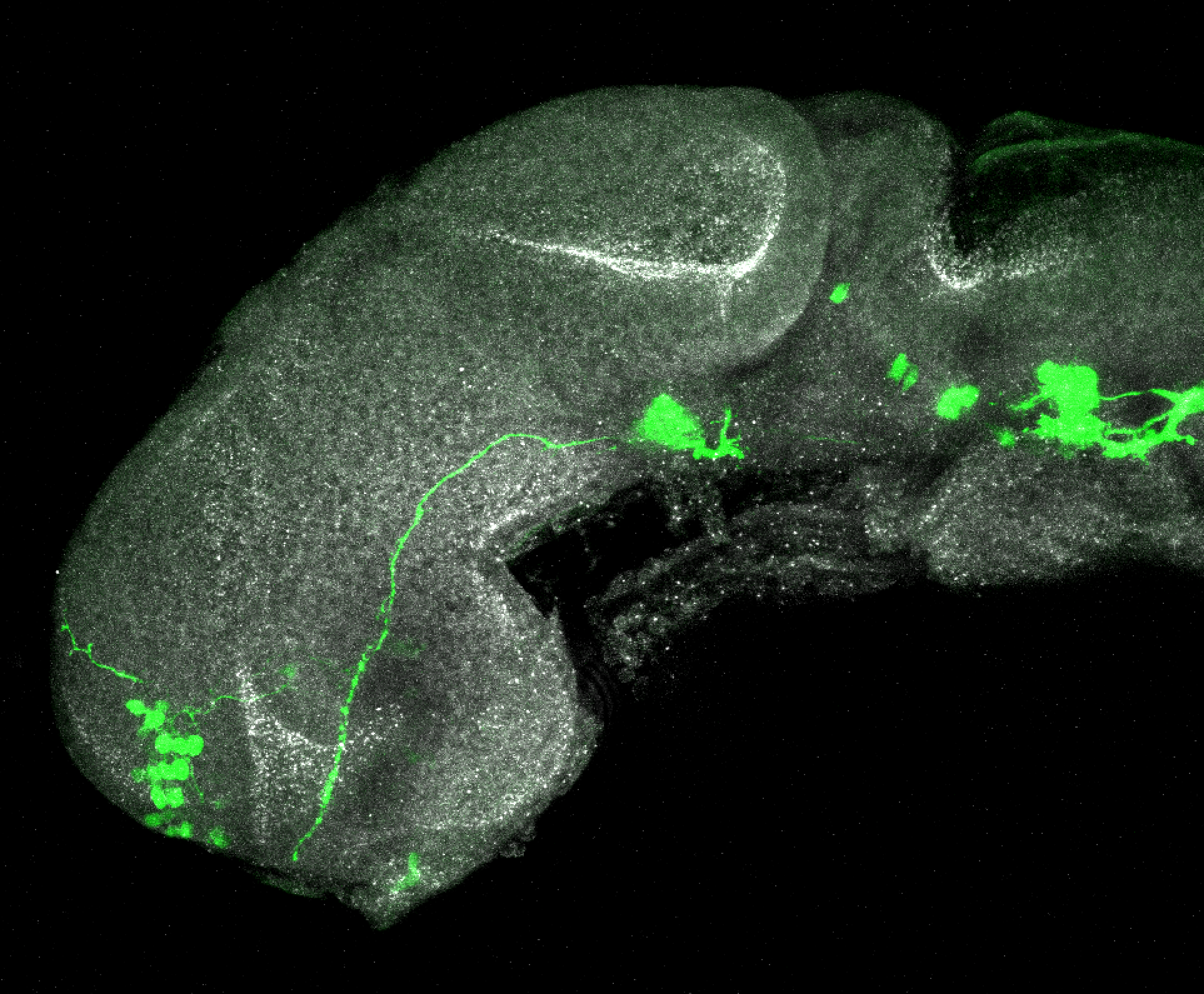
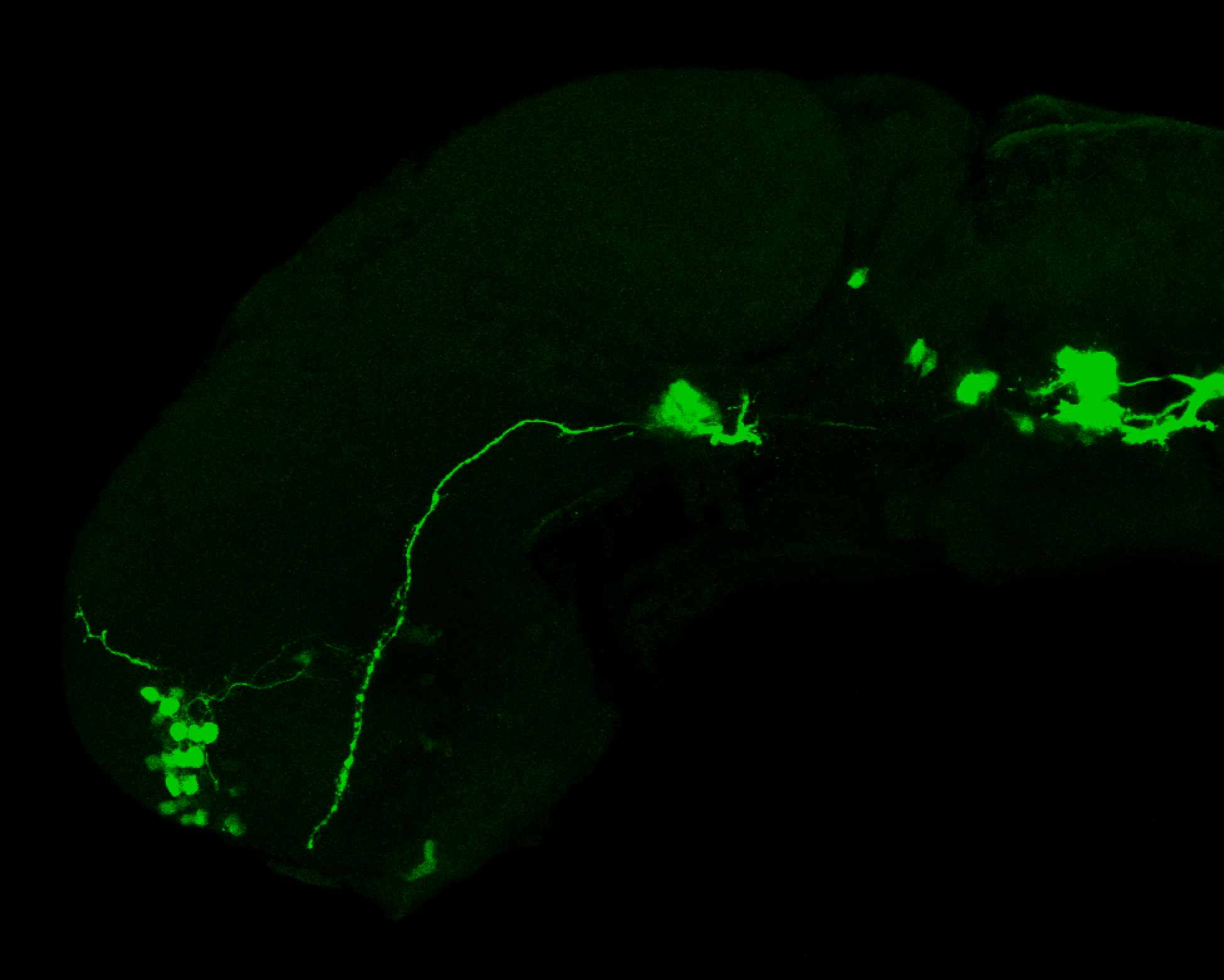
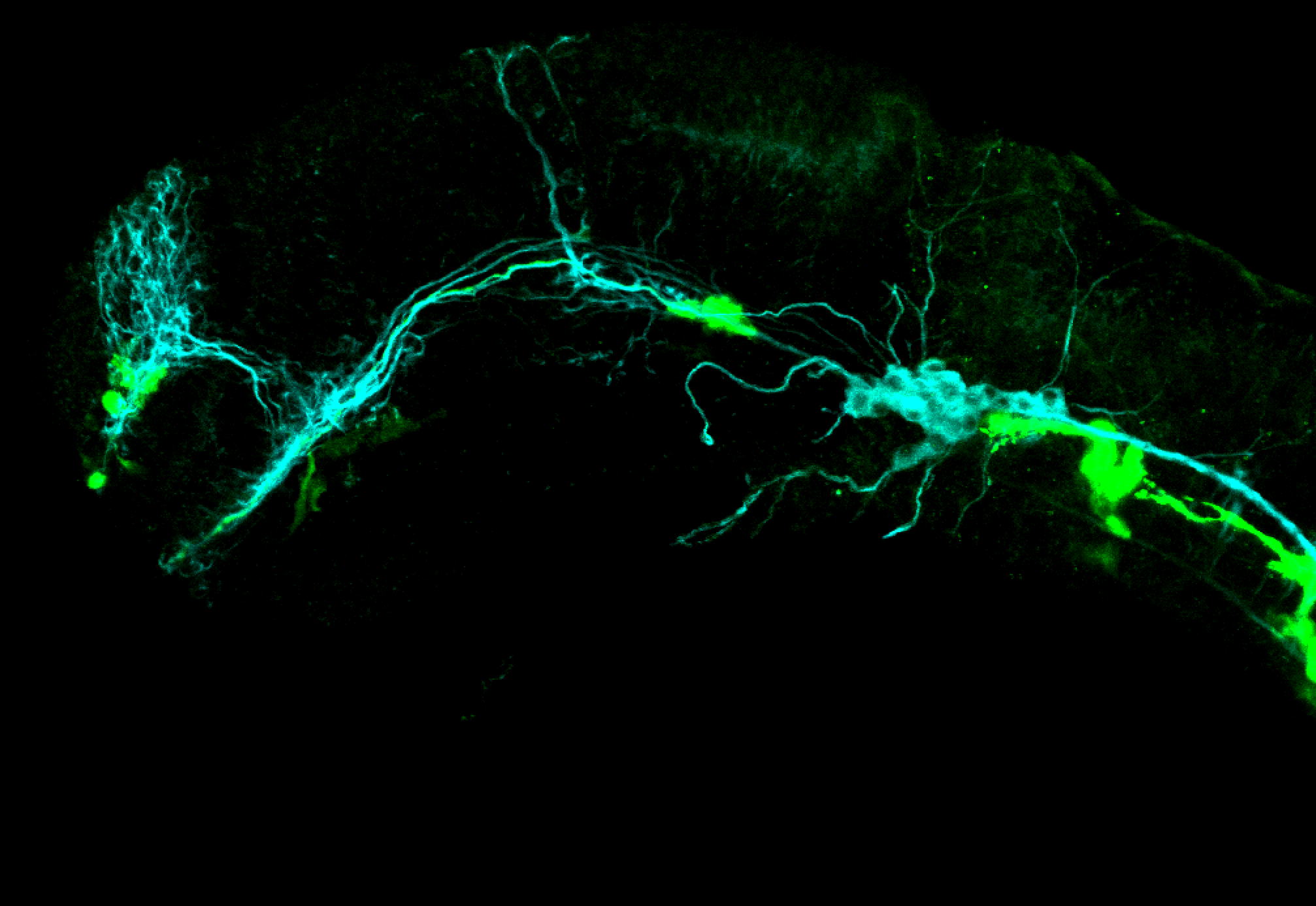
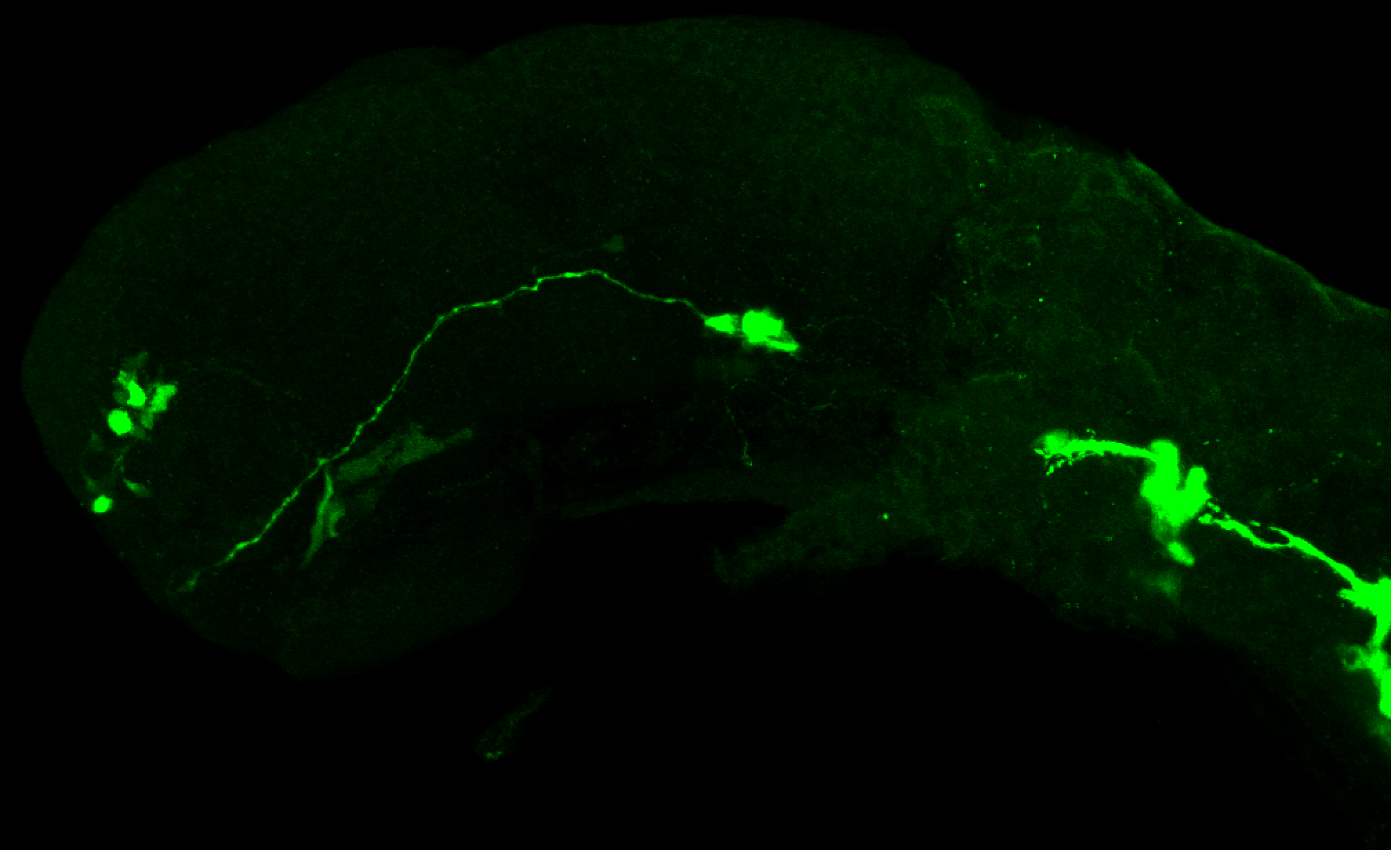
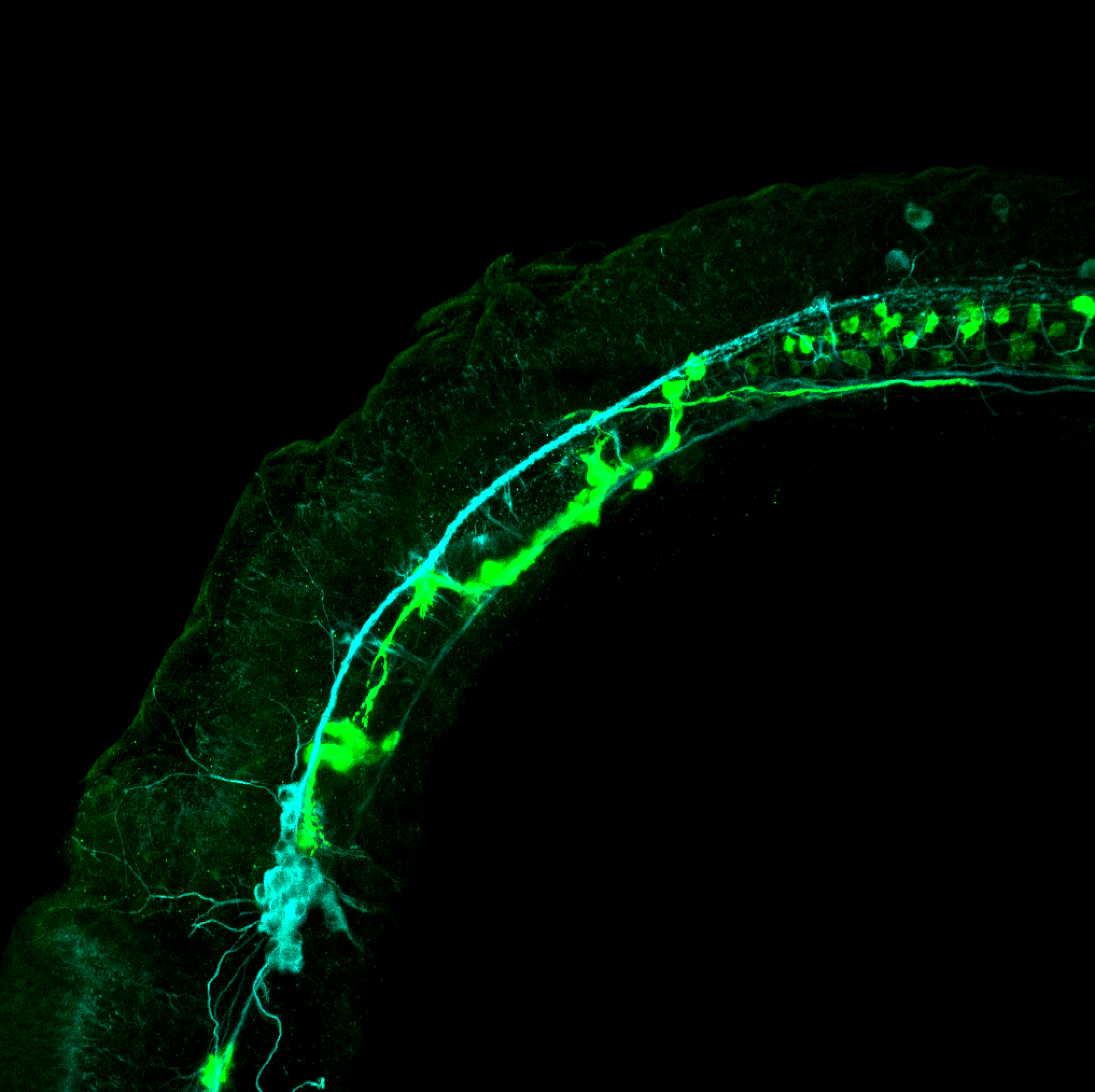
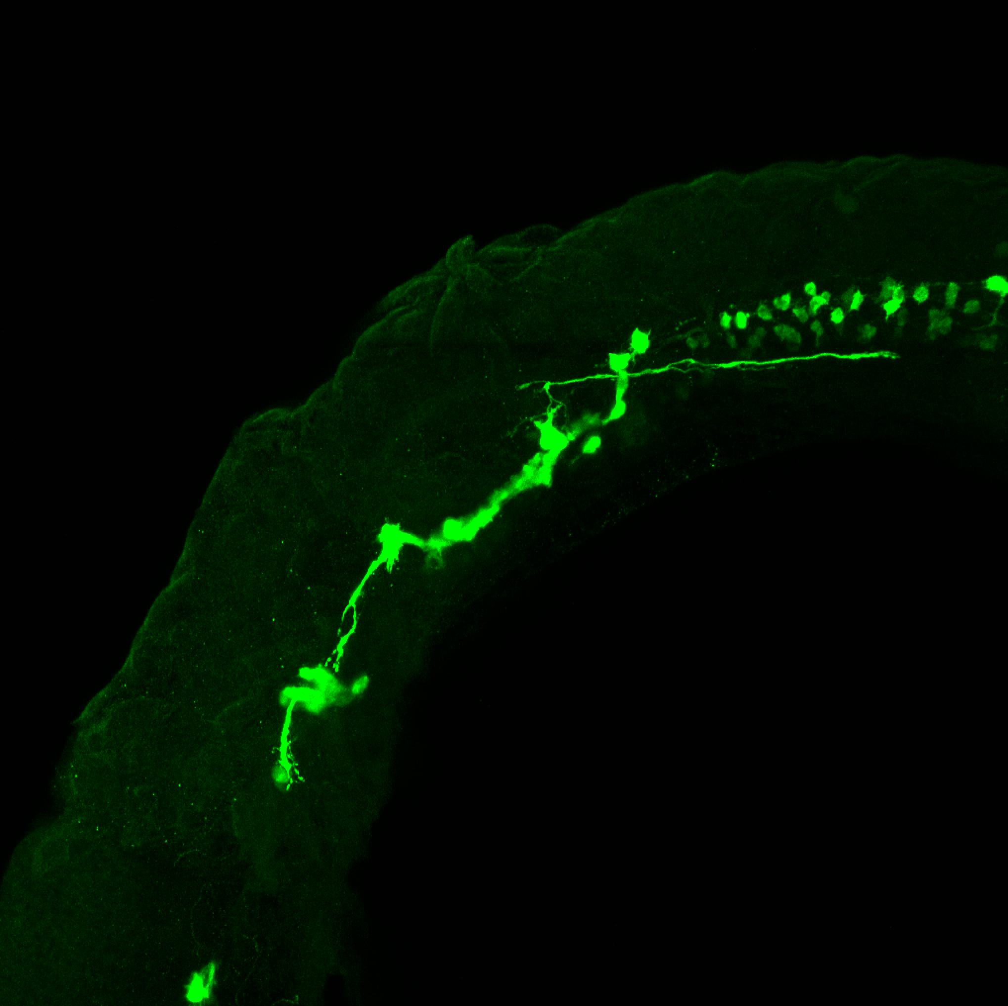
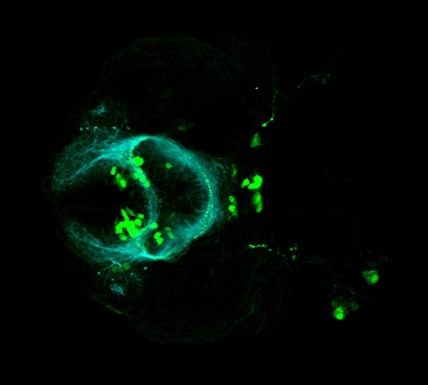
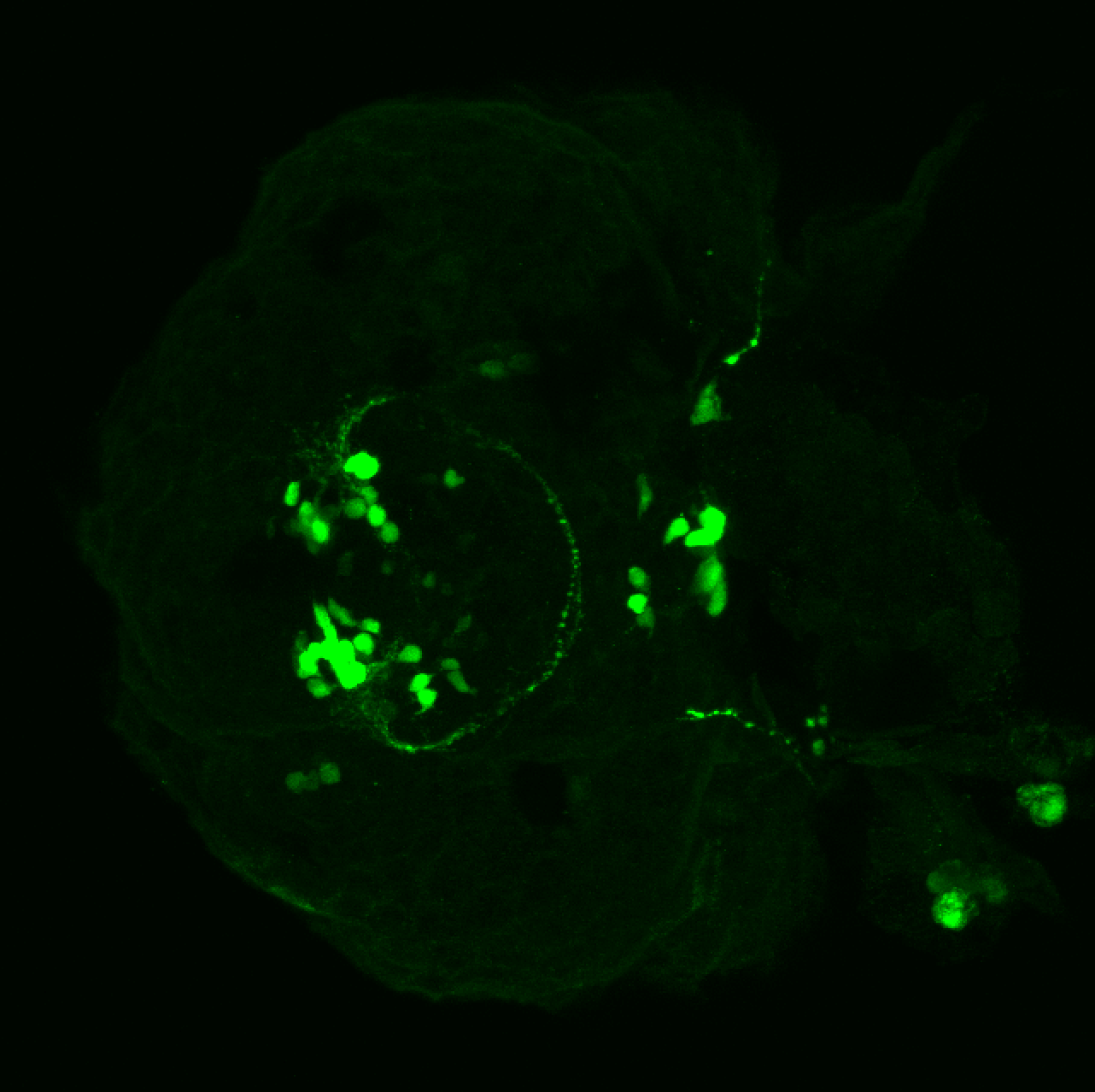
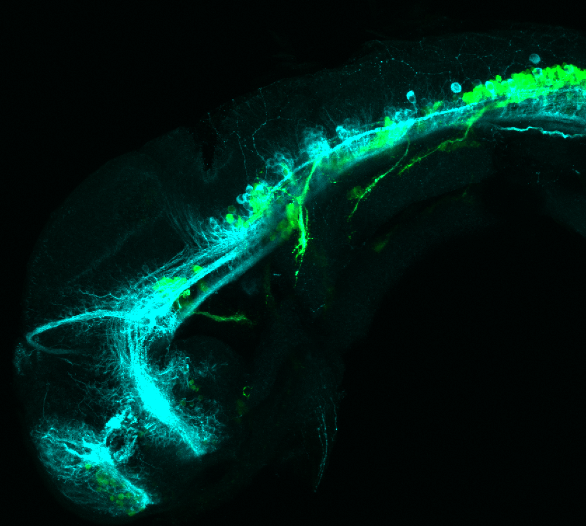
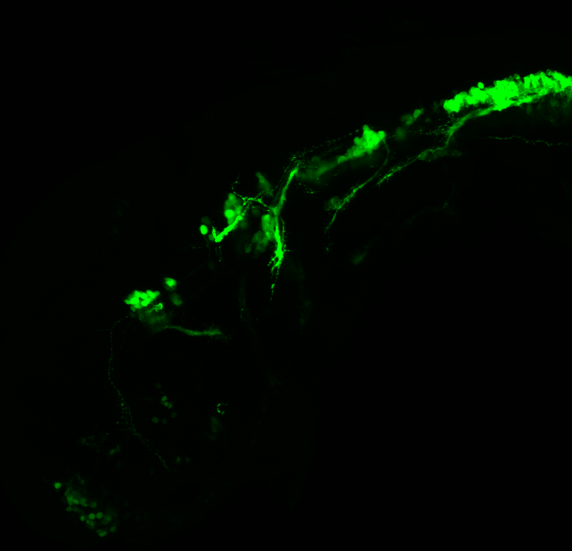
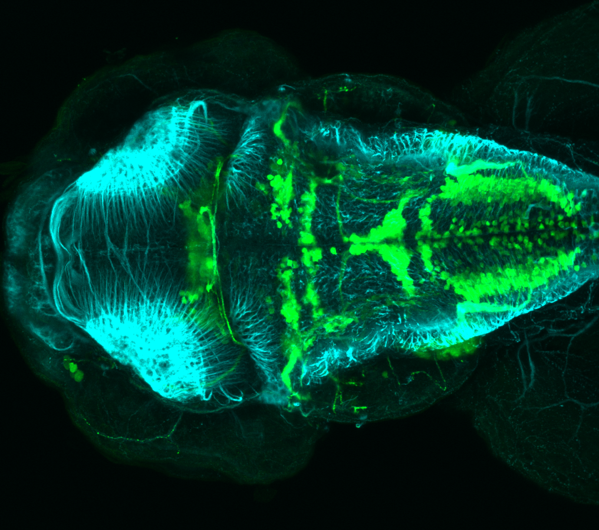
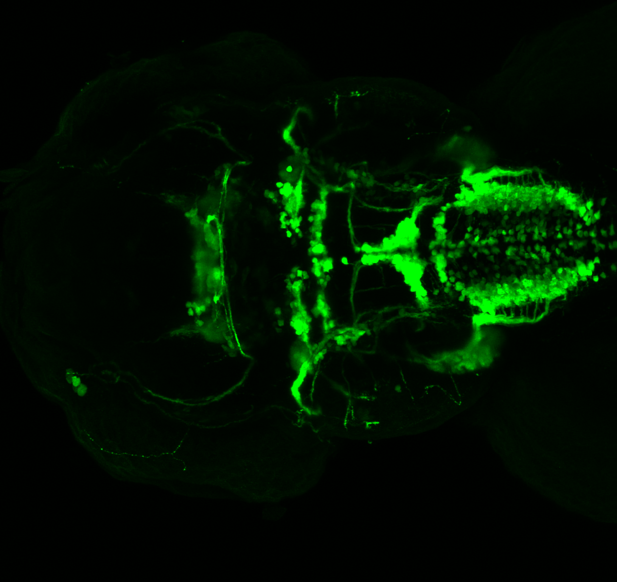
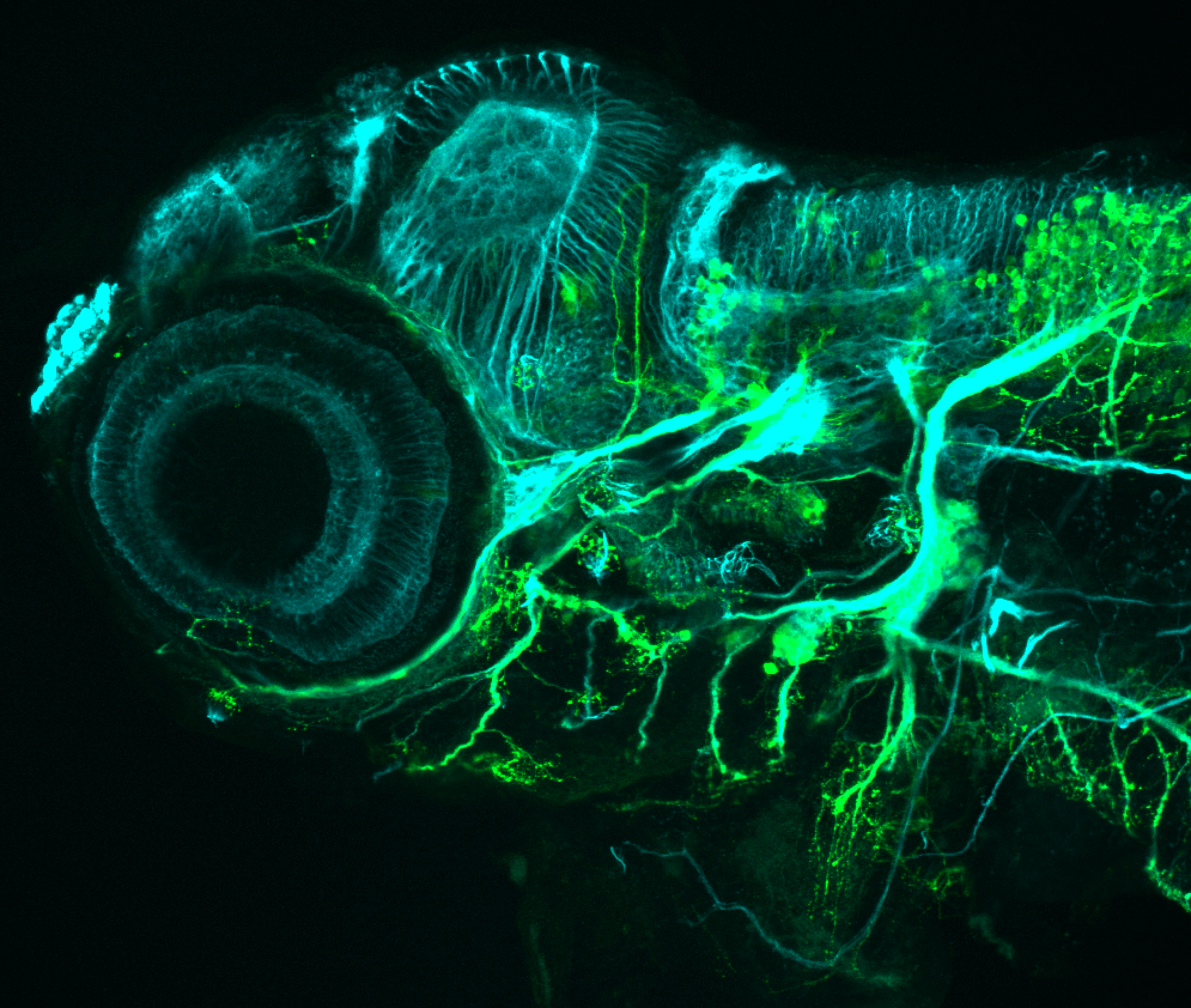
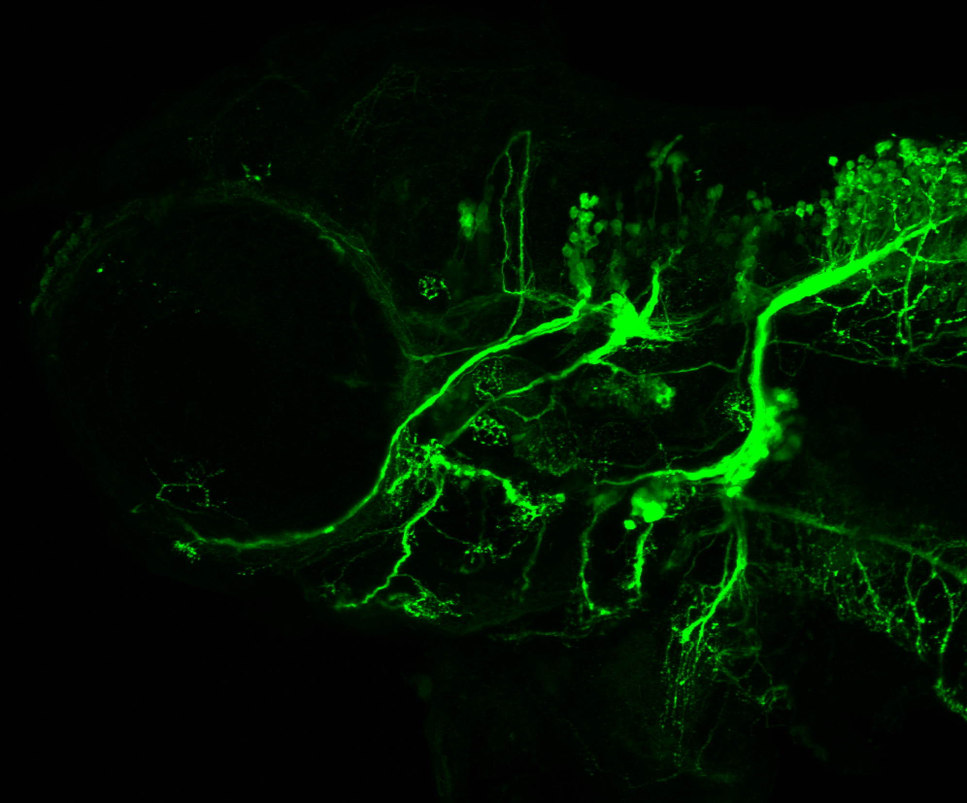
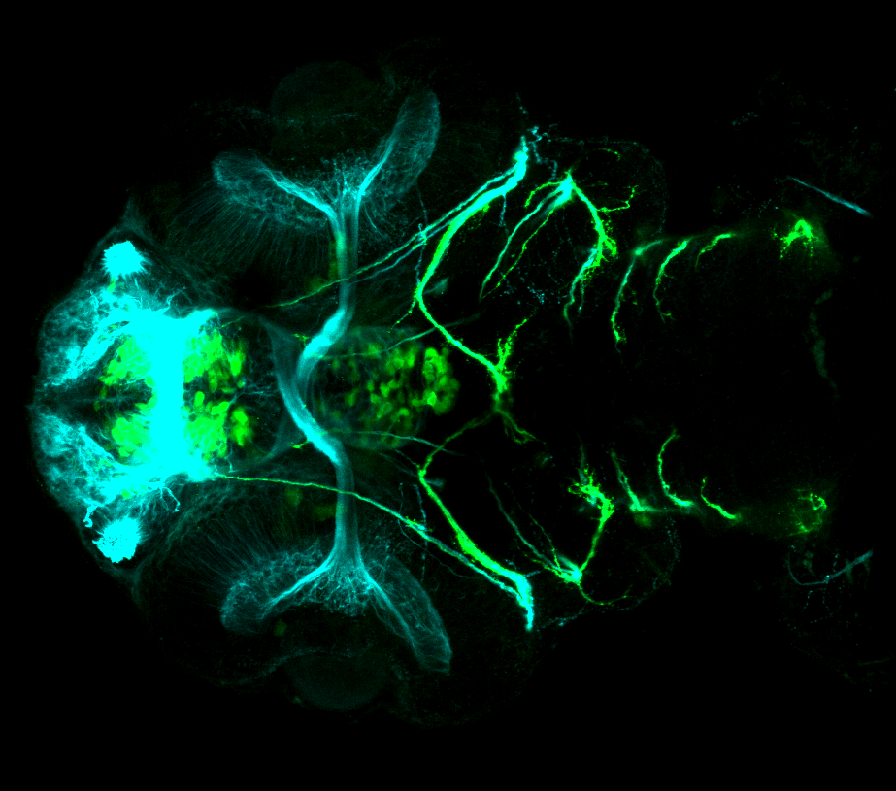
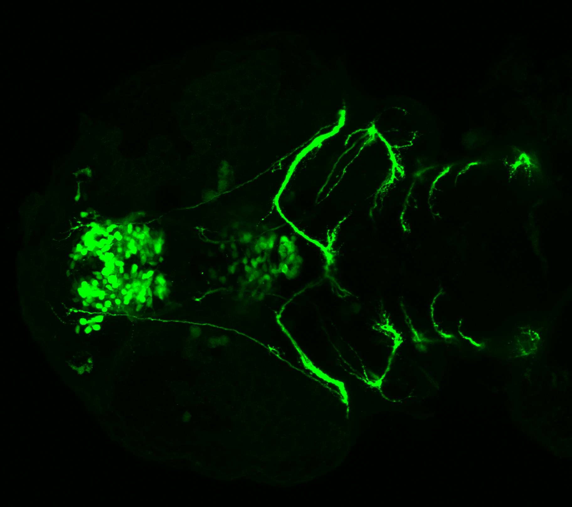
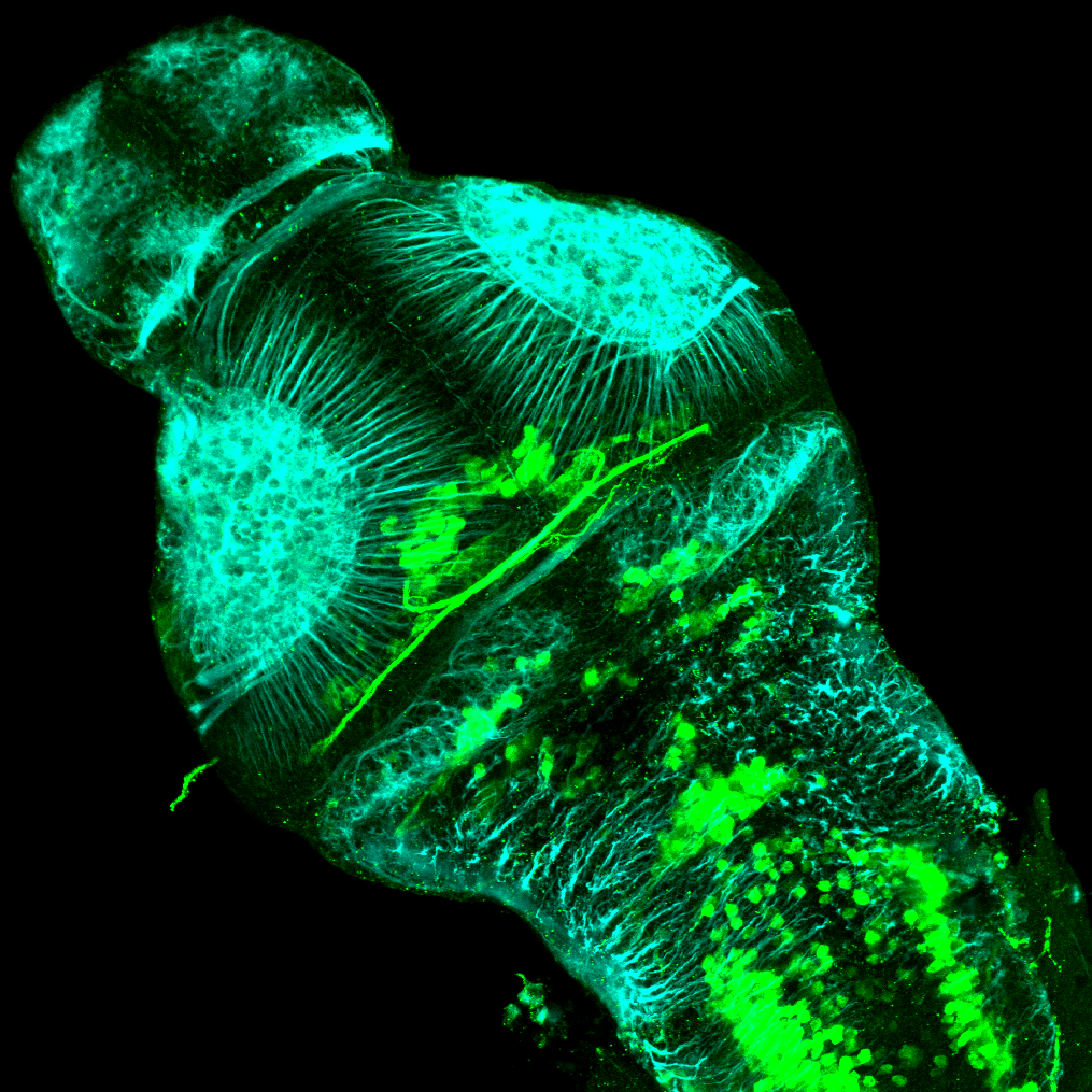
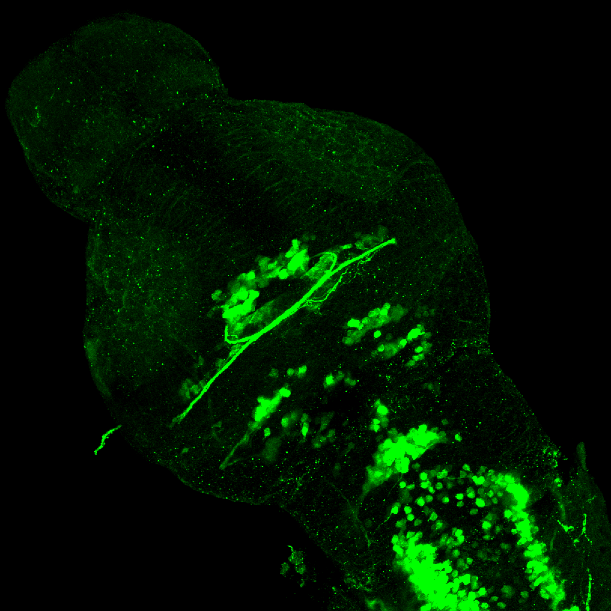
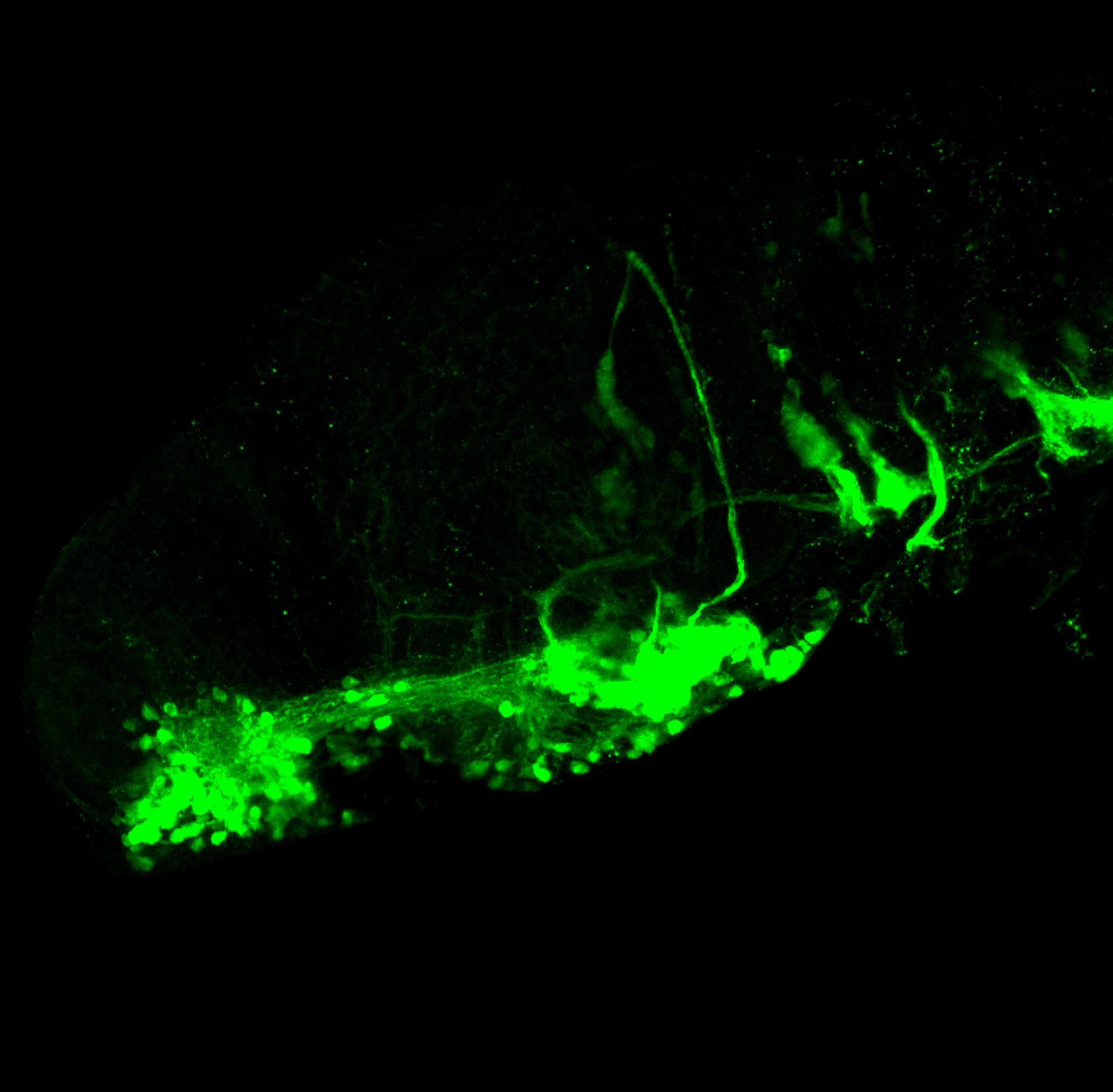
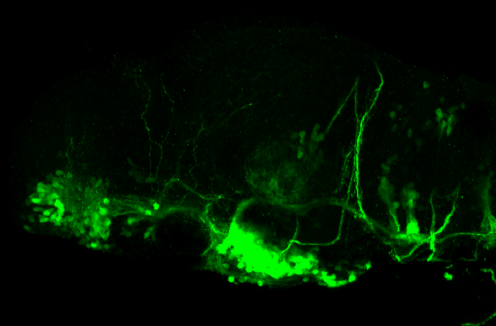
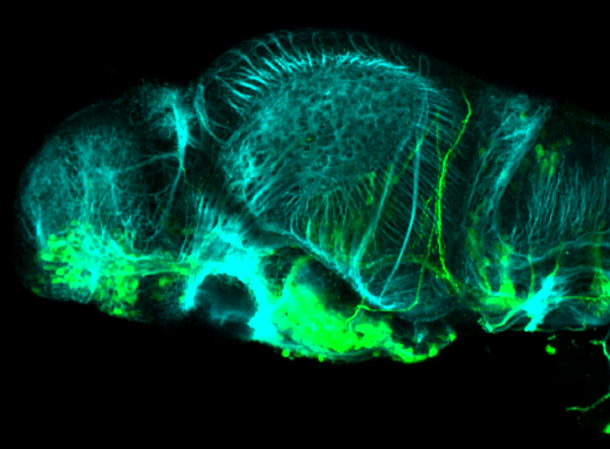
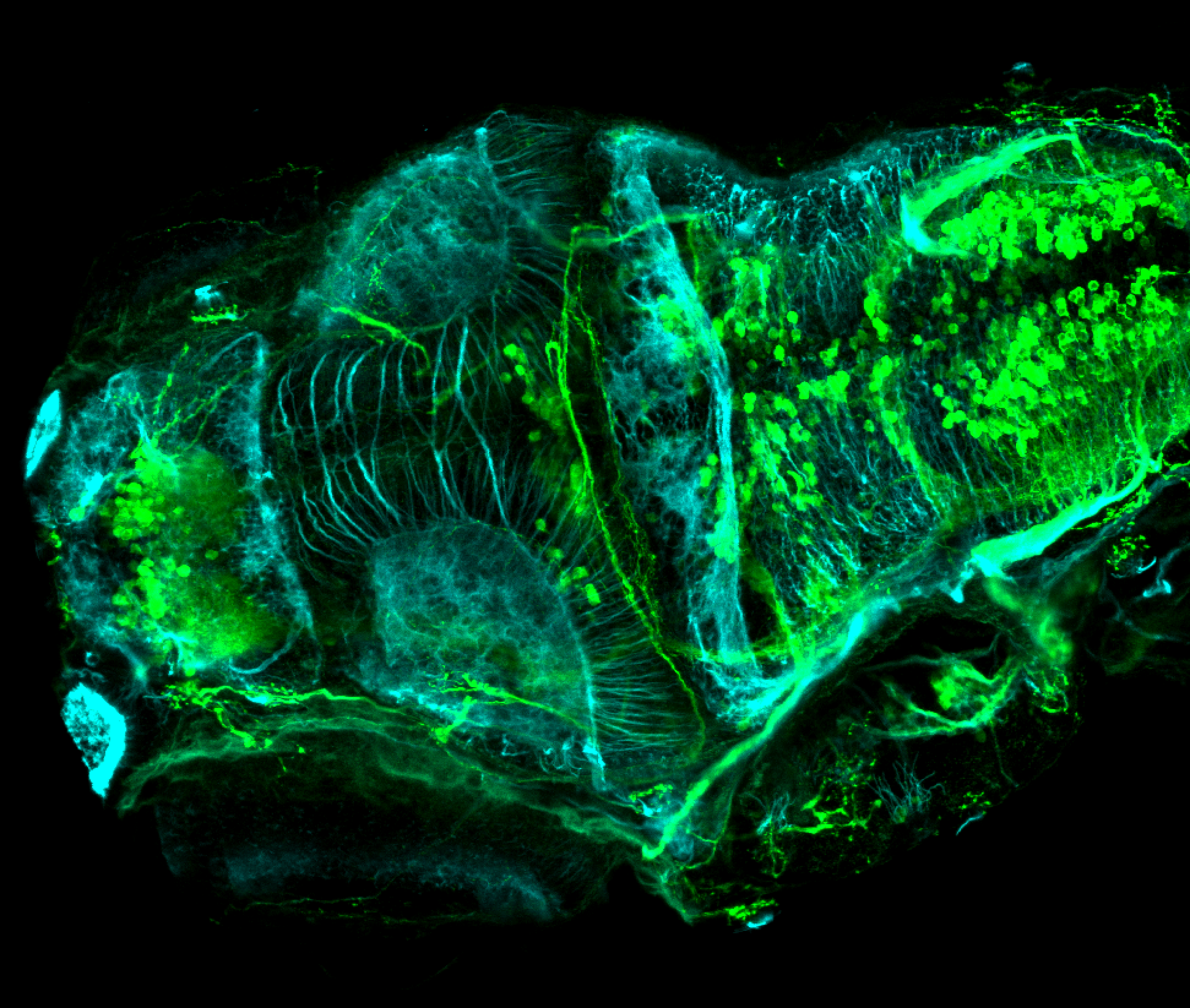
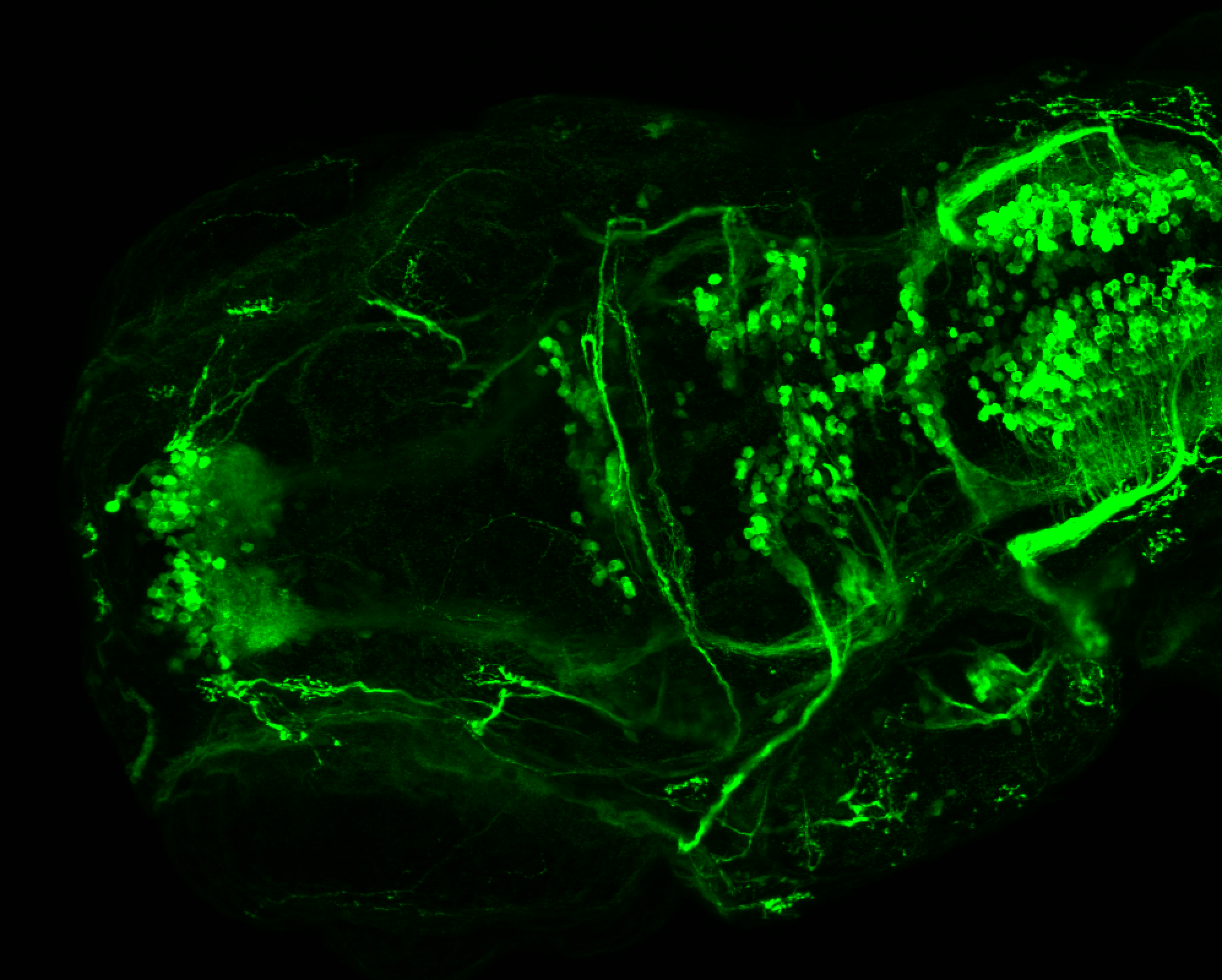
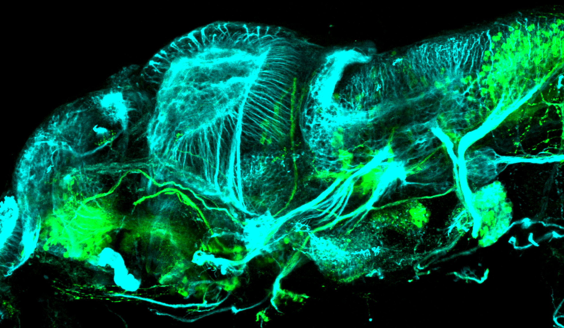
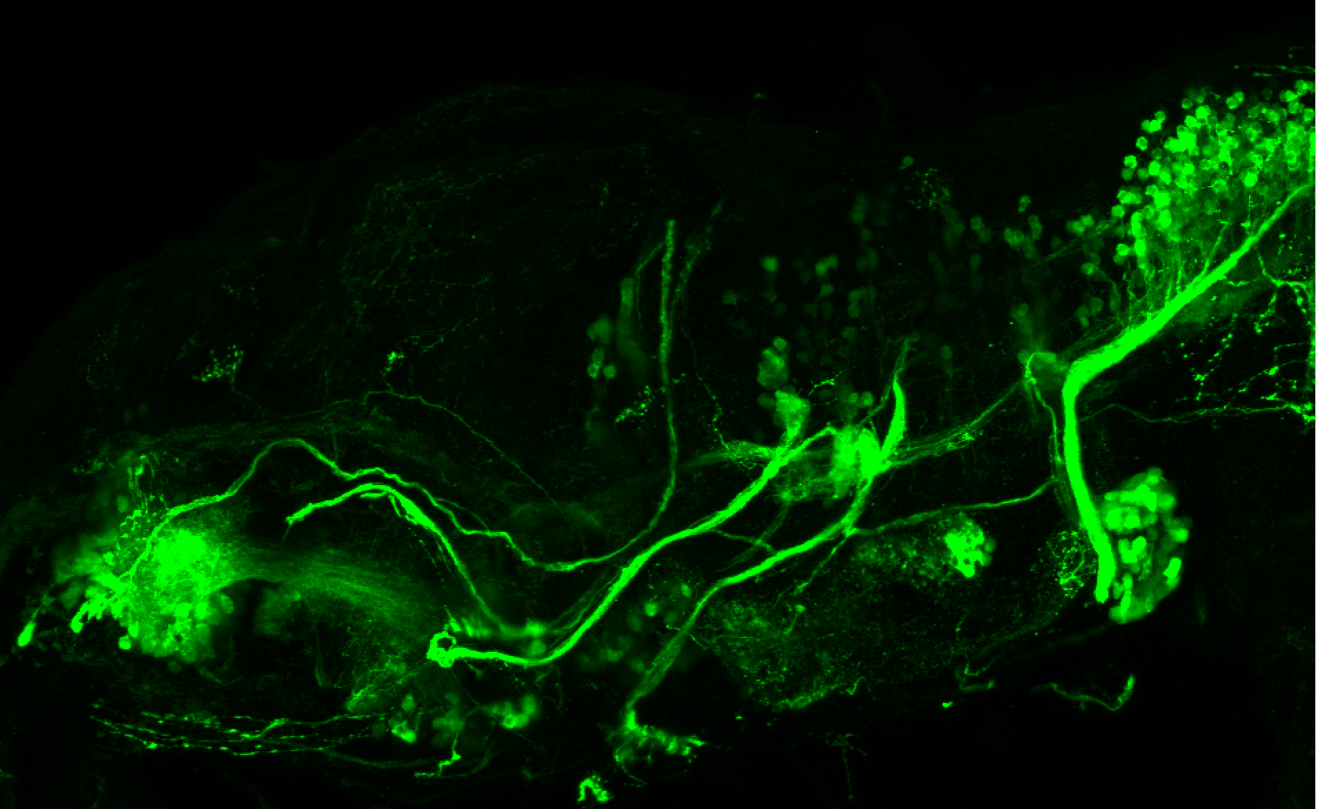
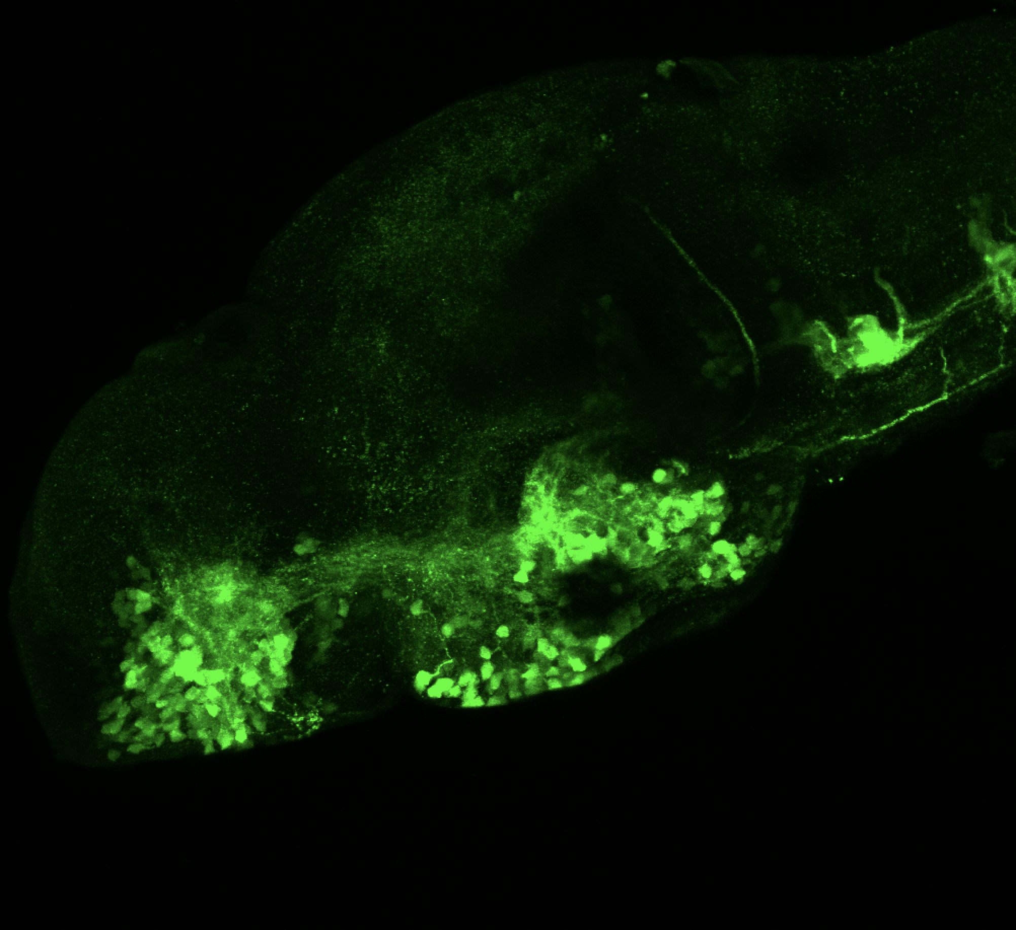
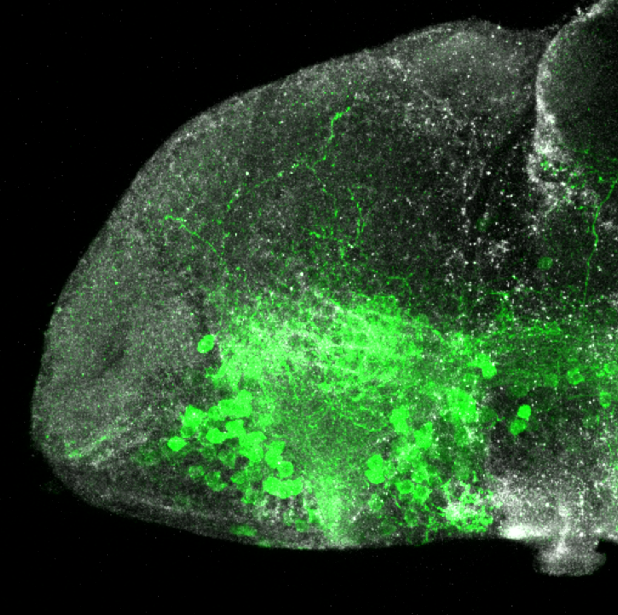
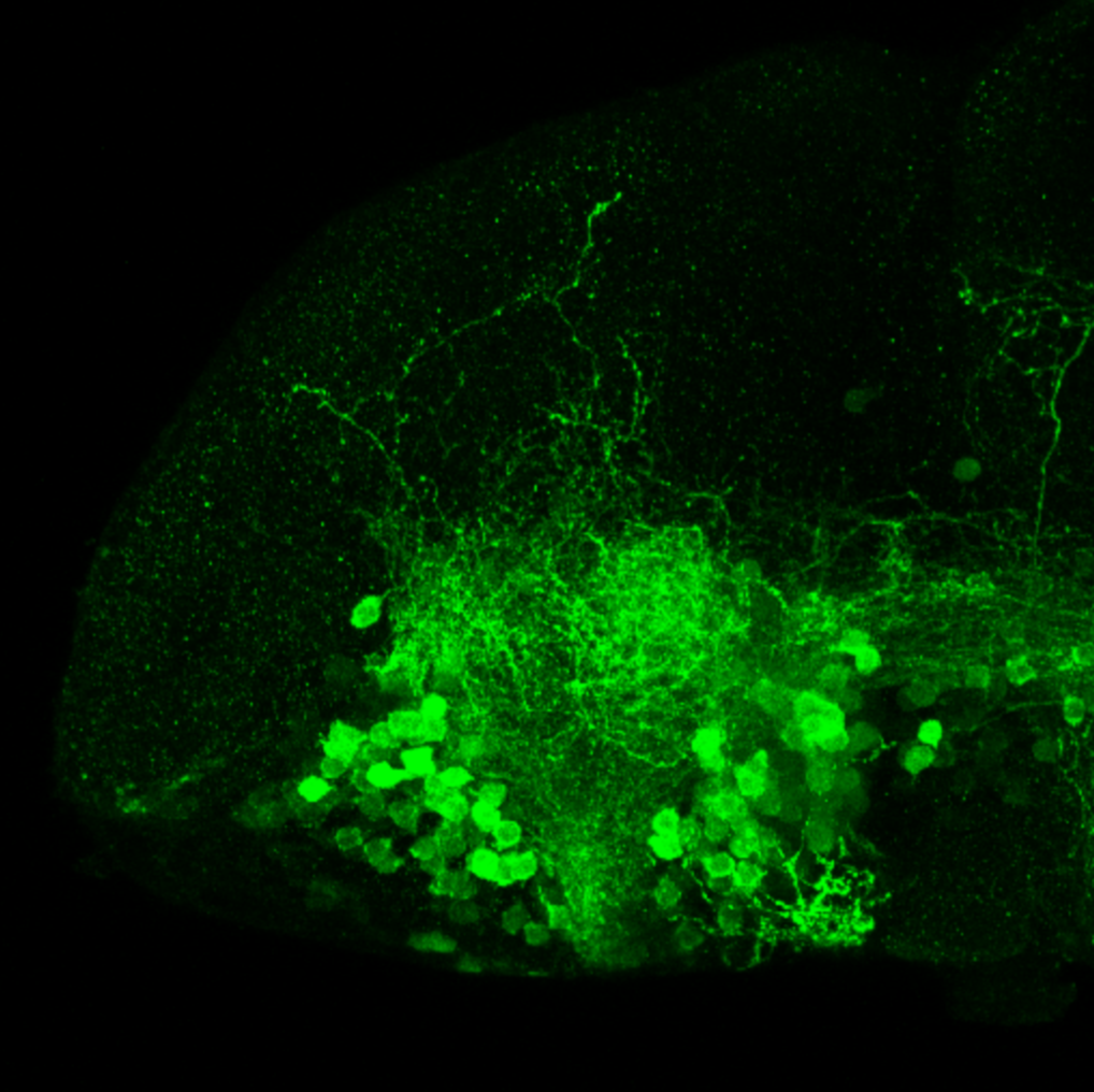
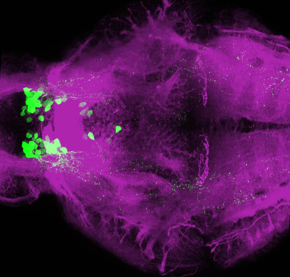
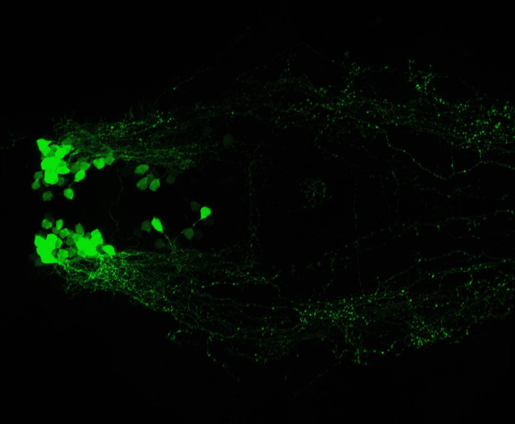
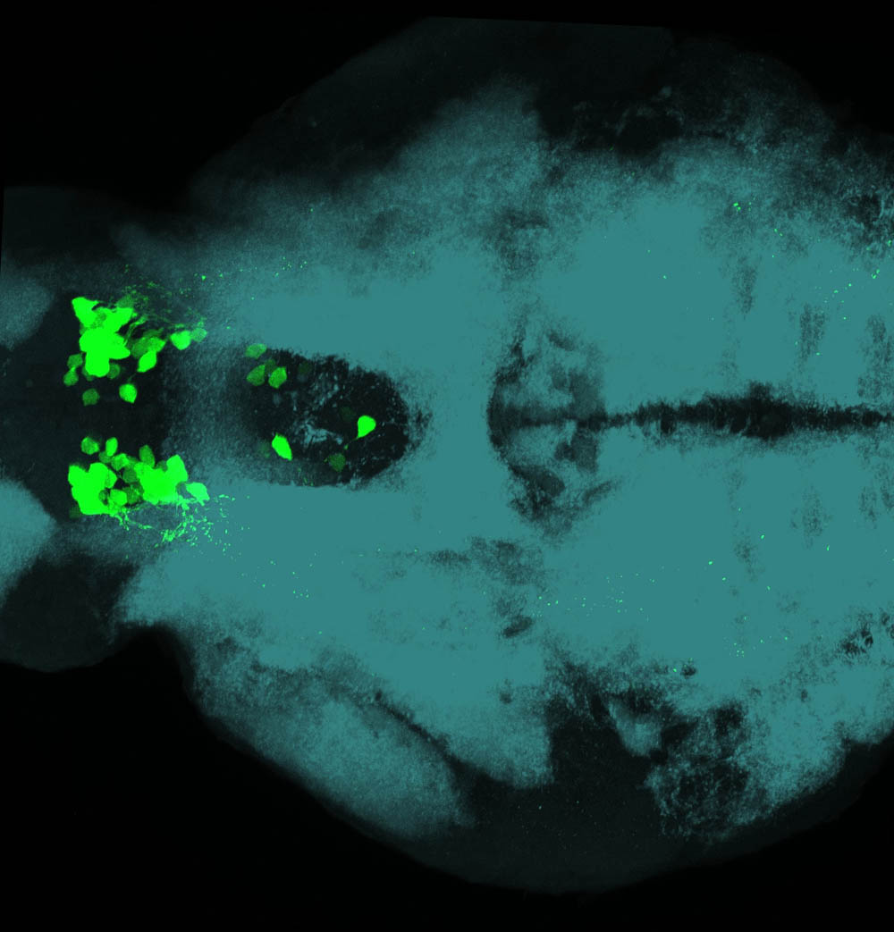
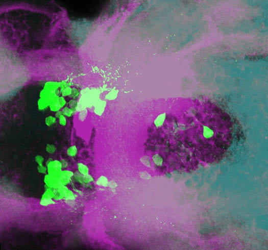
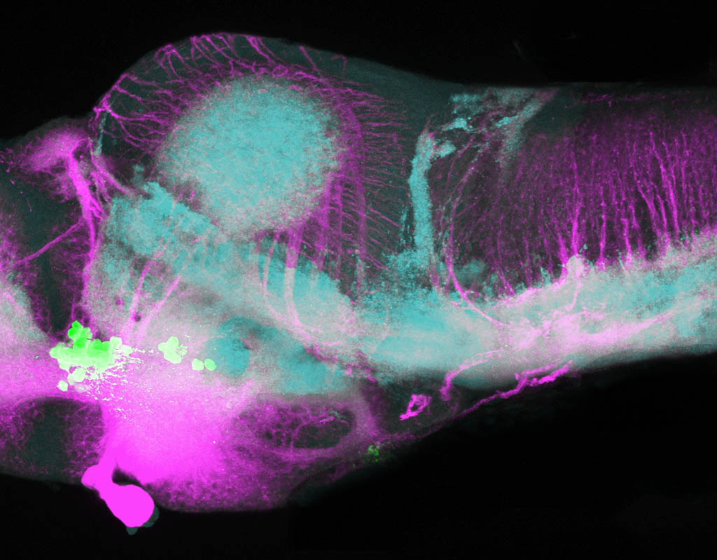
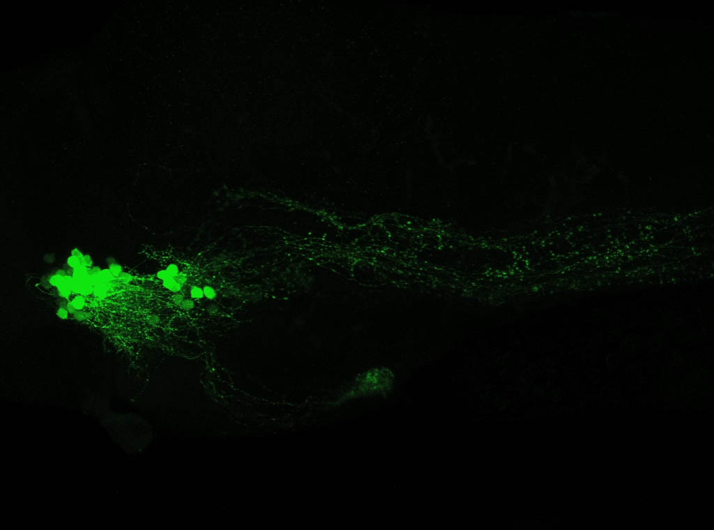
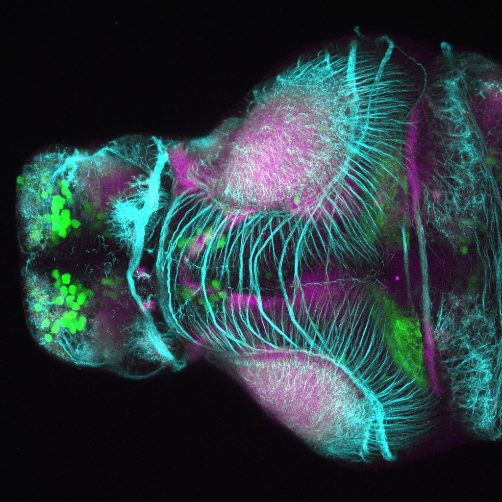
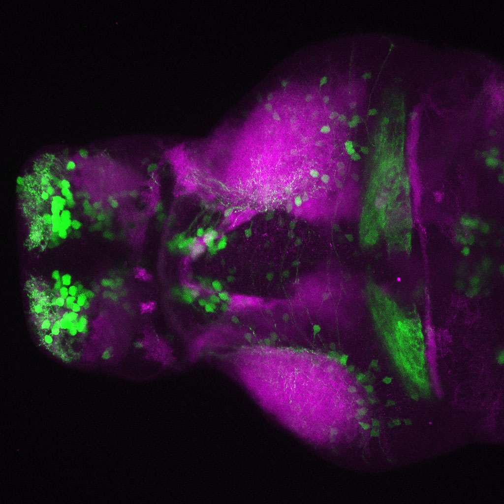
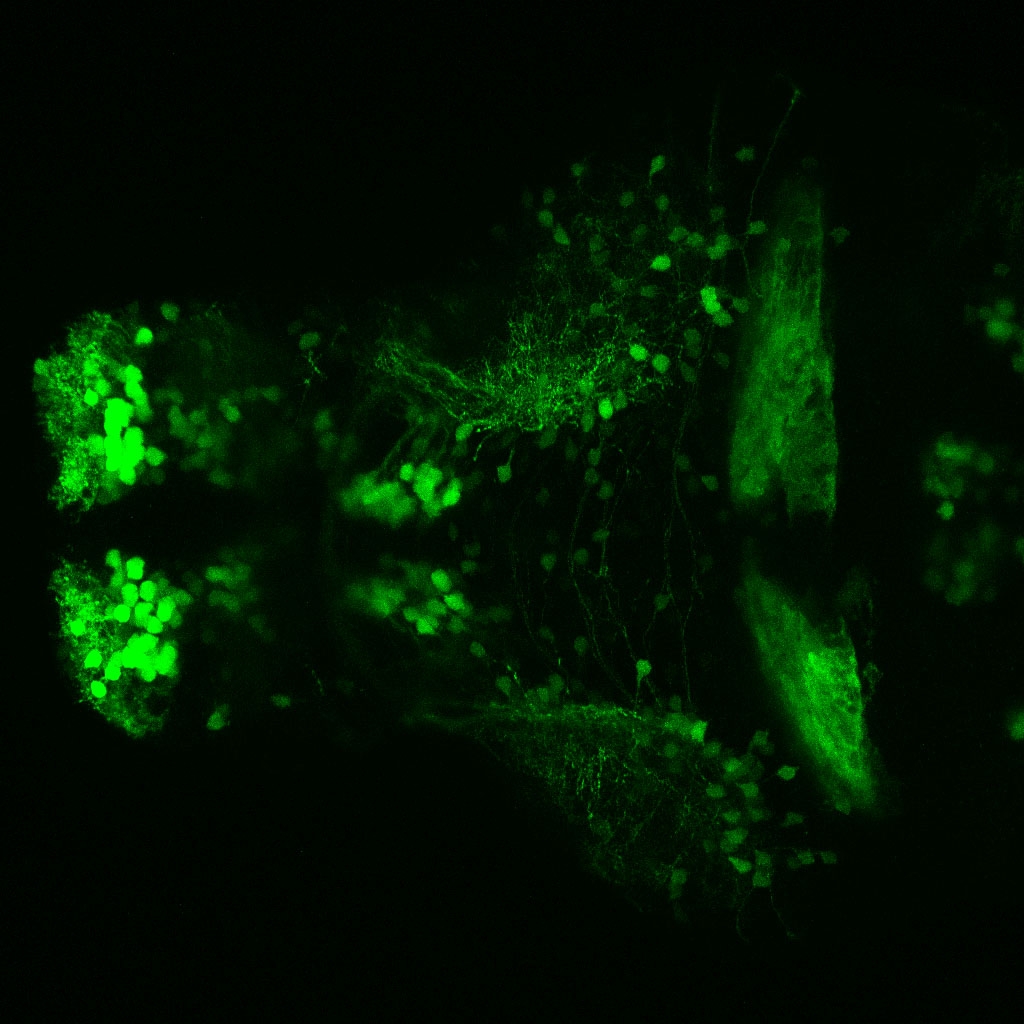
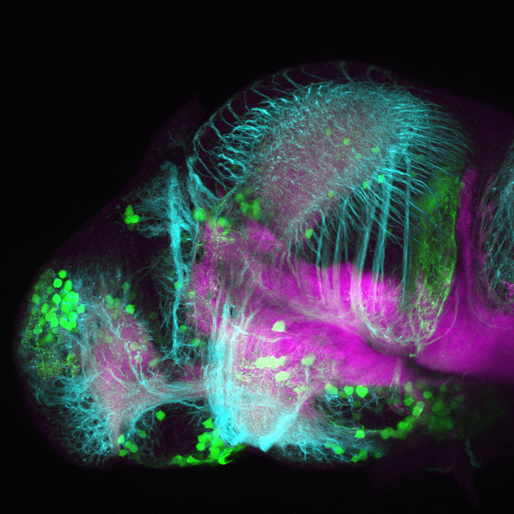
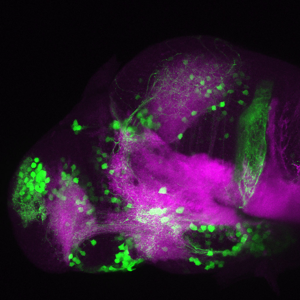
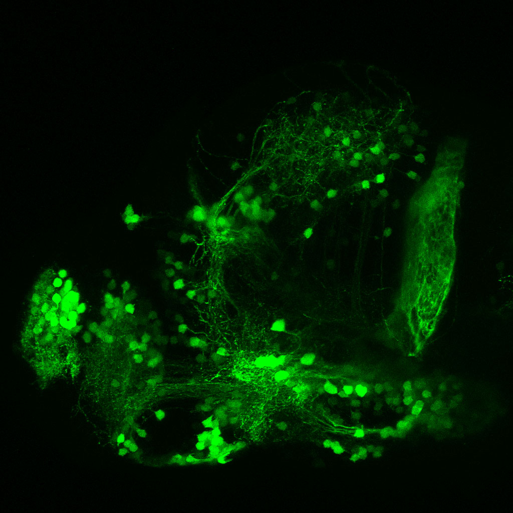
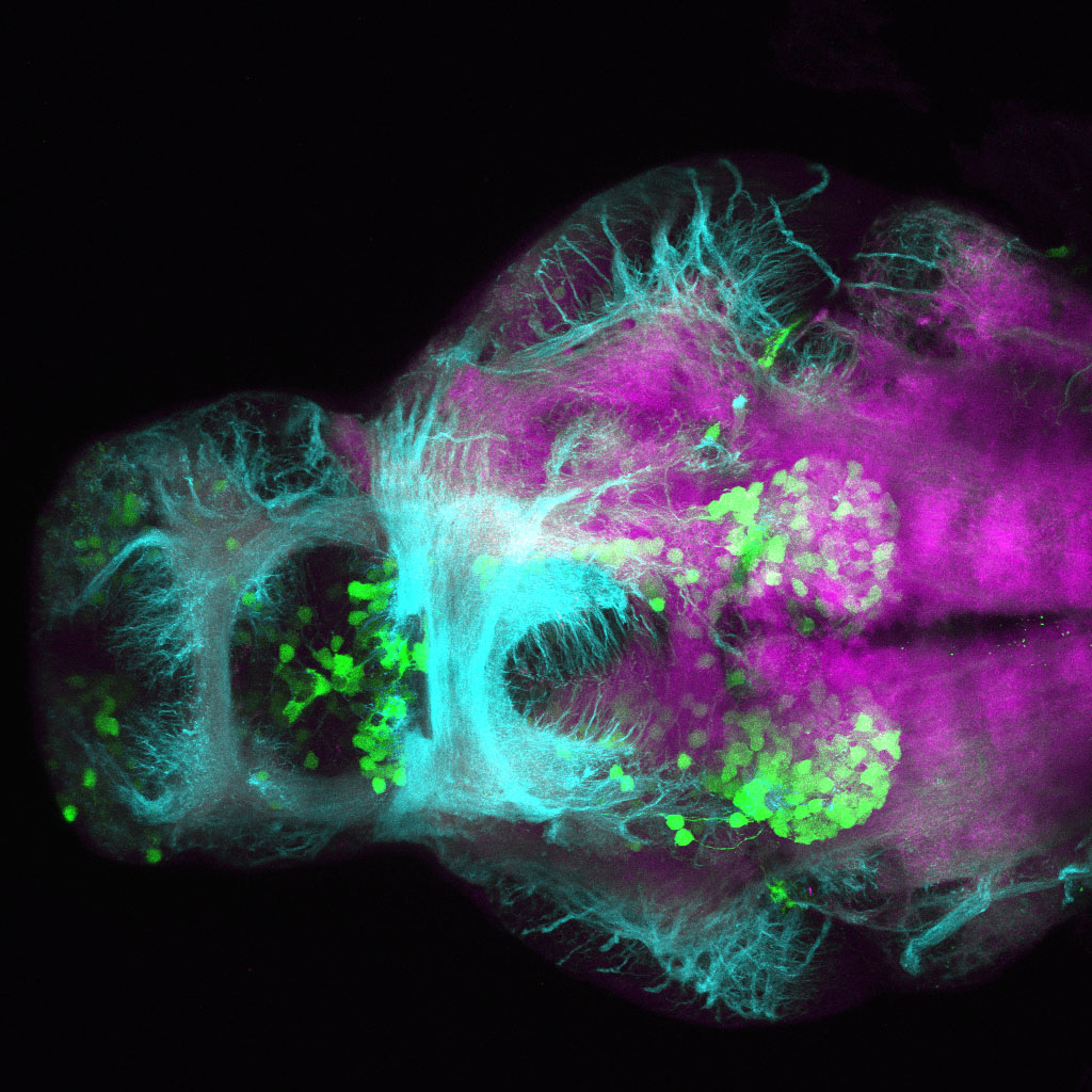
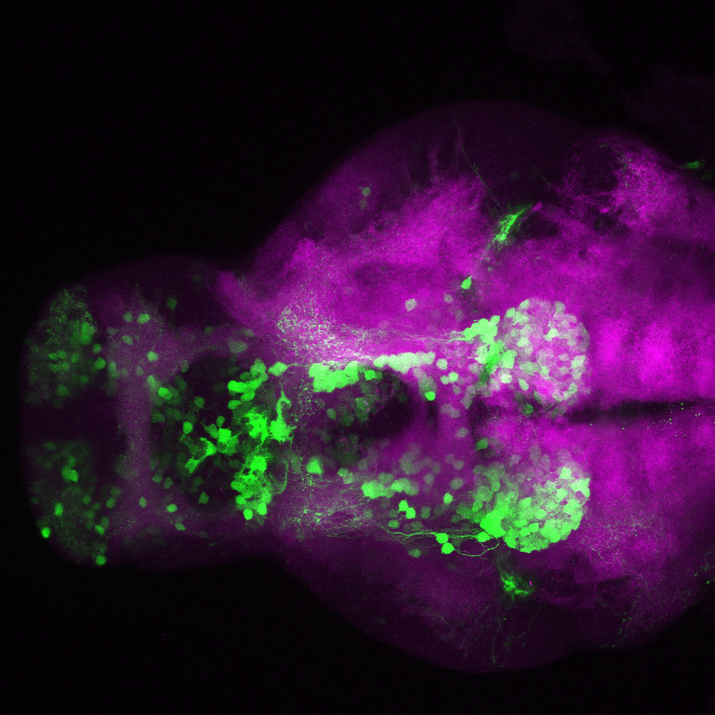
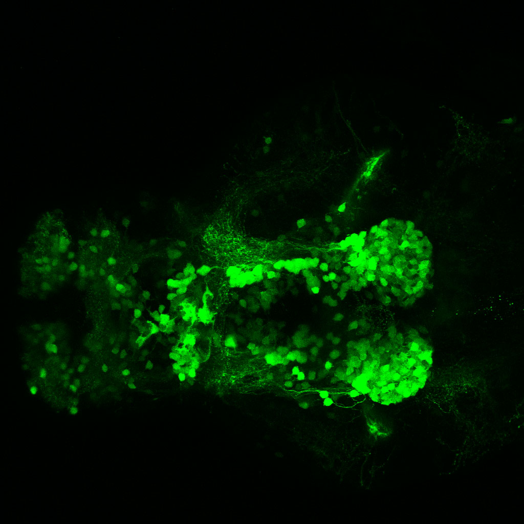
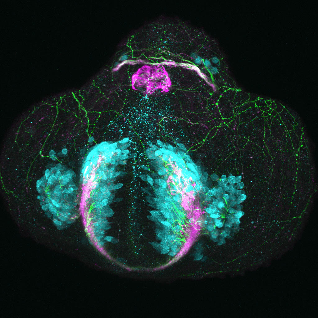
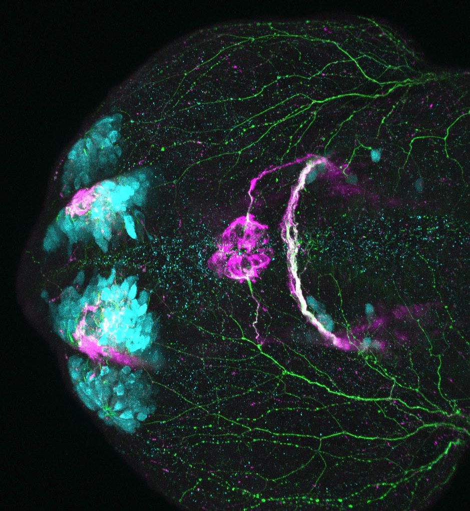
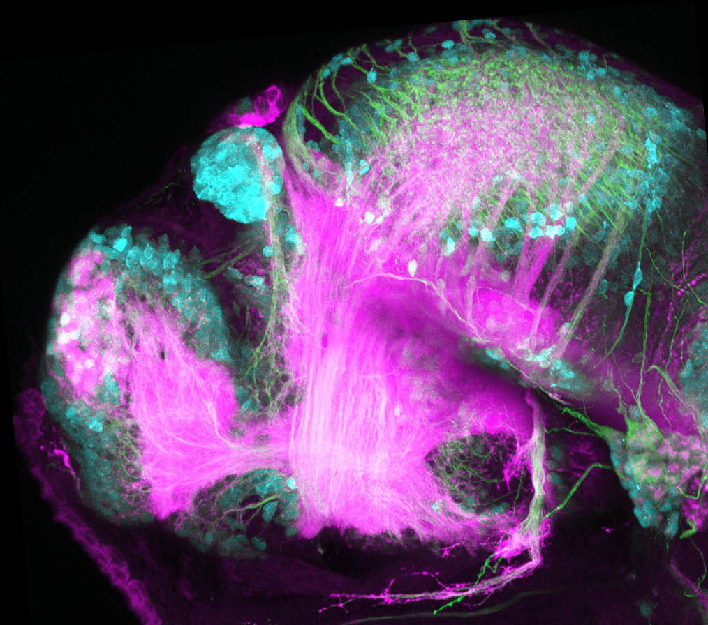
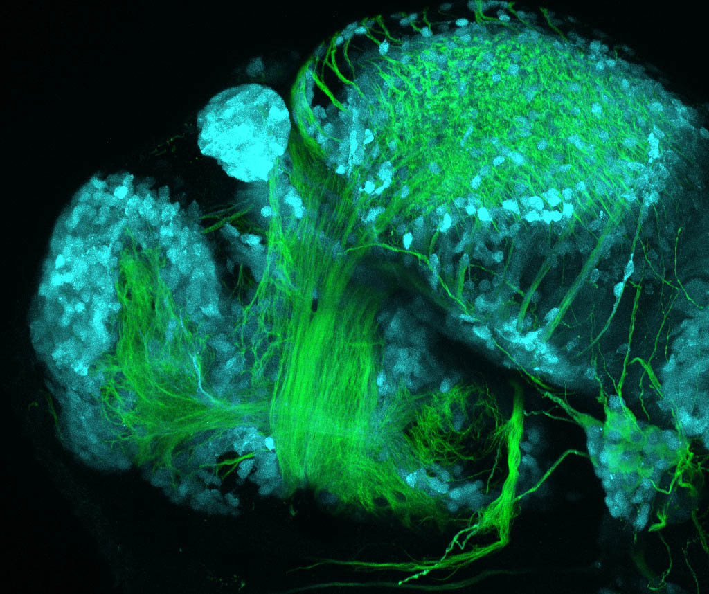
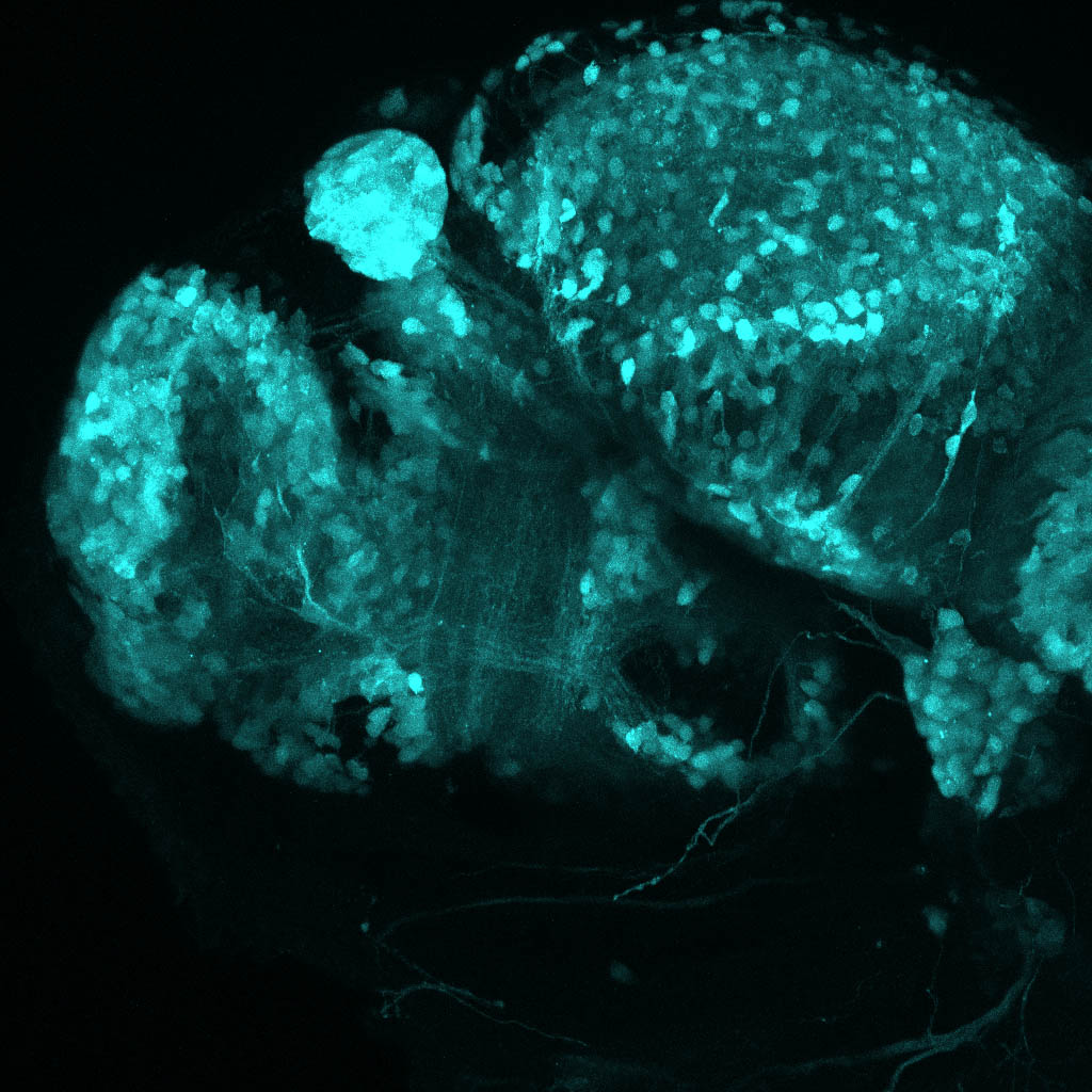
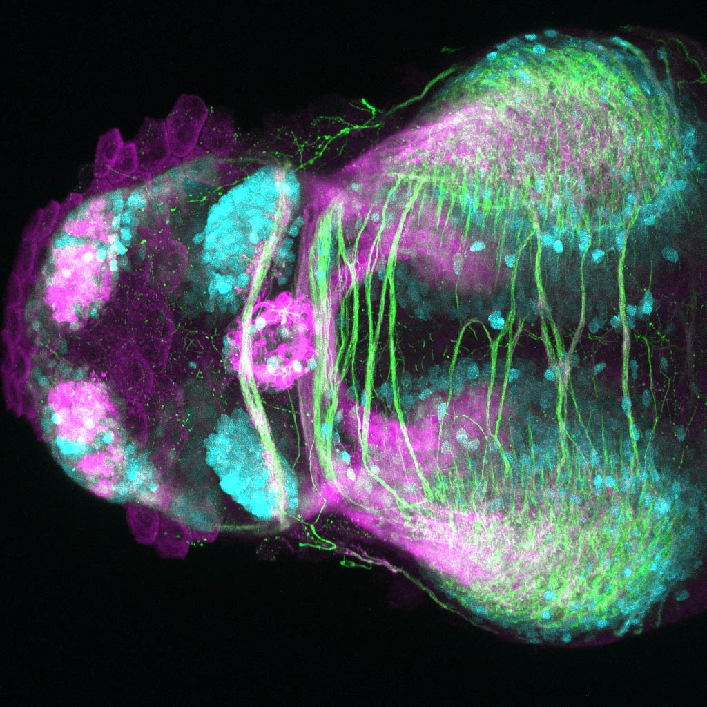
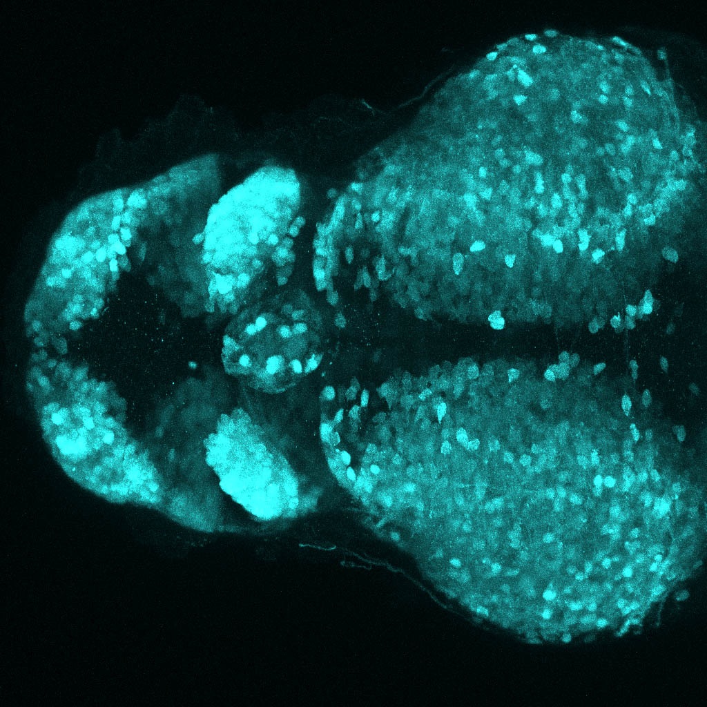
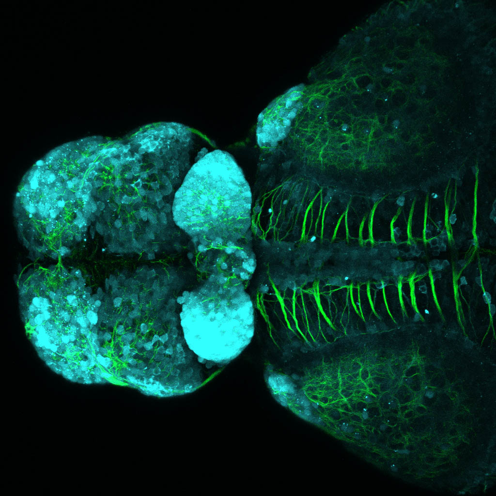
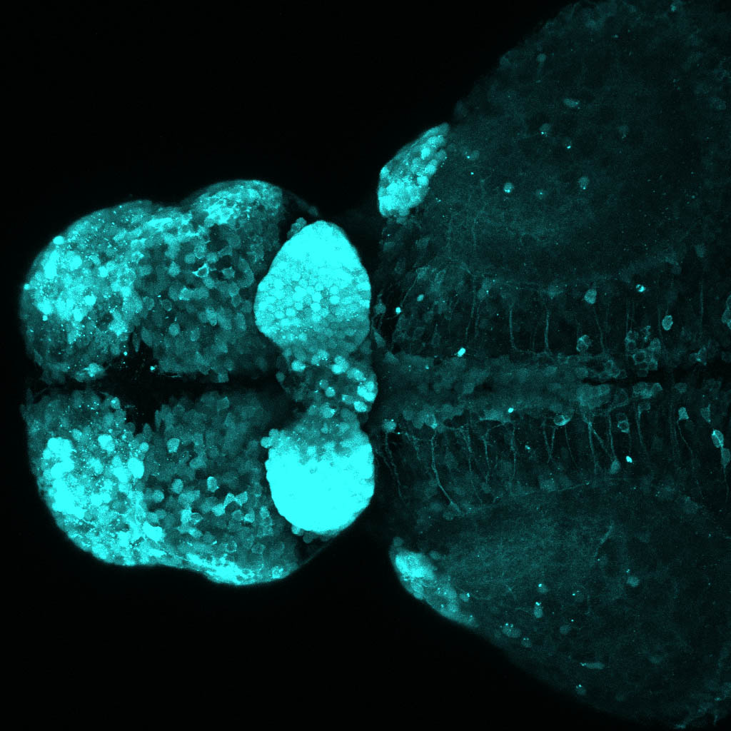
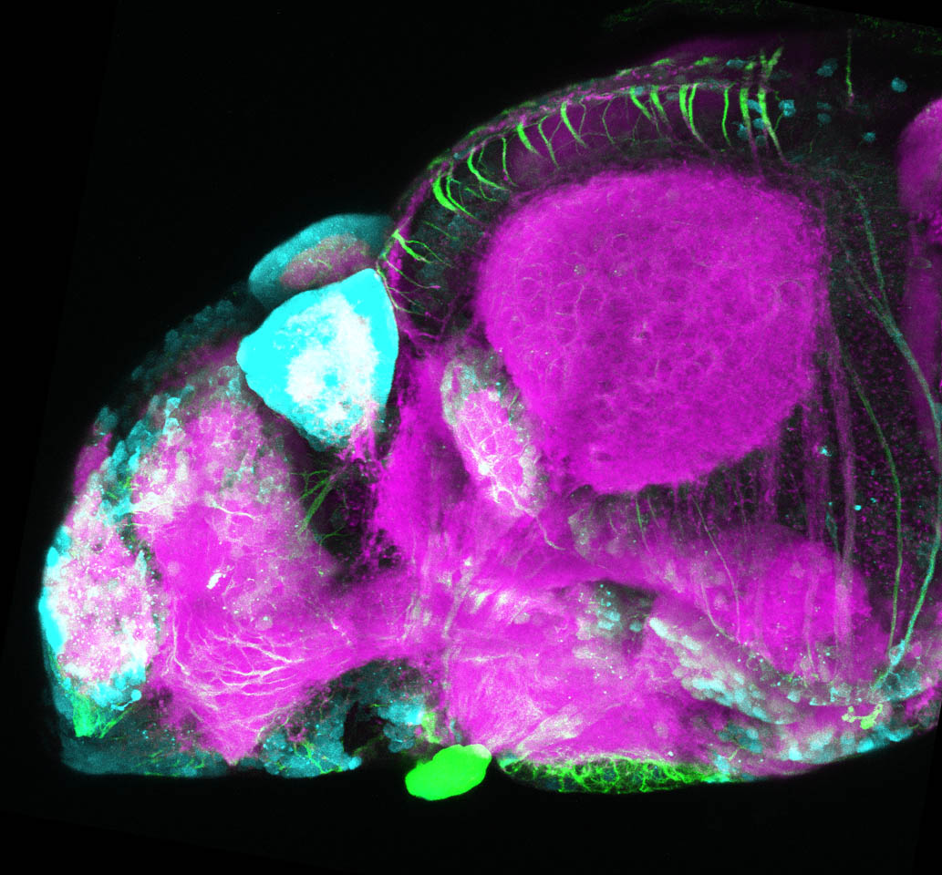
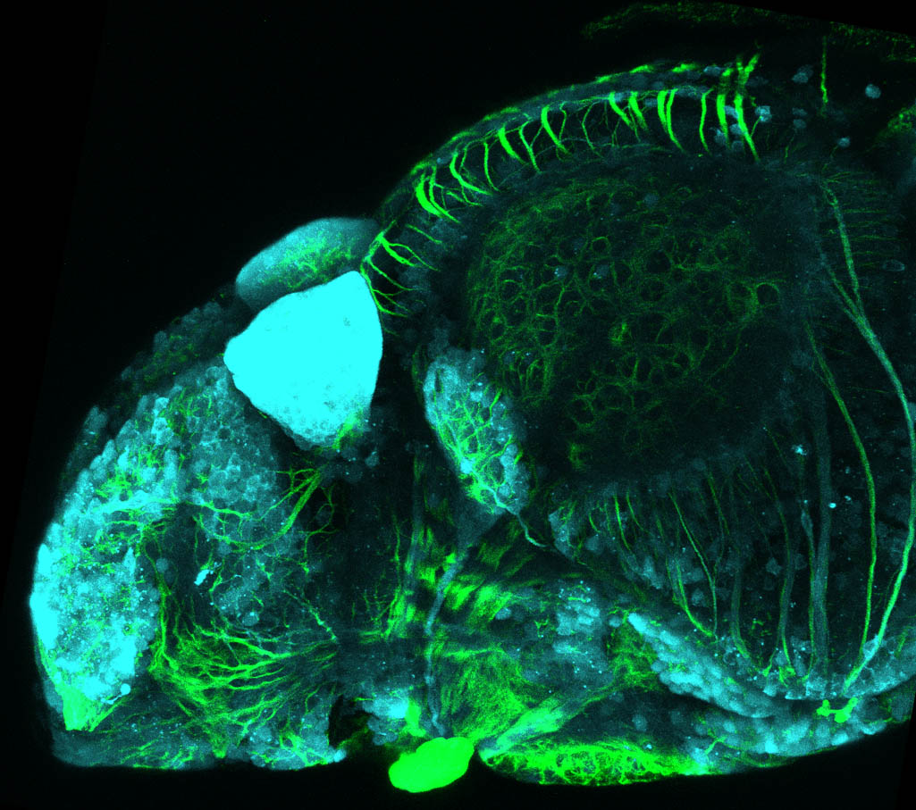
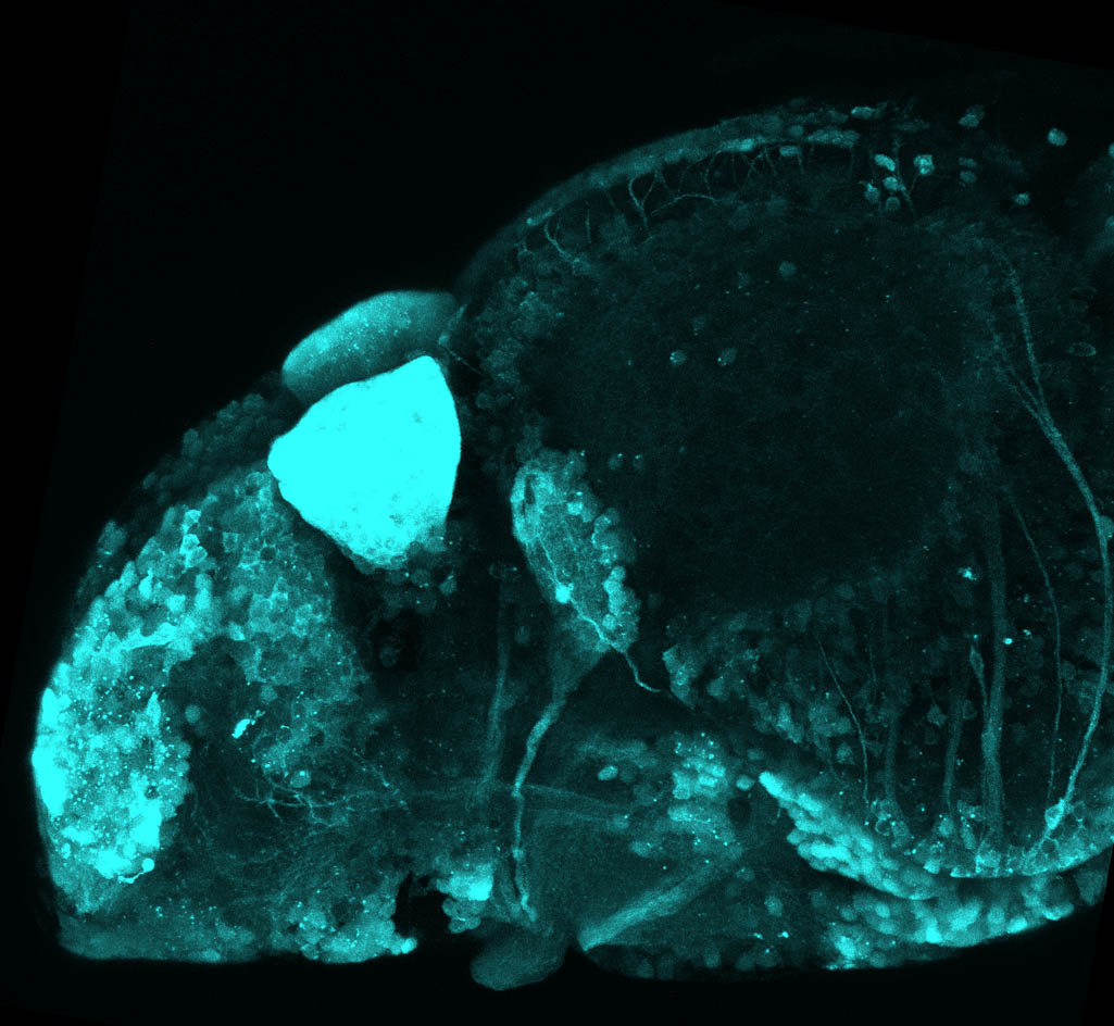
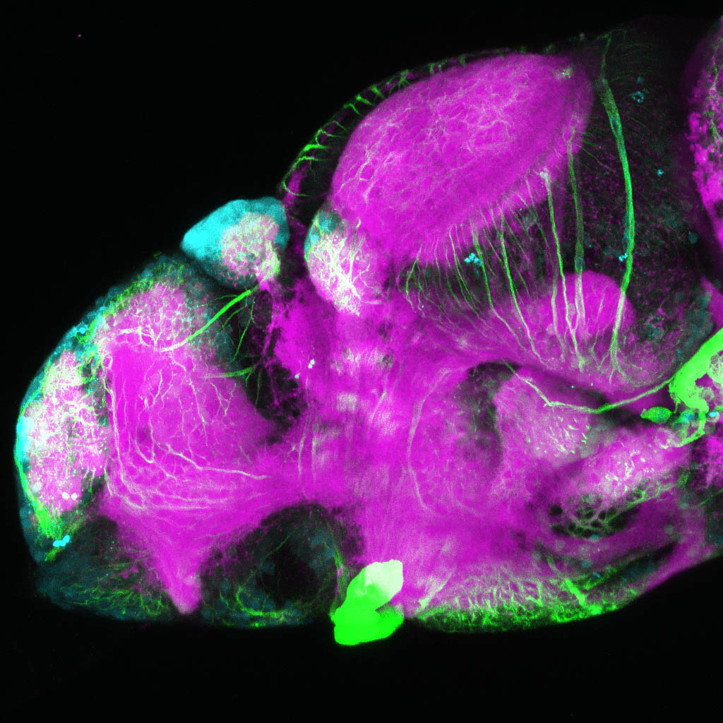
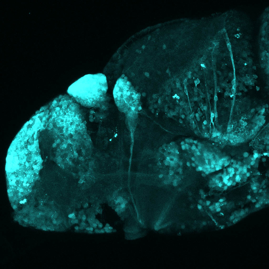
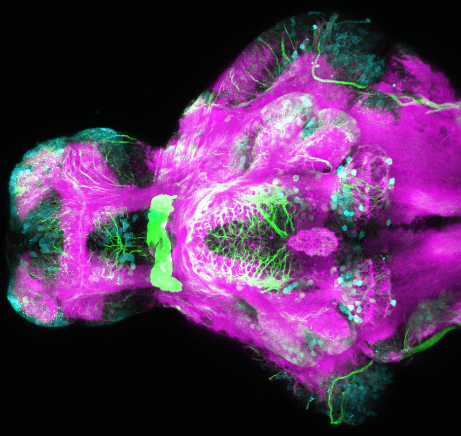
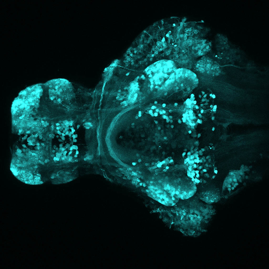
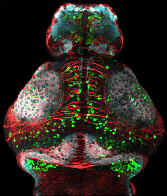
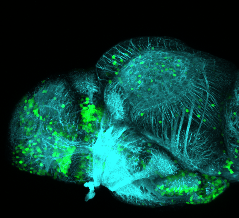
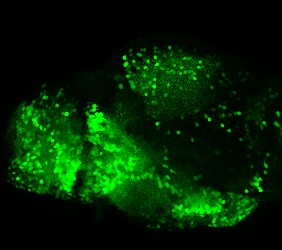
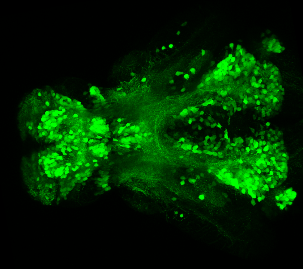
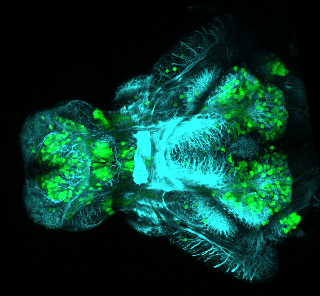
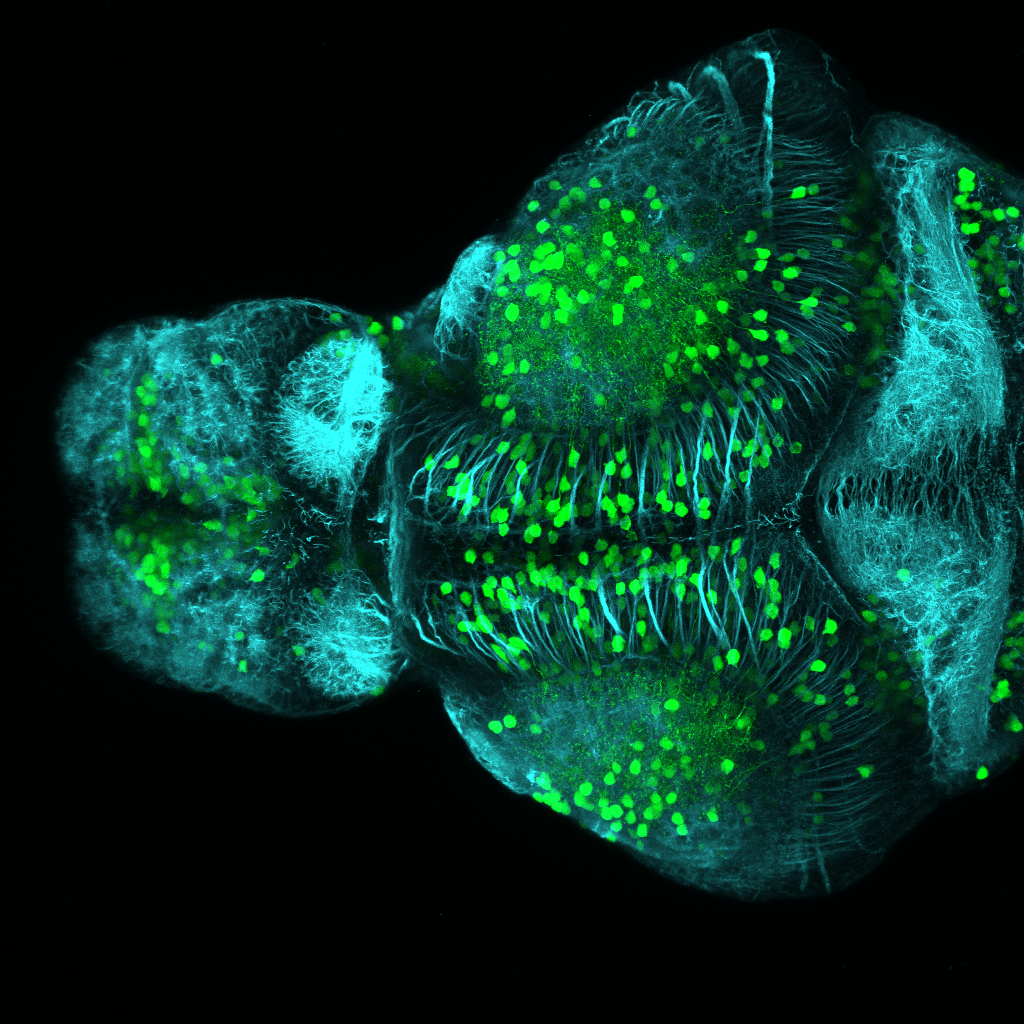
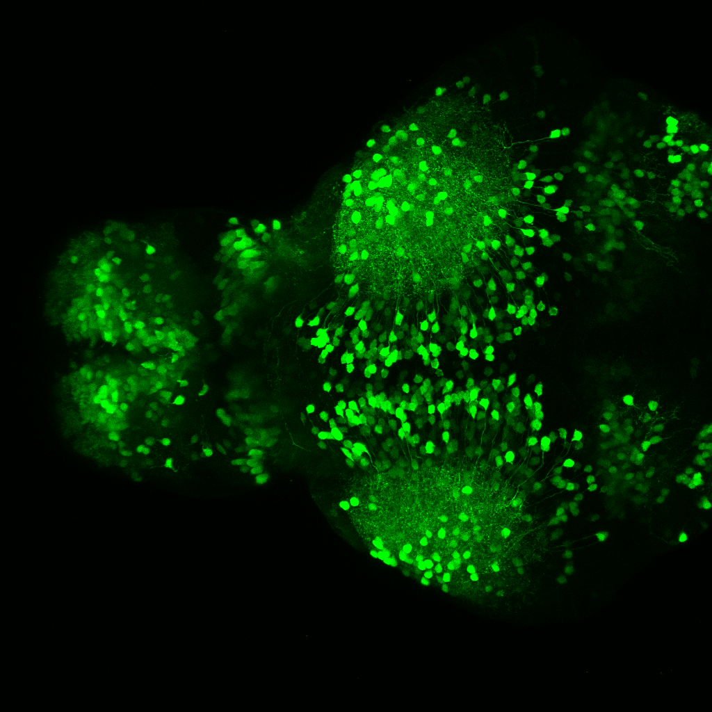
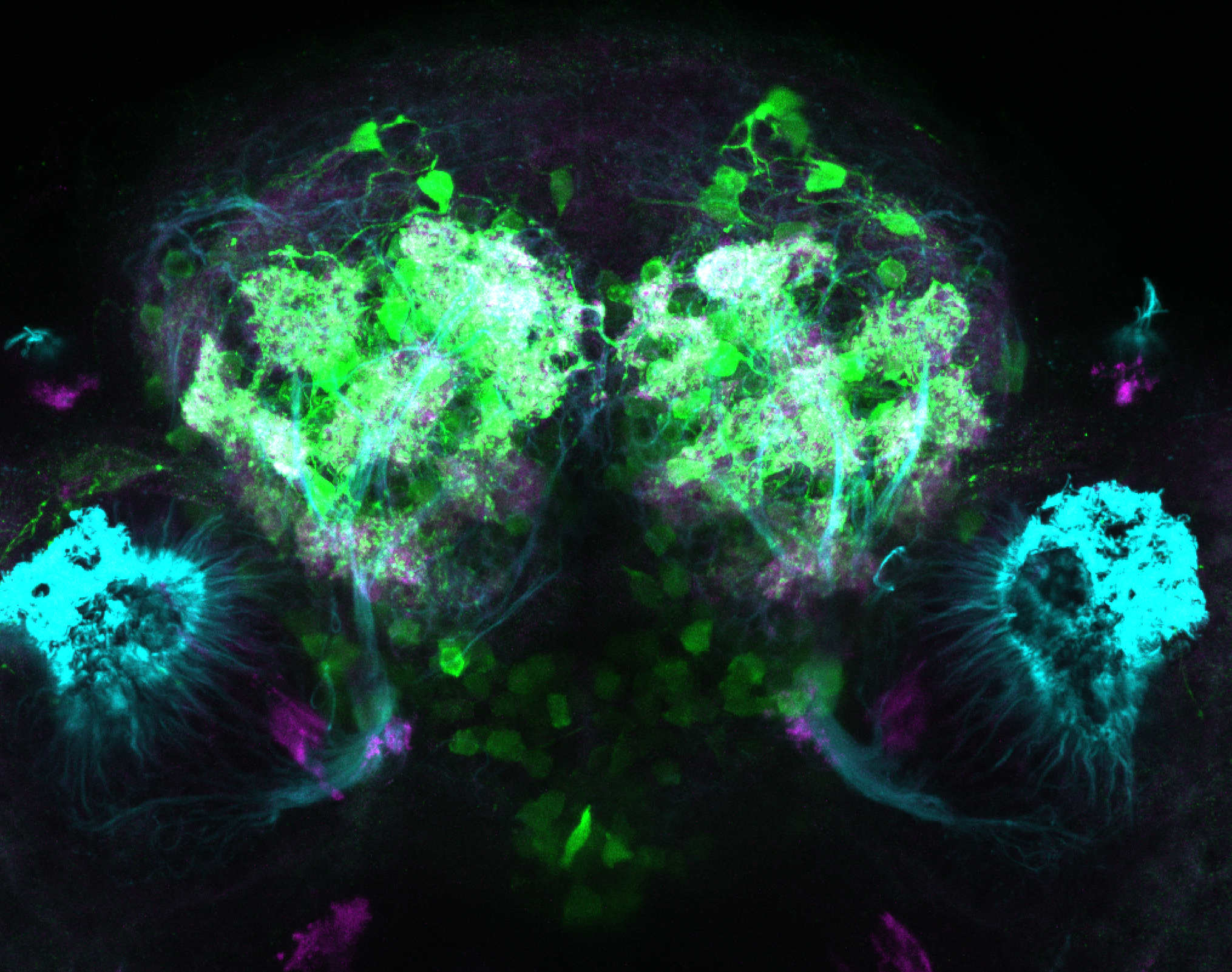
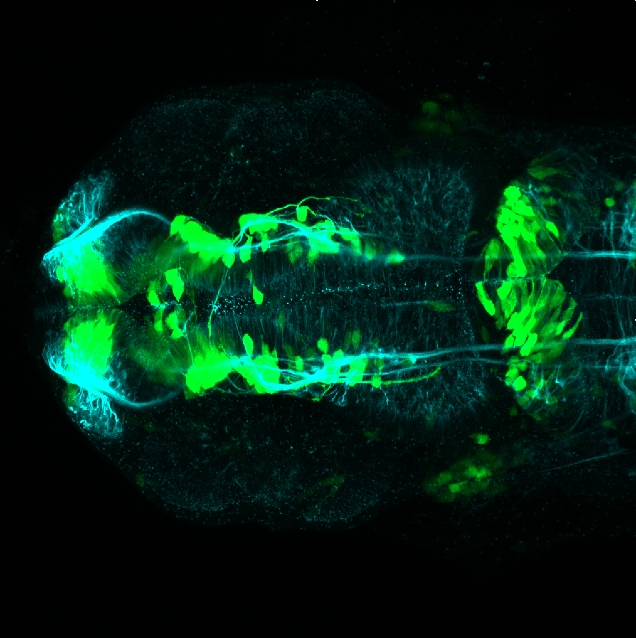
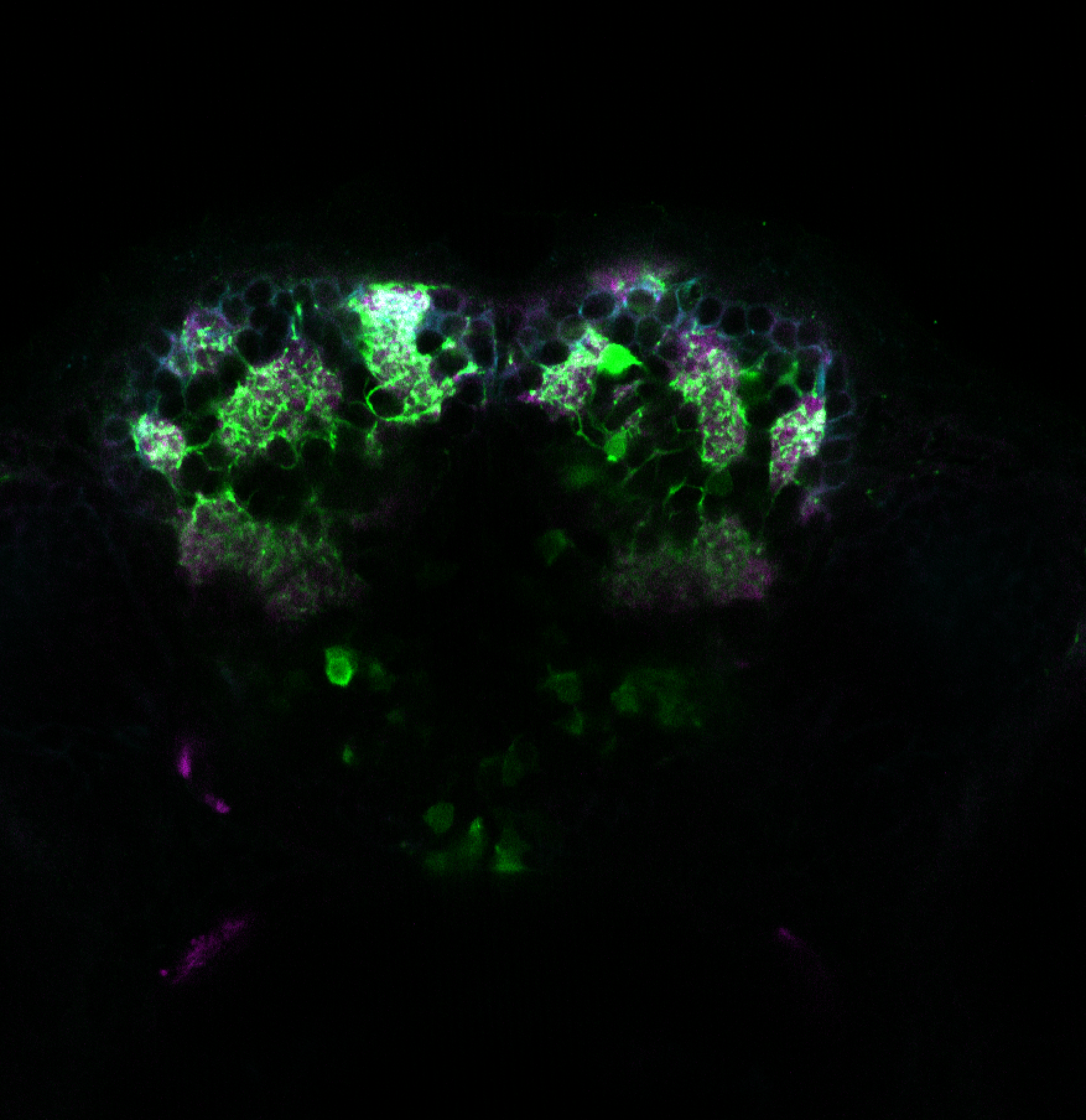
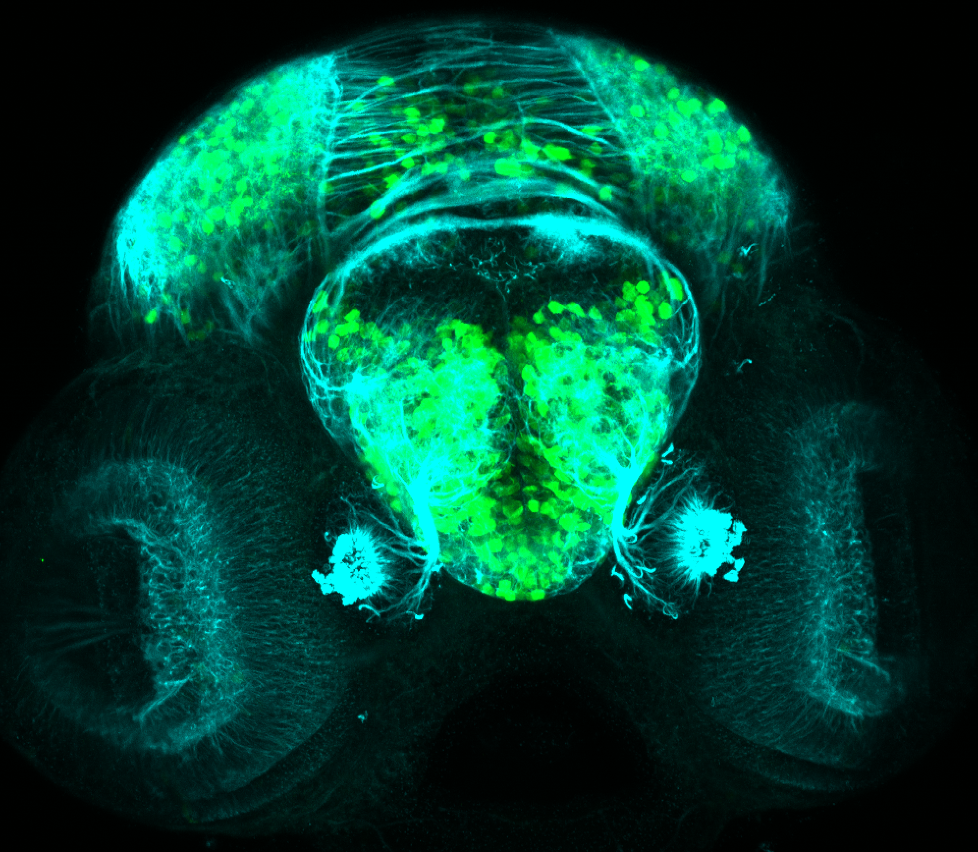
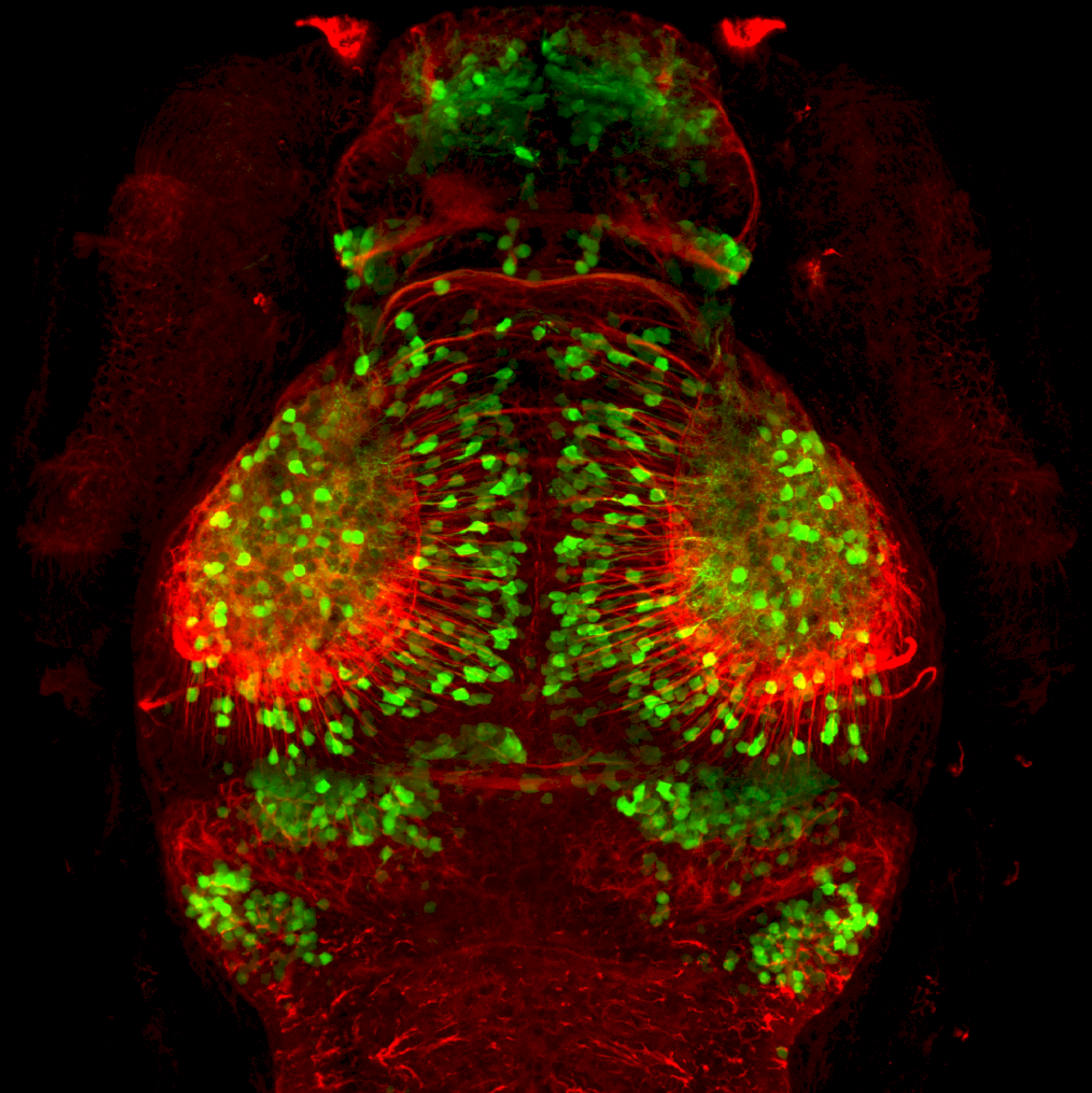
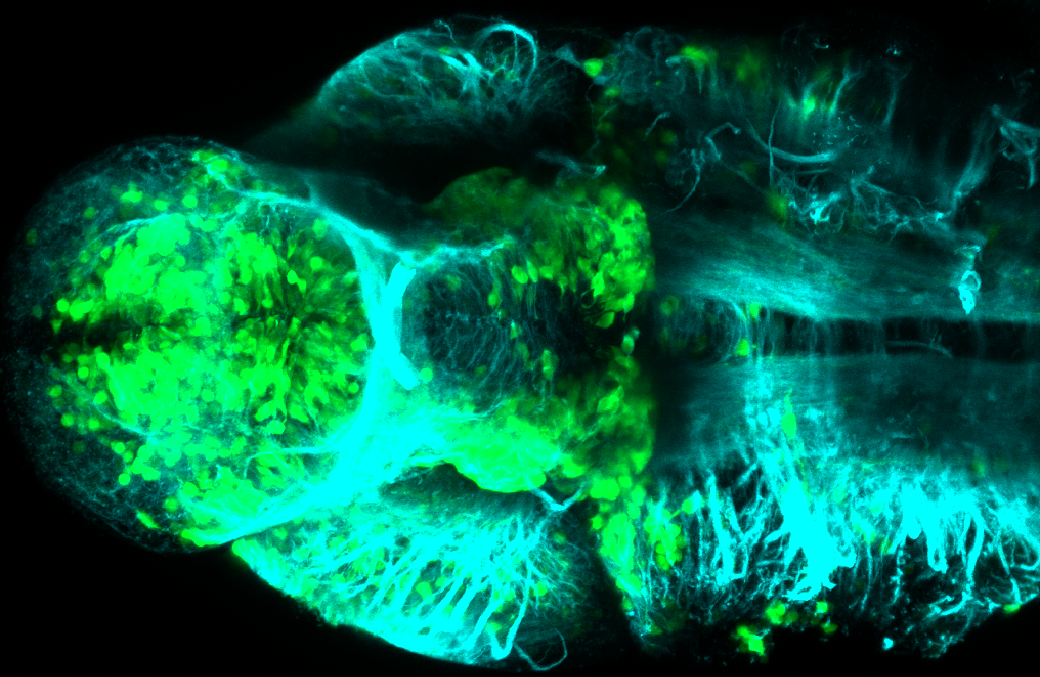
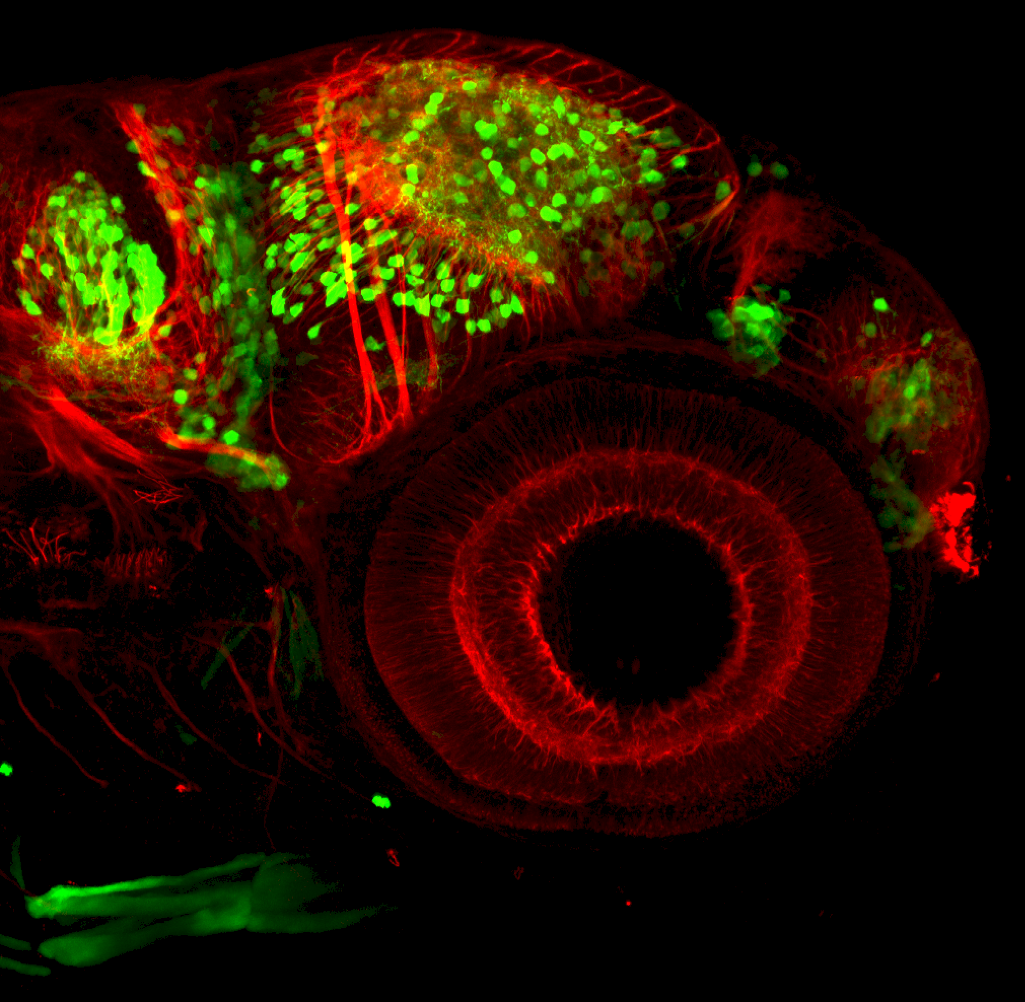
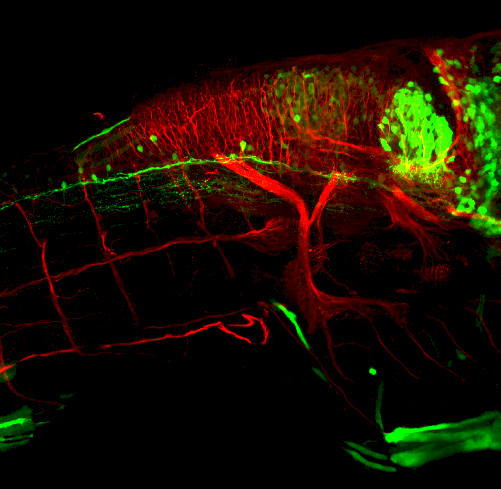
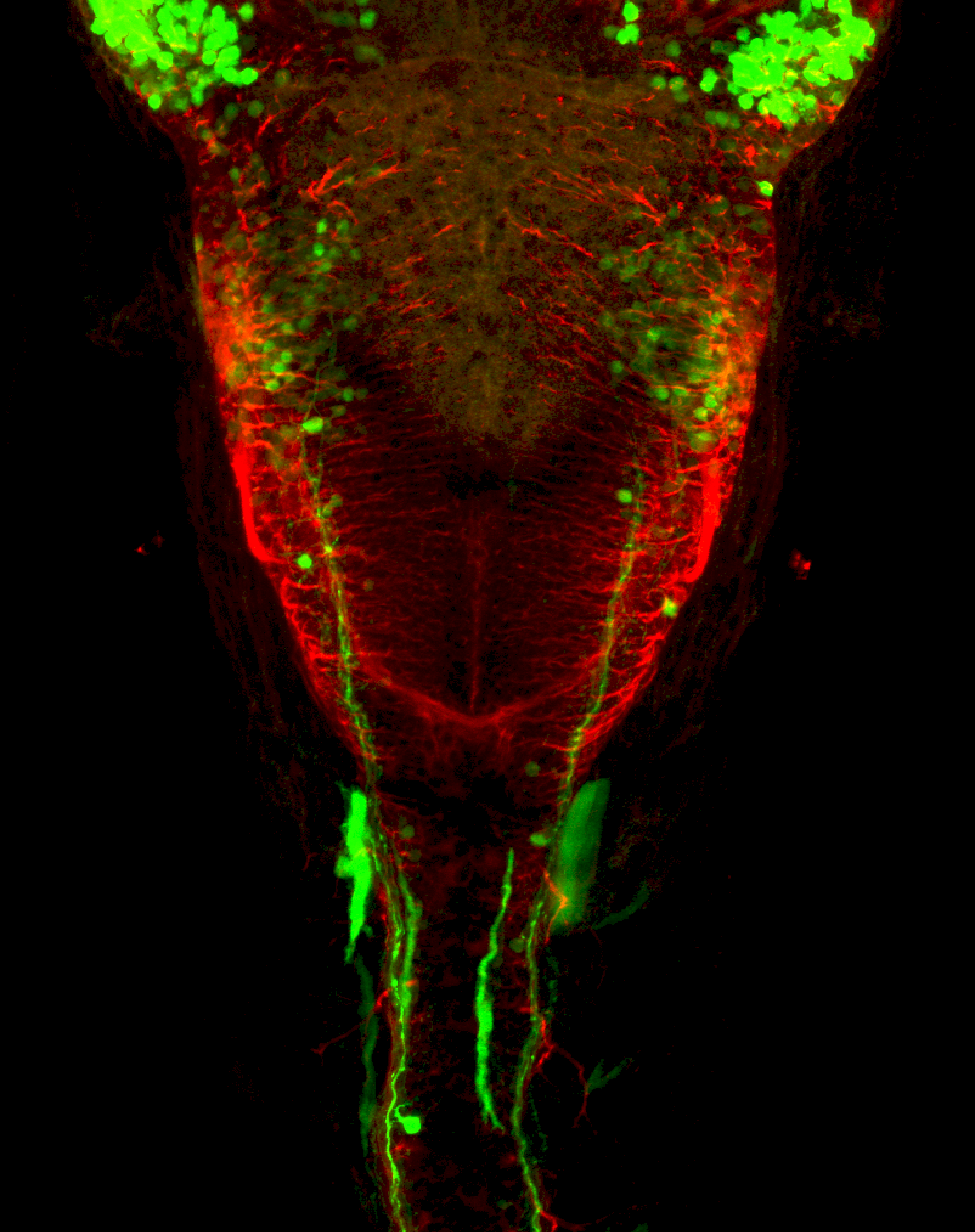
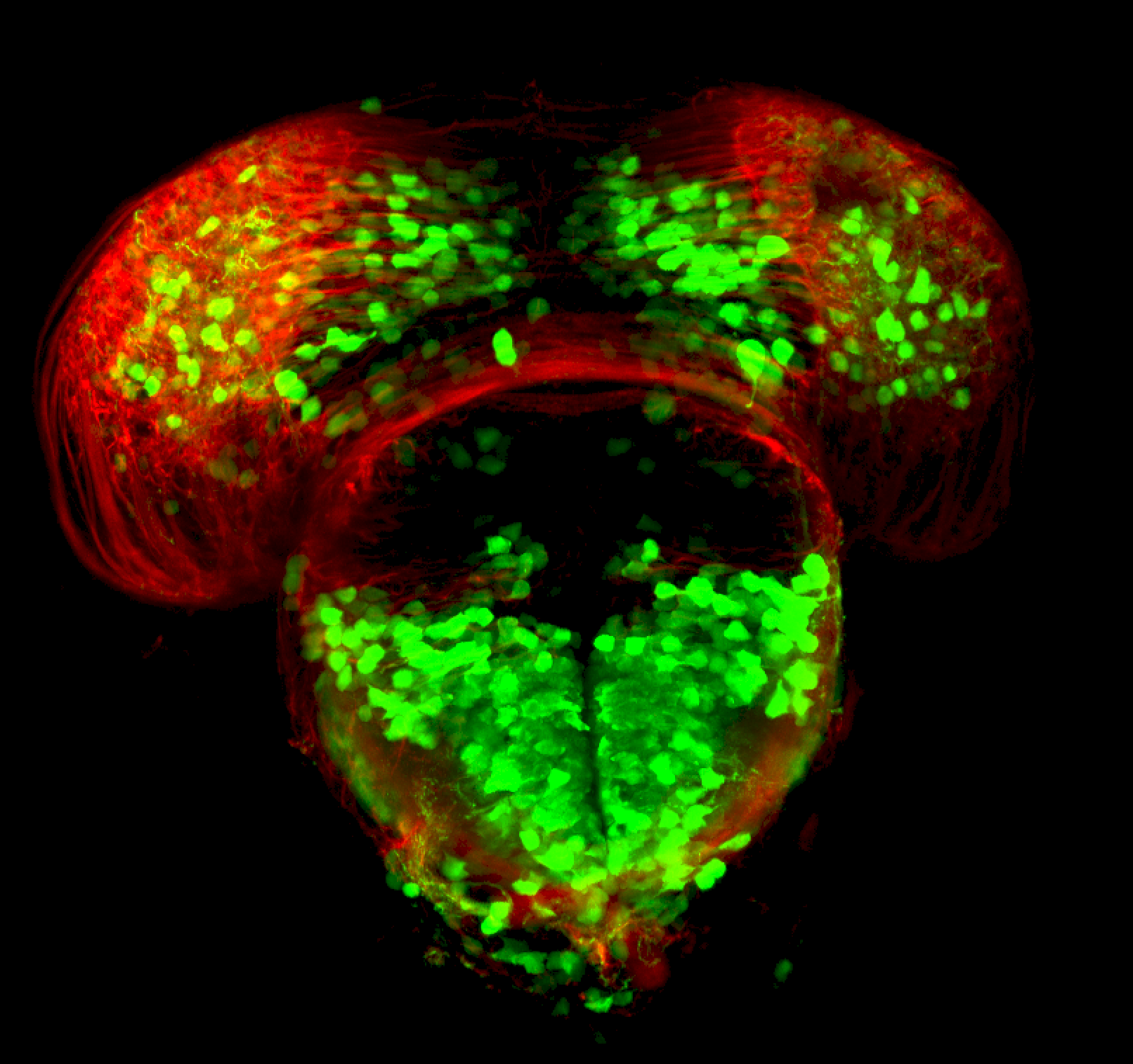
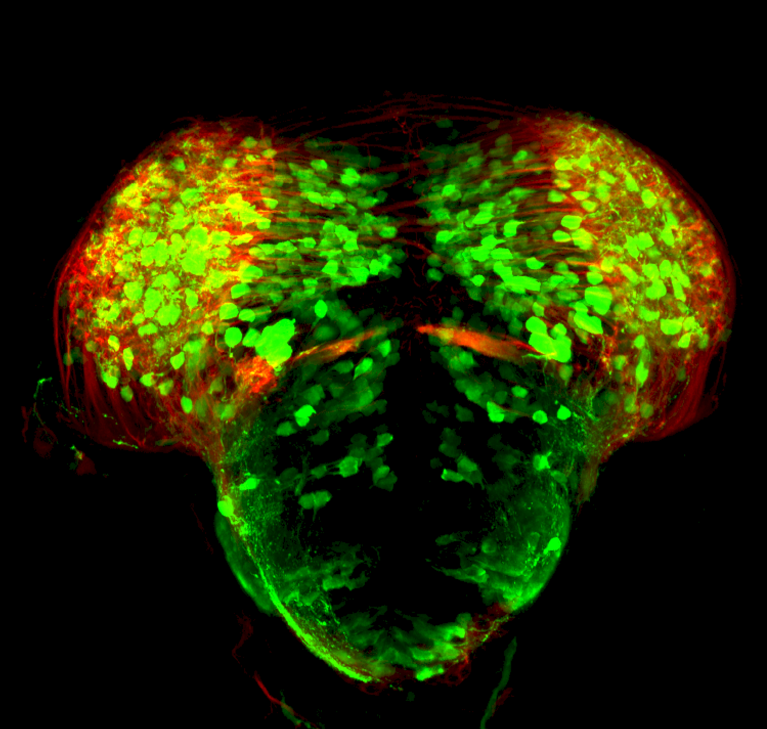
Tg(1.4dlx5a-dlx6a:GFP)ot1 larvae express GFP in subpallial neurons γ-aminobutyric acid (GABA)-expressing neurons. Several other brain regions also show GFP expression in this transgenic line such as the optic tectum and cerebellum.
 

 Dlx homeobox genes play essential roles in the differentiation, migration and survival of subpallial precursor cells that will later give rise to diverse subtypes of γ-aminobutyric acid (GABA)-expressing neurons. They also participate in the regulation of the Gad genes encoding the enzymes necessary for GABA synthesis (Yu et al., 2011).


Mouse over the different areas of GFP expression in the interactive images below to see the name of the brain area. 


More images

External Links:

 ZFIN

Lab of Origin: Mark Ekker Lab


Expressed in: 

olfactory bulb, subpallium, pallium, preoptic area, prethalamus, posterior tuberculum, hypothalamus, optic tectum, cerebellum.


Key Publications

Zerucha, T., Stuhmer, T., Hatch, G., Park, B.K., Long, Q., Yu, G., Gambarotta, A., Schultz, J.R., Rubenstein, J.L., and Ekker, M. (2000) 
A highly conserved enhancer in the Dlx5/Dlx6 intergenic region is the site of cross-regulatory interactions between dlx genes in the embryonic forebrain. 
The Journal of neuroscience : the official journal of the Society for Neuroscience. 20(2):709-721.

Yu, M., Xi, Y., Pollack, J., Debiais-Thibaud, M., Macdonald, R.B., and Ekker, M. (2011) 
Activity of dlx5a/dlx6a regulatory elements during zebrafish GABAergic neuron development. 
Int. J. Dev. Neurosci.. 29(7):681-91.

Tg(gata2:eGFP)bi105

Tg(gata2:eGFP)bi105

About

The Tg(gata2:eGFP)bi105 enhancer trap transgenic line was generated as part of an enhancer trap screen by the Becker Lab using the Tol2-transposase system. It has GFP expression throughout the pallium, in a small subpallial nucleus, parapineal organ, optic tectum, pretectum, posterior tuberculum and lateral hypothalamus. In the hindbrain, several cell bodies are labelled in the superior raphe and areas of the reticular formation and caudally in areas of the medulla oblongata


External Links:

 ZFIN

Lab of Origin: Tom Becker Lab


Transgene expressed in: 

Key Publications


Folgueira, M., Bayley, P., Navratilova, P., Becker, T.S., Wilson, S.W., and Clarke, J.D. (2012)
Morphogenesis underlying the development of the everted teleost telencephalon.
Neural Development. 7(1):32


Ragvin, A., Moro, E., Fredman, D., Navratilova, P., Drivenes, O., Engström, P.G., Alonso, M.E., Mustienes, E.D., Gomez Skarmeta, J.L., Tavares, M.J., Casares, F., Manzanares, M., van Heyningen, V., Molven, A., Njølstad, P.R., Argenton, F., Lenhard, B., and Becker, T.S. (2010)
Long-range gene regulation links genomic type 2 diabetes and obesity risk regions to HHEX, SOX4, and IRX3.
Proceedings of the National Academy of Sciences of the United States of America. 107(2):775-780

Turner, K.J., Hawkins, T.A., Yáñez, J., Anadón, R., Wilson, S.W., Folgueira, M. (2016)
Afferent Connectivity of the Zebrafish Habenulae.
Frontiers in neural circuits. 10:30


Tg(isl1:GFP)rw0

Tg(isl1:GFP)rw0

About

 This transgenic line expresses GFP in cranial motor neurons. It was made using a construct that fuses Islet-1 promotor/enhancer sequences to GFP.

isl1 cranial nerves expanded-01.png

isl1 cranial nerves expanded-02.png

More images of this transgenic

External Links:

 ZFIN

Lab or Origin: Okomoto Lab


Expressed in: 

cranial motor neurons, hindbrain, subpallium, preoptic area. 


Key Publications

Higashijima, S., Hotta, Y., and Okamoto, H. (2000)
Visualization of cranial motor neurons in live transgenic zebrafish expressing green fluorescent protein under the control of the islet-1 promoter/enhancer.
The Journal of neuroscience. 20(1):206-218

Suli, A., Mortimer, N., Shepherd, I., and Chien, C.B. (2006)
Netrin/DCC signaling controls contralateral dendrites of octavolateralis efferent neurons.
The Journal of neuroscience. 26(51):13328-13337.

Schoppik, D., Bianco, I.H., Prober, D.A., Douglass, A.D., Robson, D.N., Li, J.M.B., Greenwood, J.S.F., Soucy, E., Engert, F., Schier, A.F. (2017)
Gaze-stabilizing central vestibular neurons project asymmetrically to extraocular motoneuron pools. The Journal of neuroscience. 37(47):11353-11365.

Rebman, J.K., Kirchoff, K.E., Walsh, G.S. (2016)
Cadherin-2 Is Required Cell Autonomously for Collective Migration of Facial Branchiomotor Neurons.
PLoS One. 11:e0164433.

Barsh, G.R., Isabella, A.J., Moens, C.B. (2017)
Vagus Motor Neuron Topographic Map Determined by Parallel Mechanisms of hox5 Expression and Time of Axon Initiation.
Current biology : CB. 27(24):3812-3825.e3.

Cox, J.A., Lamora, A., Johnson, S.L., and Voigt, M.M. (2011)
Diverse mechanisms for assembly of branchiomeric nerves.
Developmental Biology. 357(2):305-17.

Tg(oxt:EGFP)

Tg(oxt:EGFP)

About

 This transgenic construct drives GFP expression in oxytocin expressing neurons in the neuro-secretary pre-optic area(NPO) part of the neuroendocrine system.


External Links:

 ZFIN

Lab or Origin: Gil Levkowitz Lab


Expressed in: 

preoptic area, neurosecretory preoptic area (NPO).  


Key Publications

Gutnick, A., Blechman, J., Kaslin, J., Herwig, L., Belting, H.G., Affolter, M., Bonkowsky, J.L., and Levkowitz, G. (2011)
The hypothalamic neuropeptide oxytocin is required for formation of the neurovascular interface of the pituitary.
Developmental Cell. 21(4):642-654.


Machluf, Y., Gutnick, A., and Levkowitz, G. (2011)
Development of the zebrafish hypothalamus.
Annals of the New York Academy of Sciences. 1220(1):93-105.

Tg(slc6a3:EGFP)ot80

Tg(slc6a3:EGFP)ot80

About

The Tg(slc6a3:EGFP)ot80 transgenic line expresses GFP under the control of
cis-regulatory elements of the dopamine transporter (DAT) gene slc6a3.


External Links:

 ZFIN

Lab of Origin: Mark Ekker Lab


Transgene expressed in: 

olfactory bulb, subpallium, preoptic region, pretectum, posterior tuberculum, hypothalamus, optic tectum, cerebellum

Key Publications


Xi Y1, Yu M, Godoy R, Hatch G, Poitras L, Ekker M.
Transgenic zebrafish expressing green fluorescent protein in dopaminergic neurons of the ventral diencephalon.
Dev Dyn. 2011 Nov;240(11):2539-47. doi: 10.1002/dvdy.22742. Epub 2011 Sep 19.


Tg(slc17a6b: DsRed)nns9Tg

Tg(slc17a6b: DsRed)nns9Tg

About

 Slc17a6b is a vesicular glutamate transporter that that mediates the uptake of the excitatory neurotransmitter glutamate into vesicles in the presynaptic terminals of excitatory neurons. This BAC transgenic line from the Yoshihara lab drives the expression of DSRed in glutamatergic neurons. Expression can be seen in many neurons throughout the brain. Strong expression in the dorsal and ventral habenular subnuclei, olfactory bulbs, the pallium and optic tectum


External Links:

 ZFIN

Lab or Origin: Yoshihara Lab


Expressed in: 

 glutamatergic neurons, olfactory epithelium, olfactory bulb, pallium, subpallium, dorsal habenula, ventral habenula, preoptic area, prethalamus, pretectum (AF9), optic tectum, trigeminal sensory ganglion.


Key Publications

 
Miyasaka, N., Morimoto, K., Tsubokawa, T., Higashijima, S., Okamoto, H., and Yoshihara, Y. (2009)
From the olfactory bulb to higher brain centers: genetic visualization of secondary olfactory pathways in zebrafish.
The Journal of neuroscience. 29(15):4756-4767.

Kani, S., Bae, Y.K., Shimizu, T., Tanabe, K., Satou, C., Parsons, M.J., Scott, E., Higashijima, S.I., and Hibi, M. (2010) Proneural gene-linked neurogenesis in zebrafish cerebellum.
Developmental Biology. 343(1-2):1-17.