Viewing entries tagged
tegmentum

Et(fos:Gal4-VP16)s1168t

Et(fos:Gal4-VP16)s1168t

About

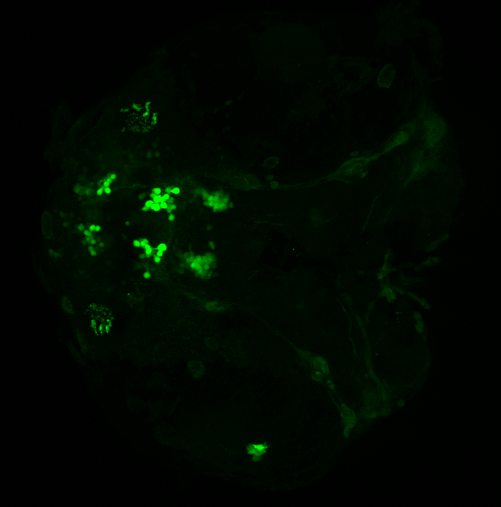
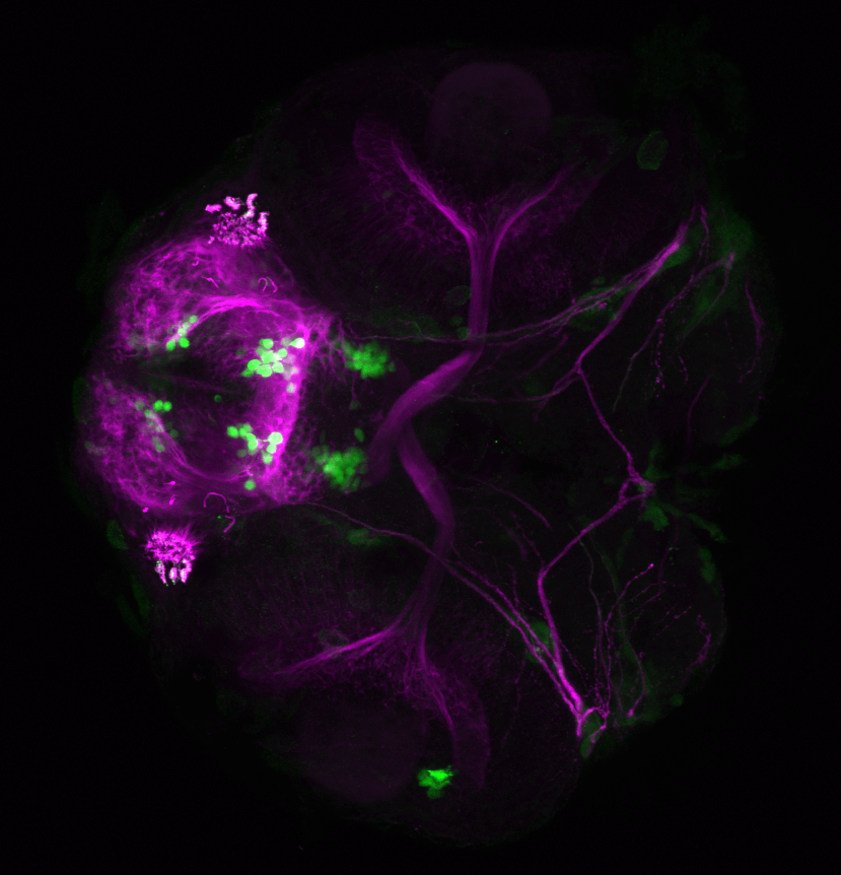
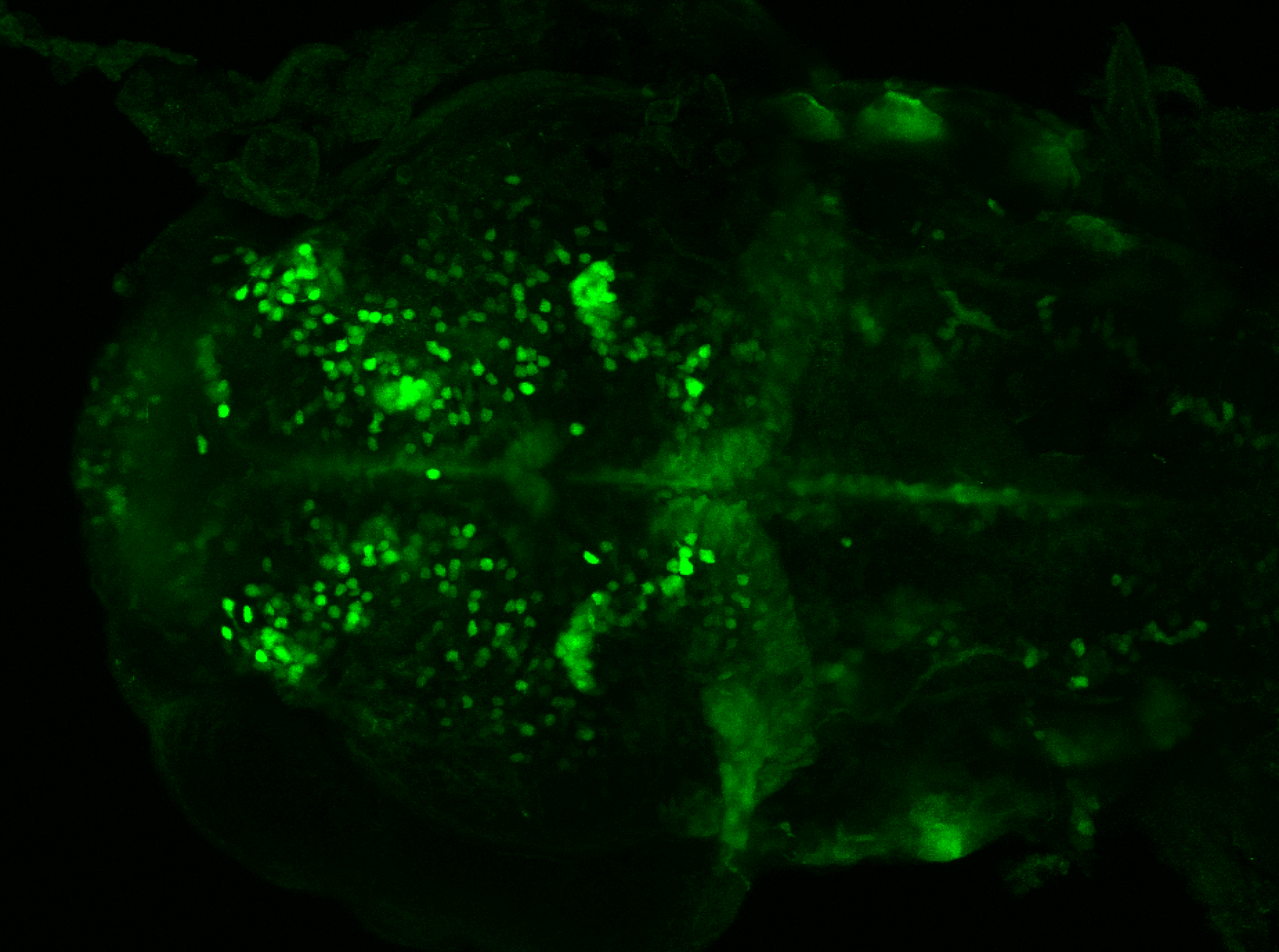
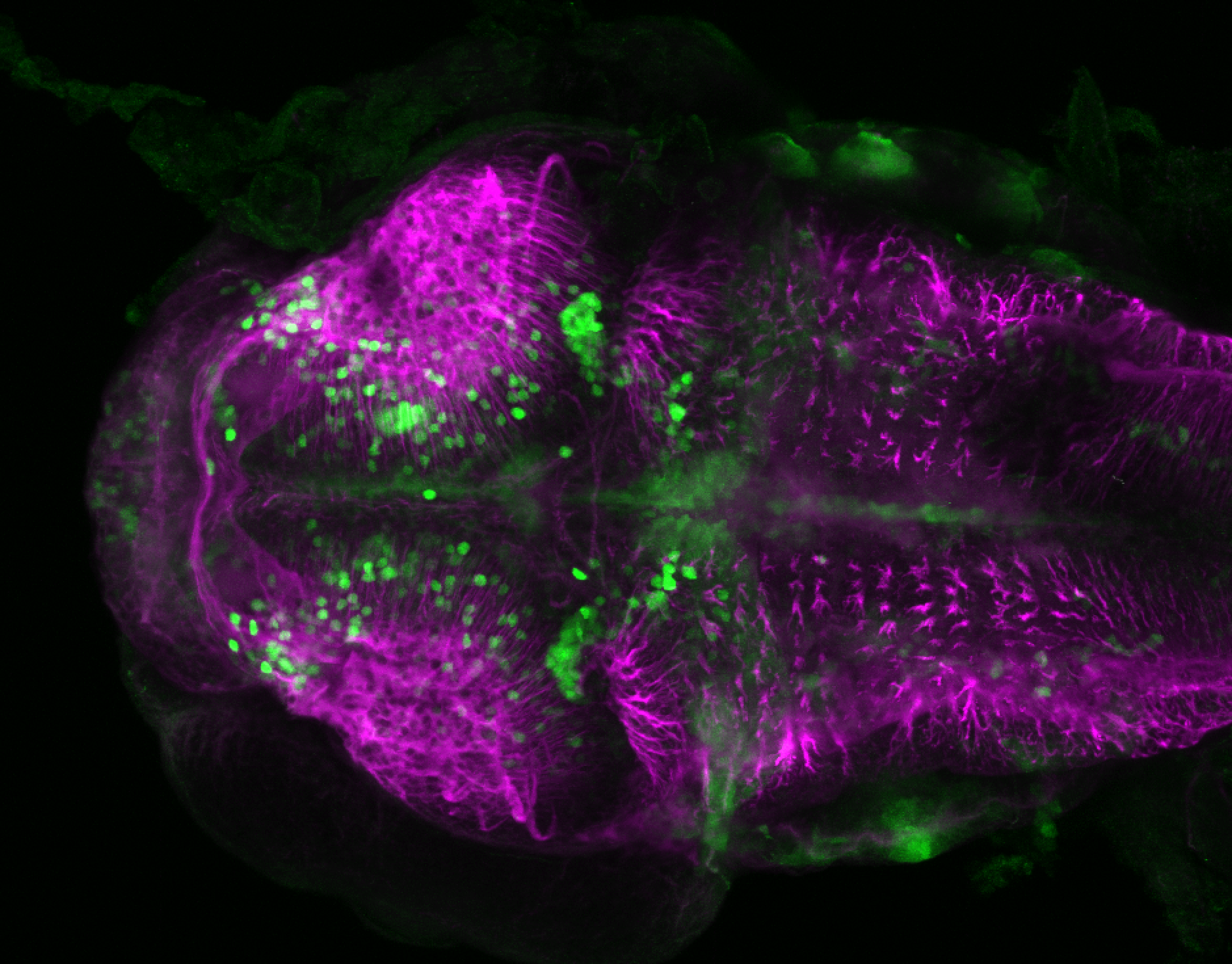
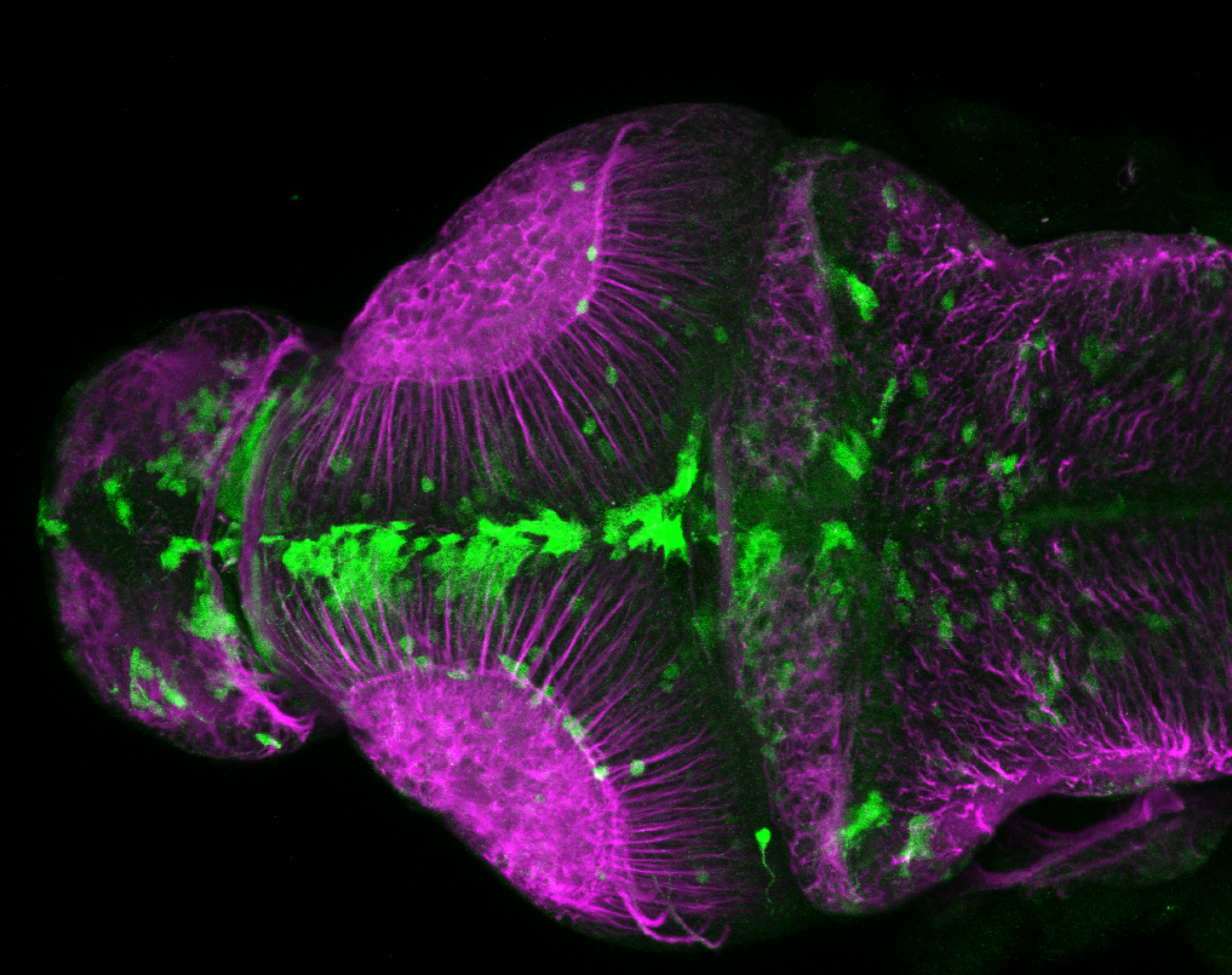
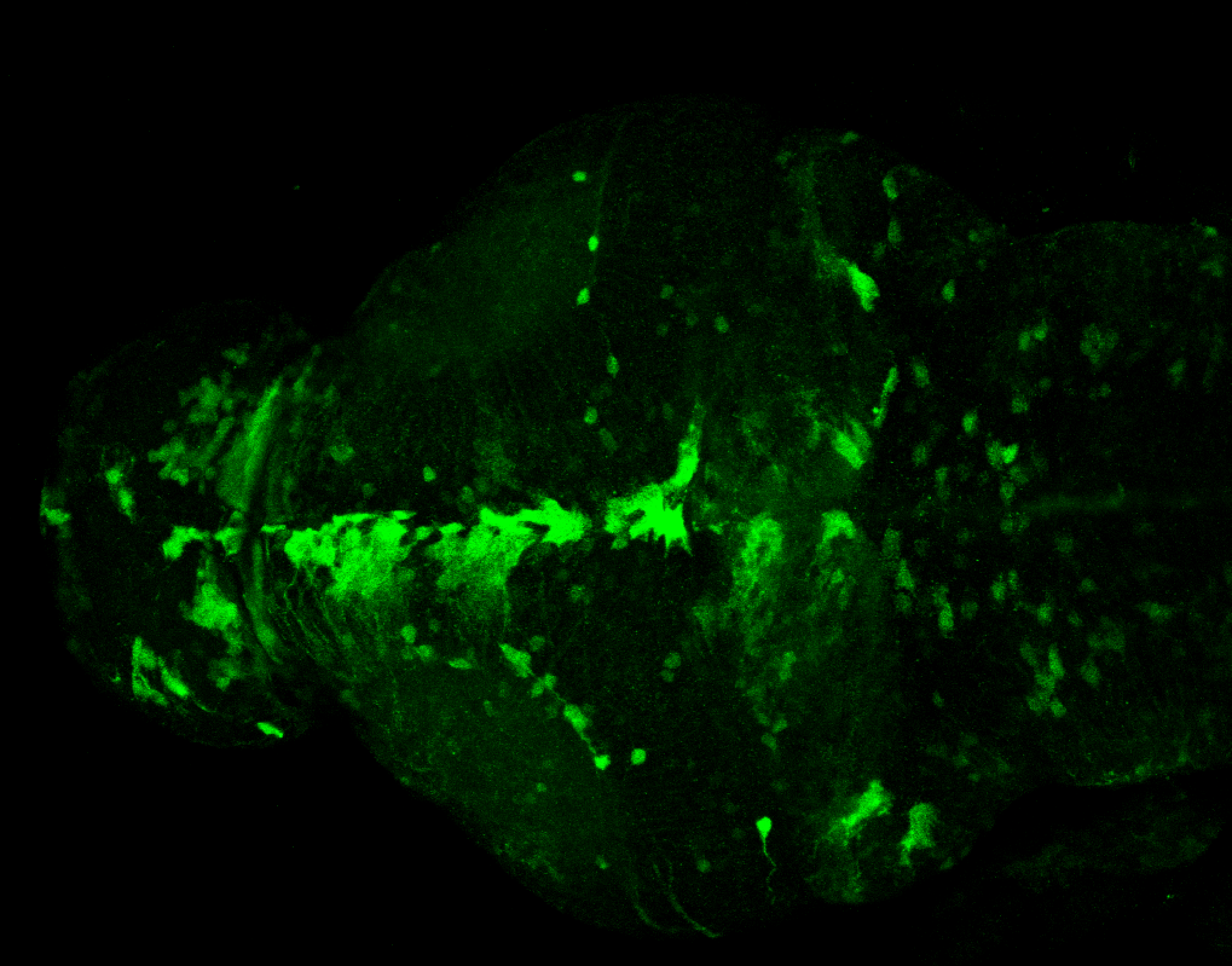
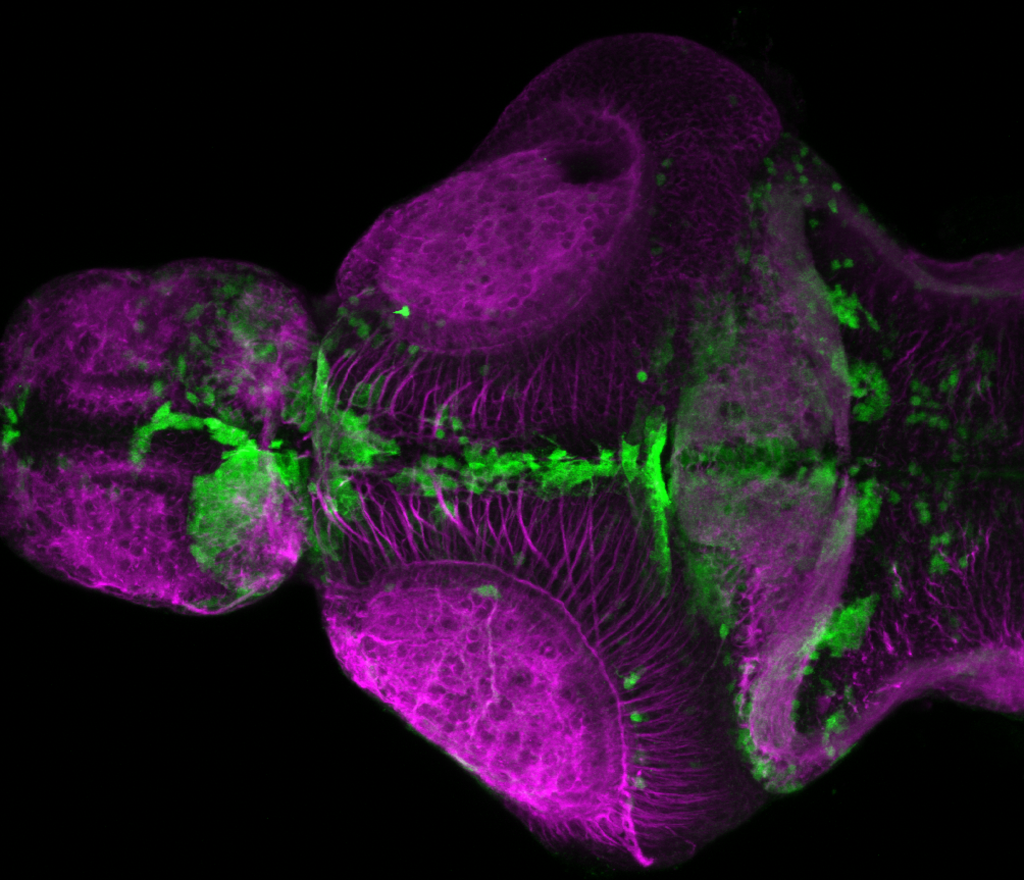
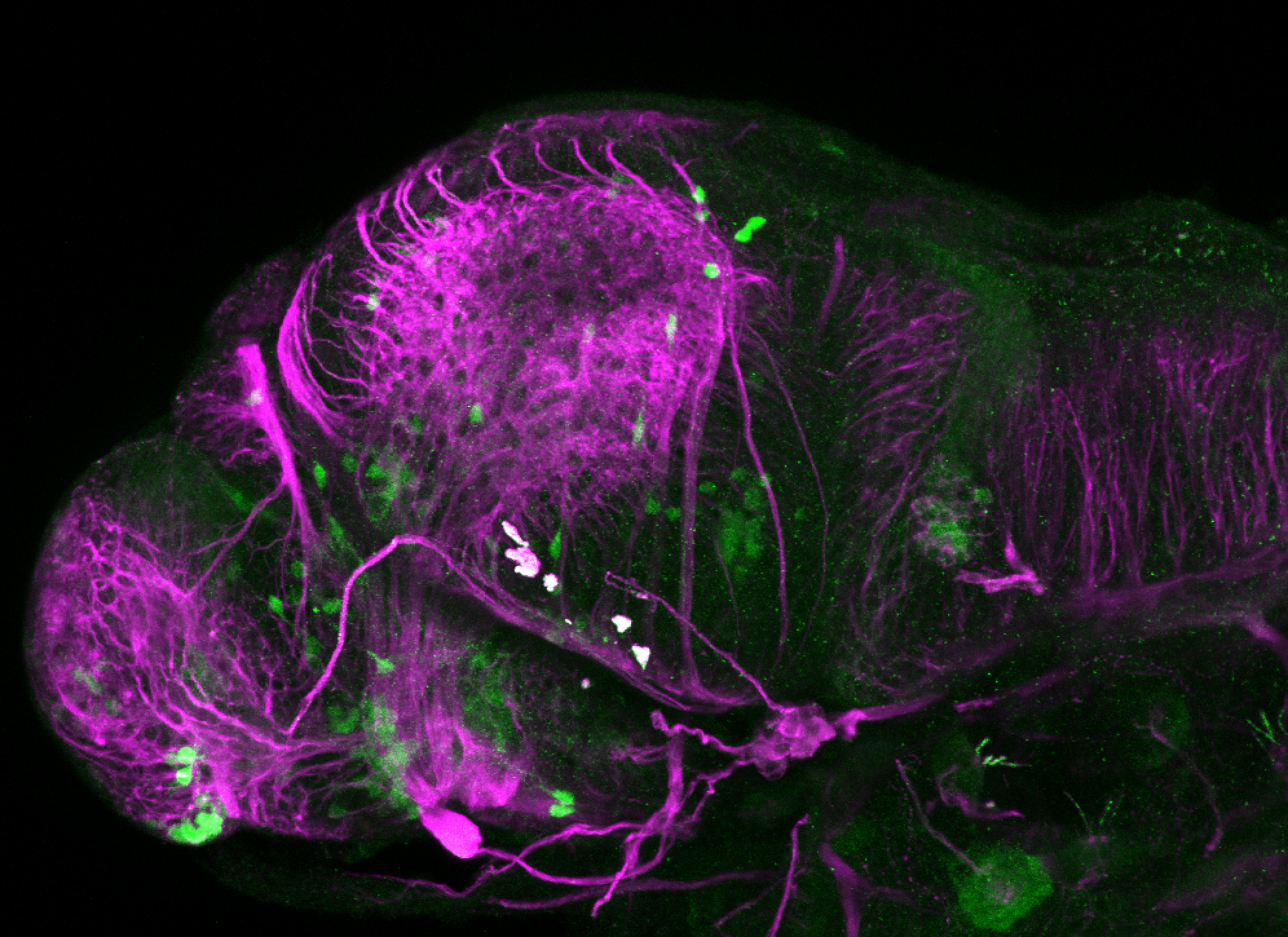
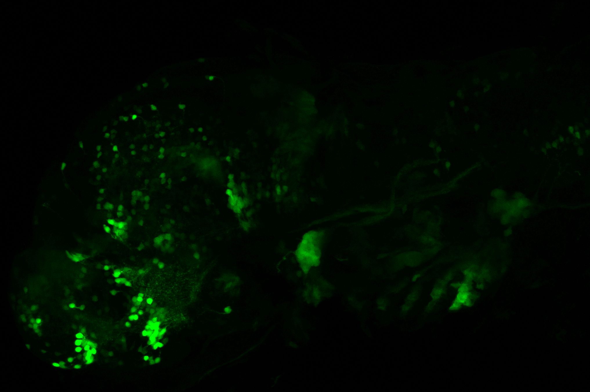
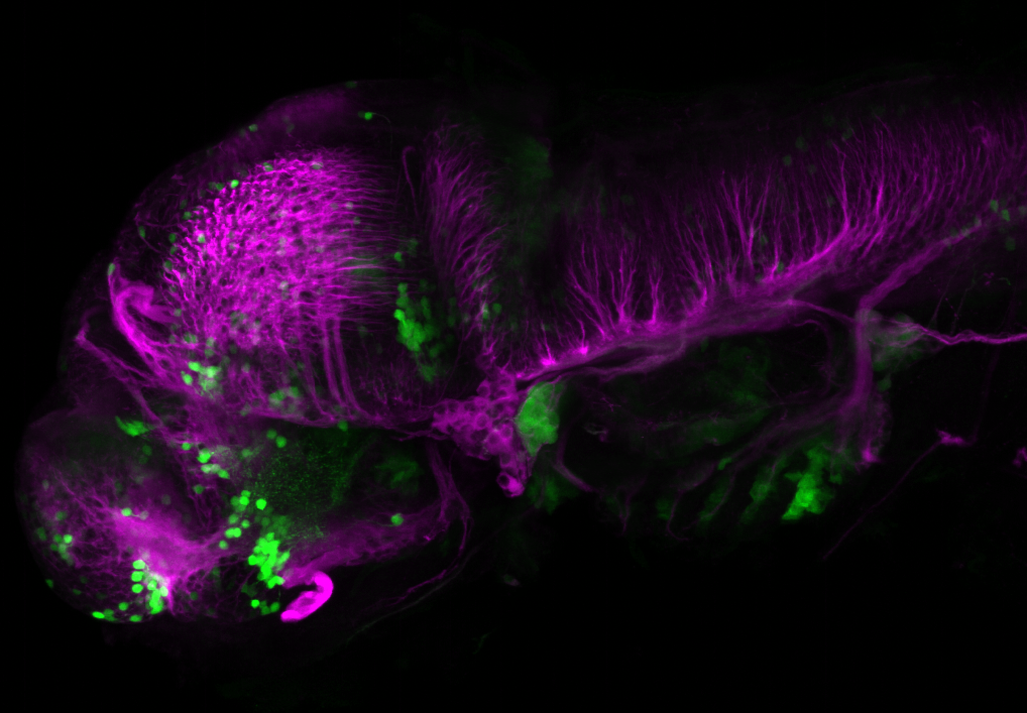
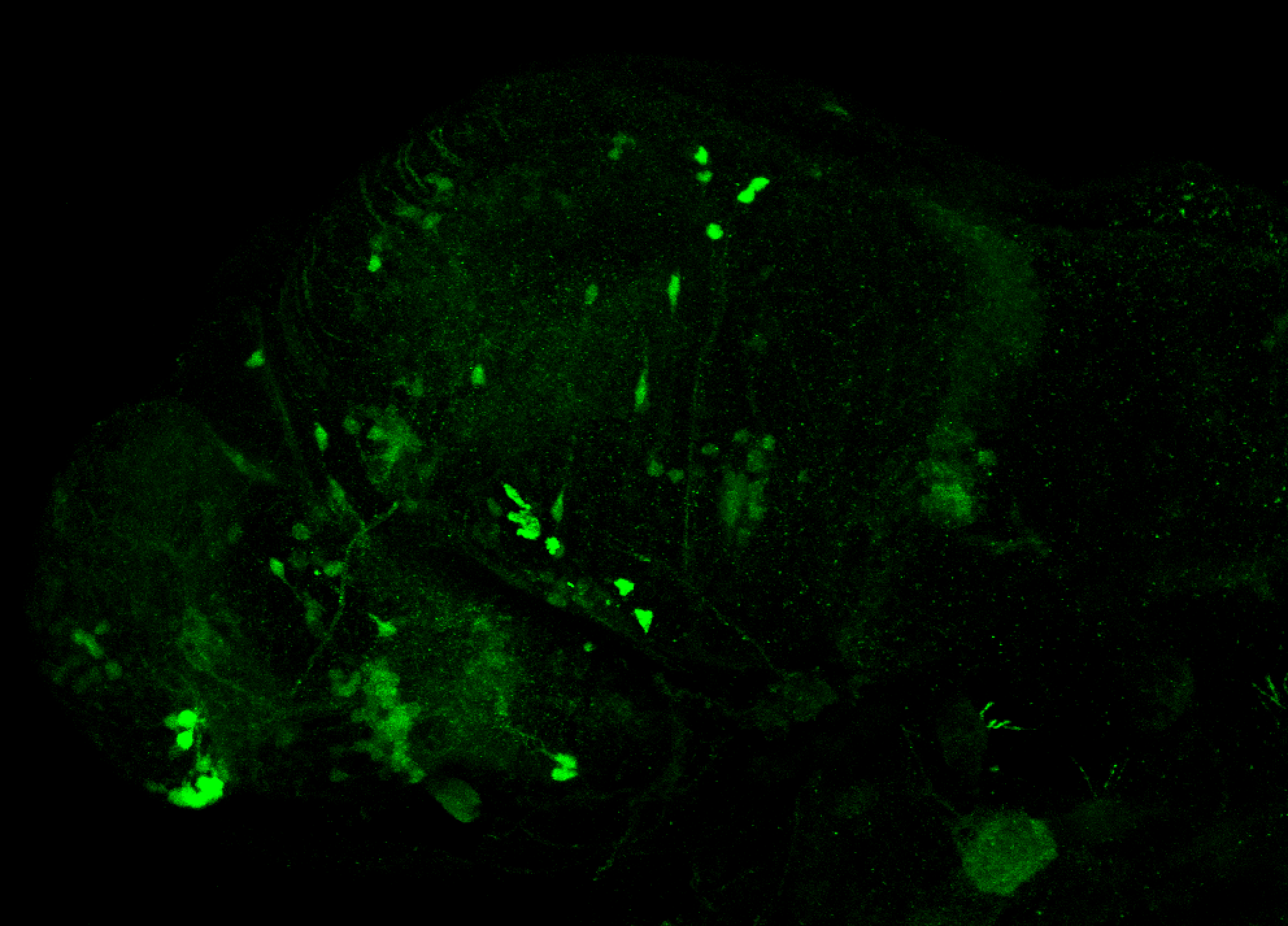
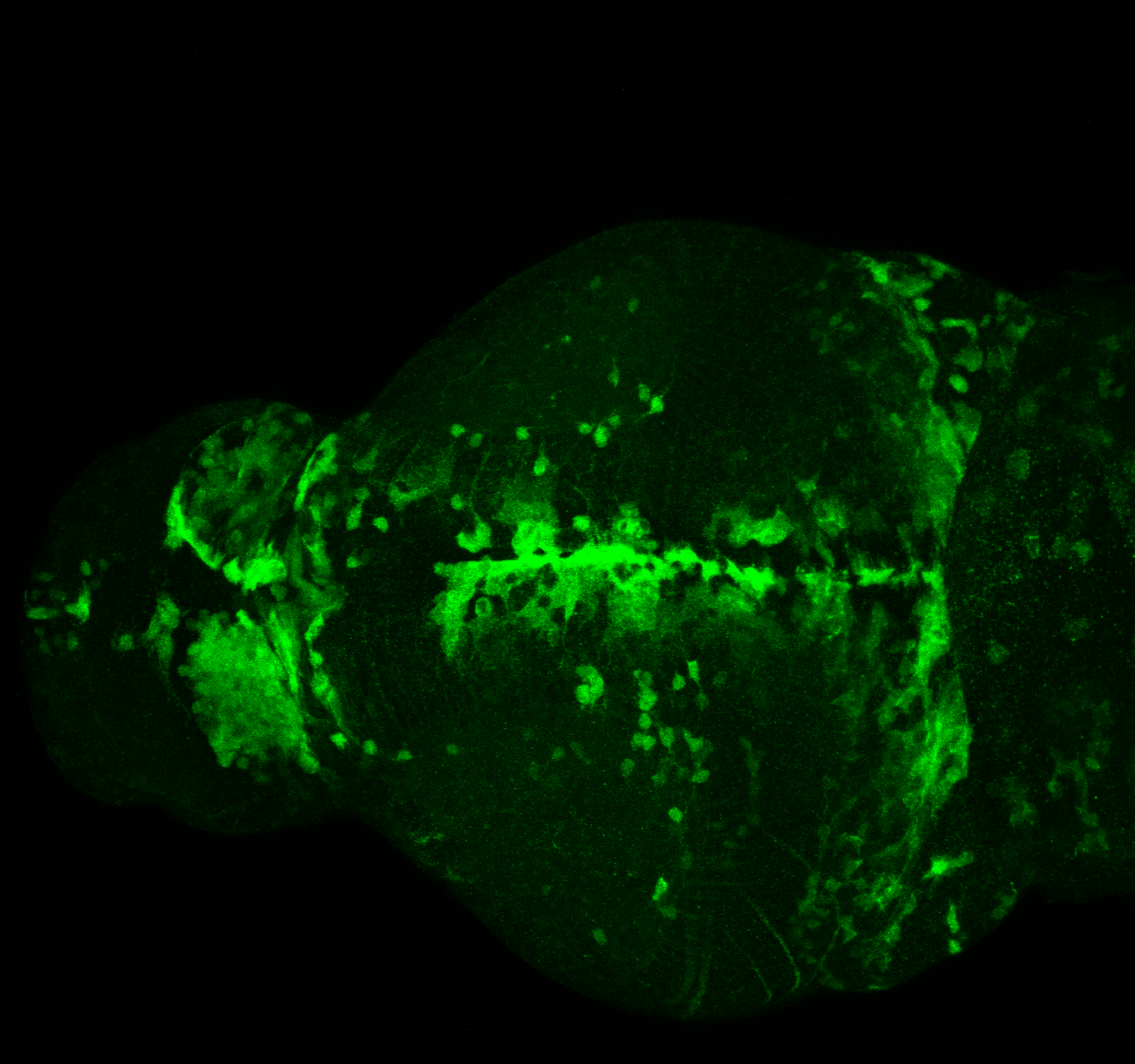
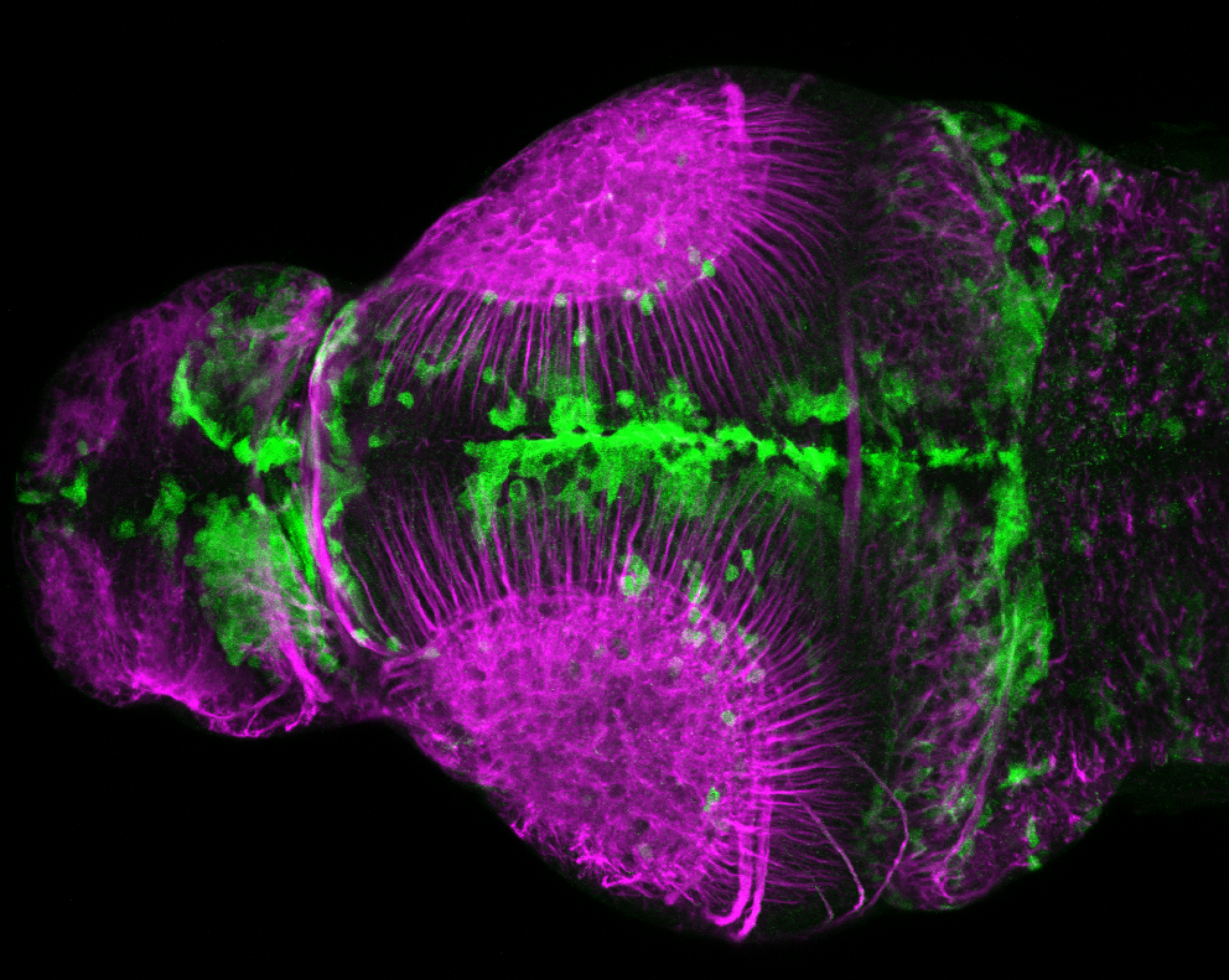
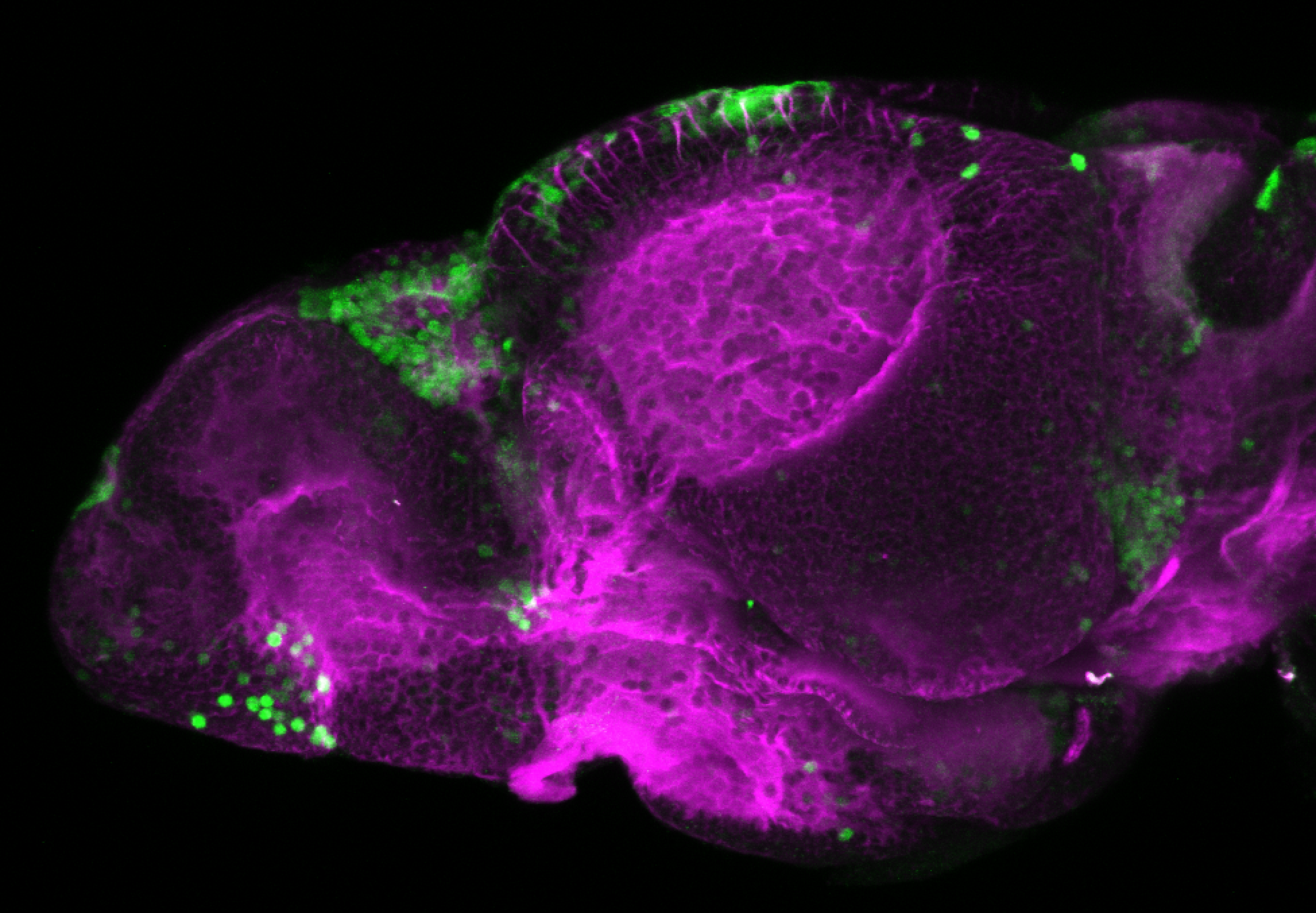
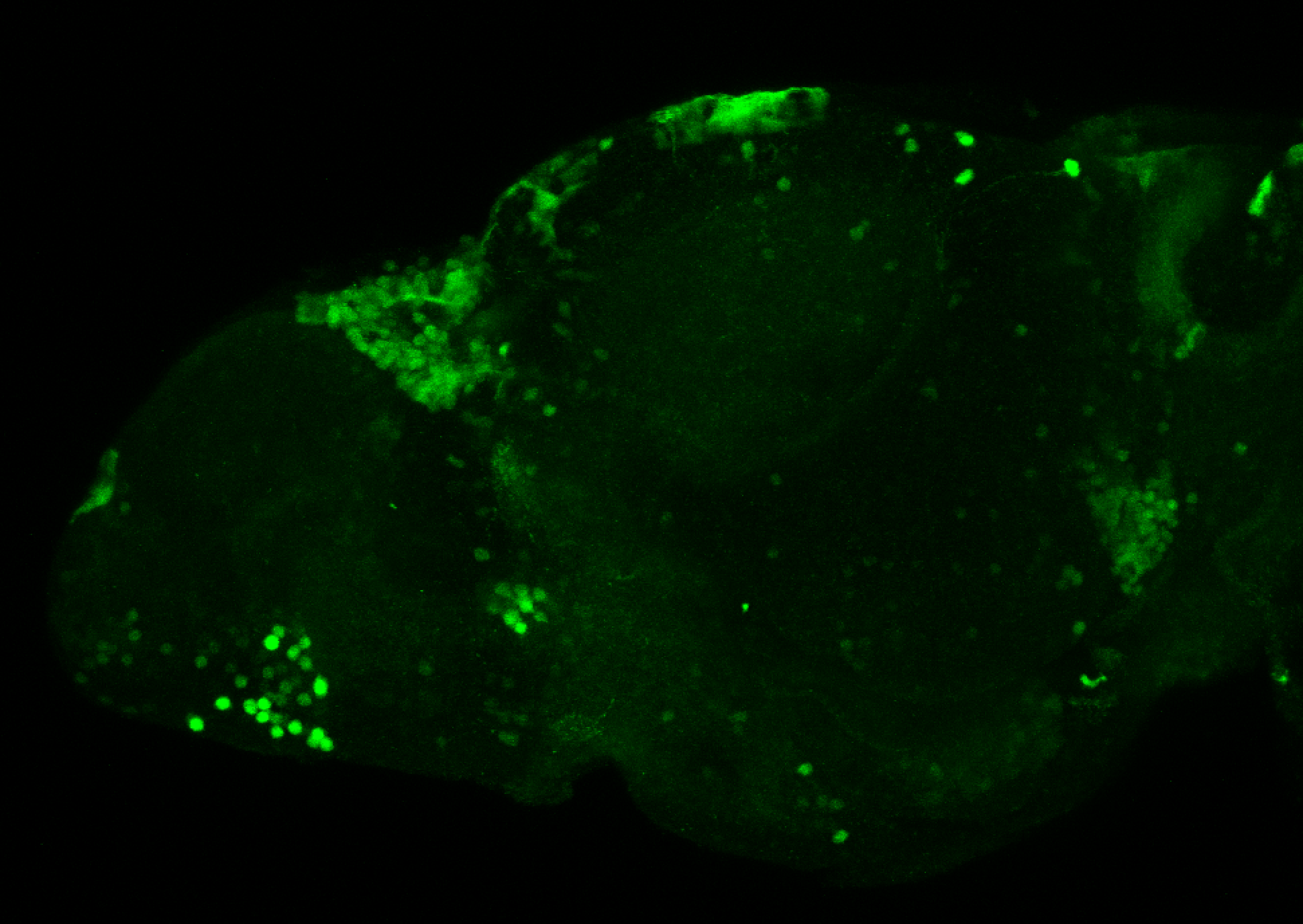
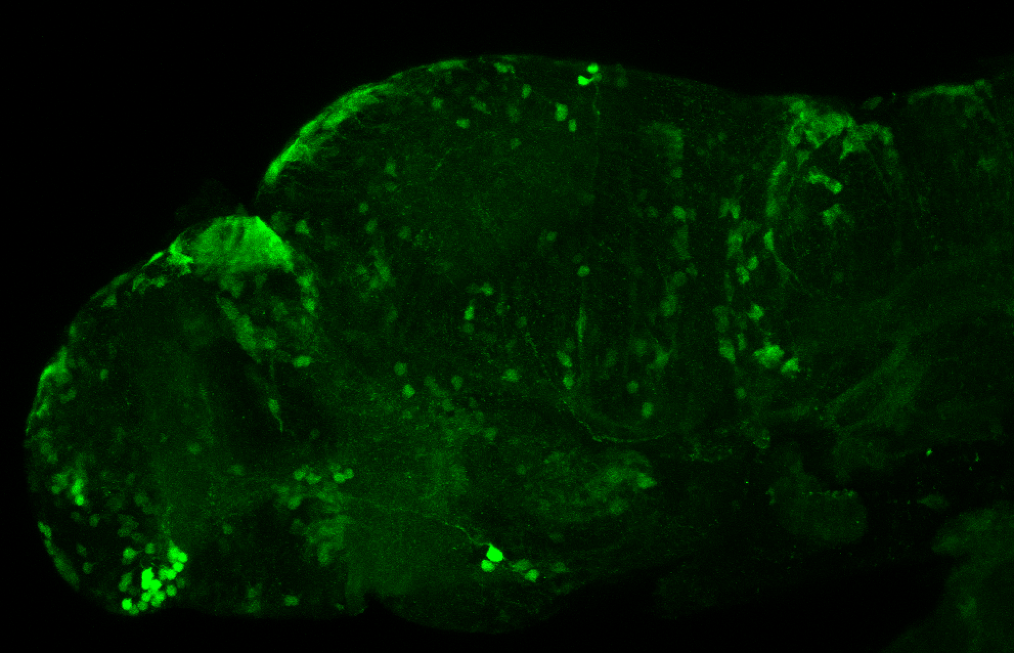
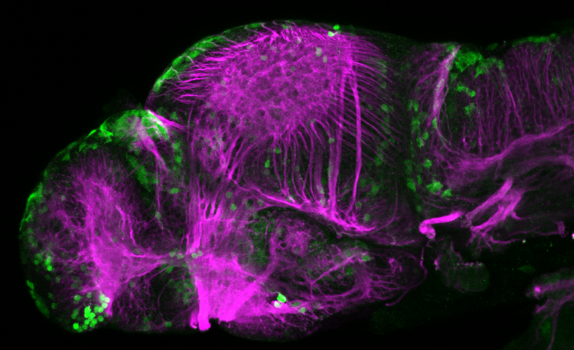
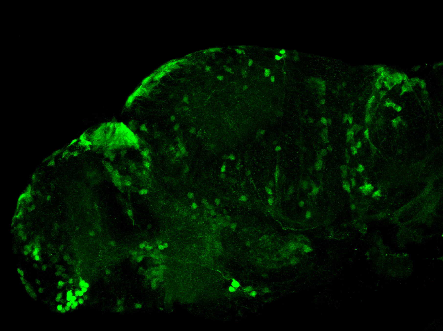
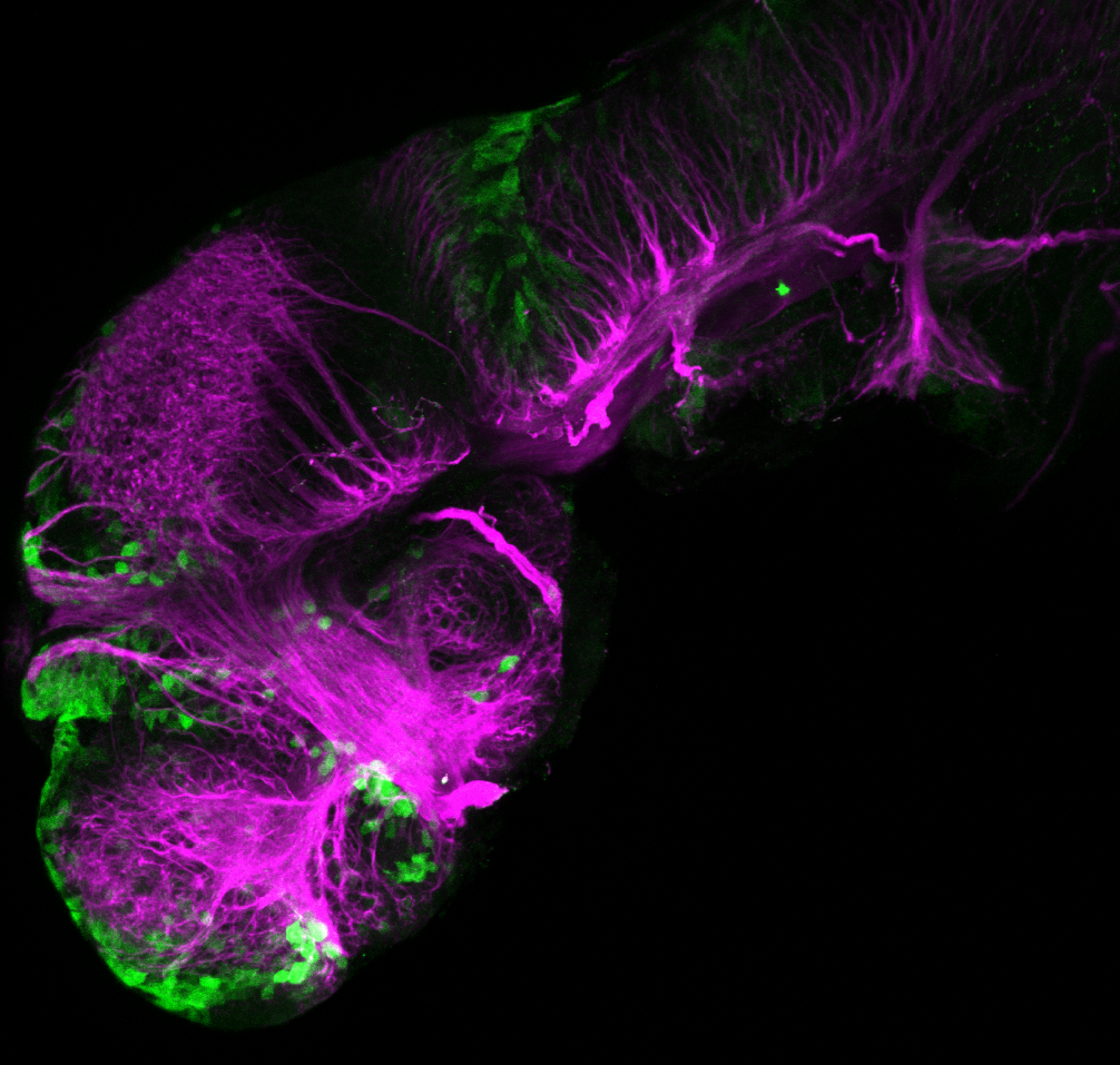
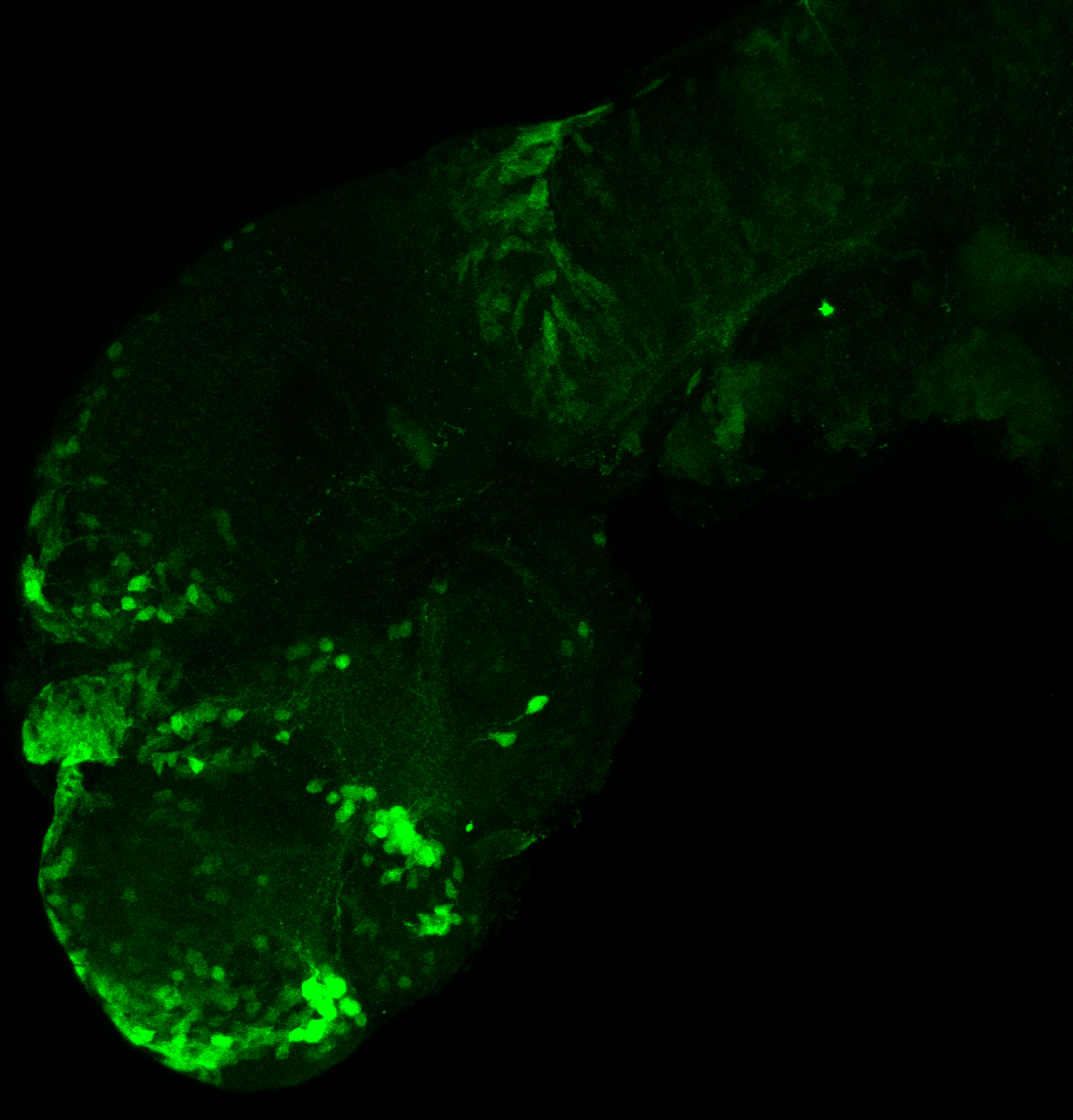
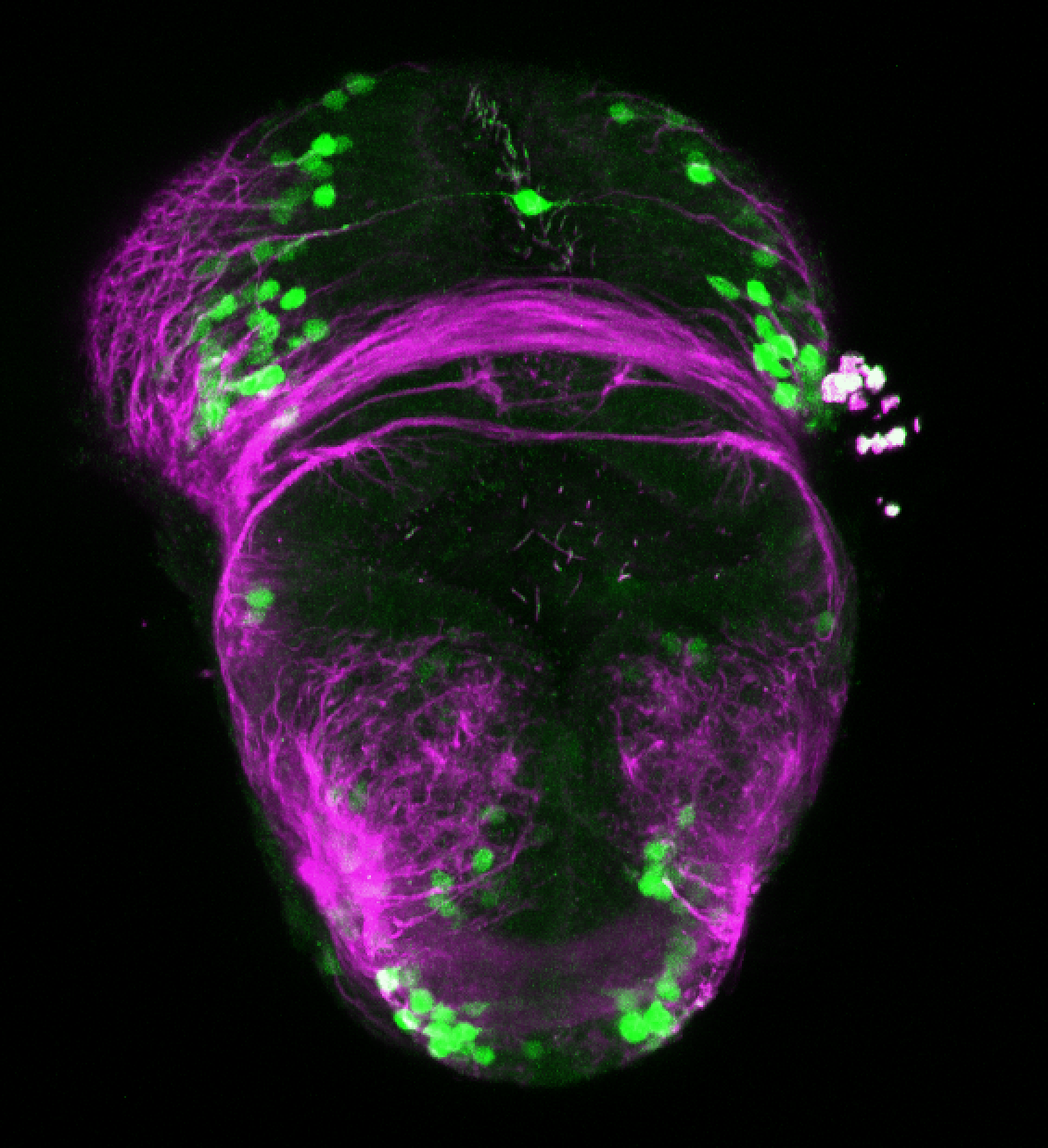
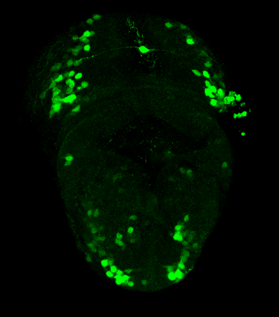
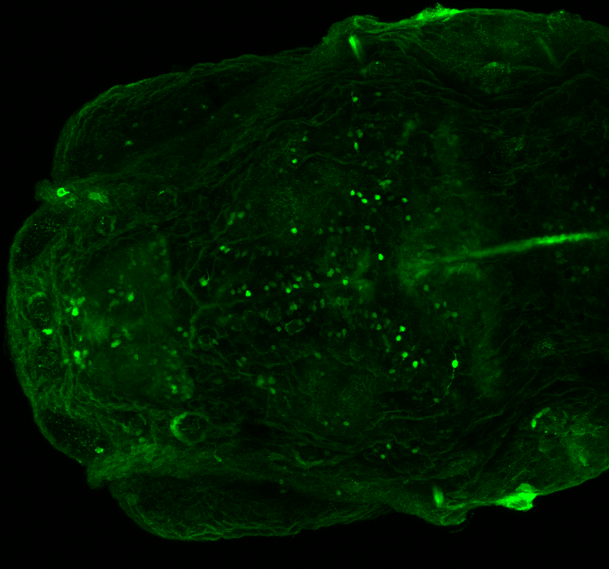
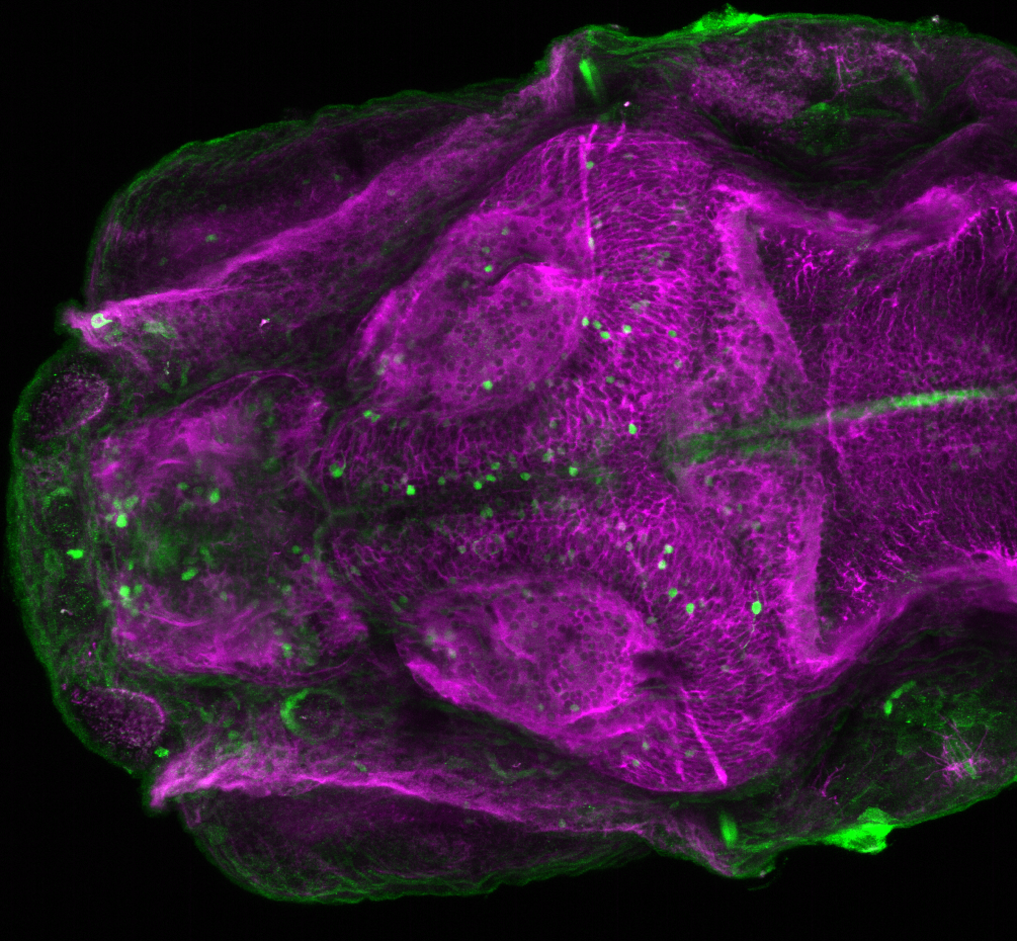
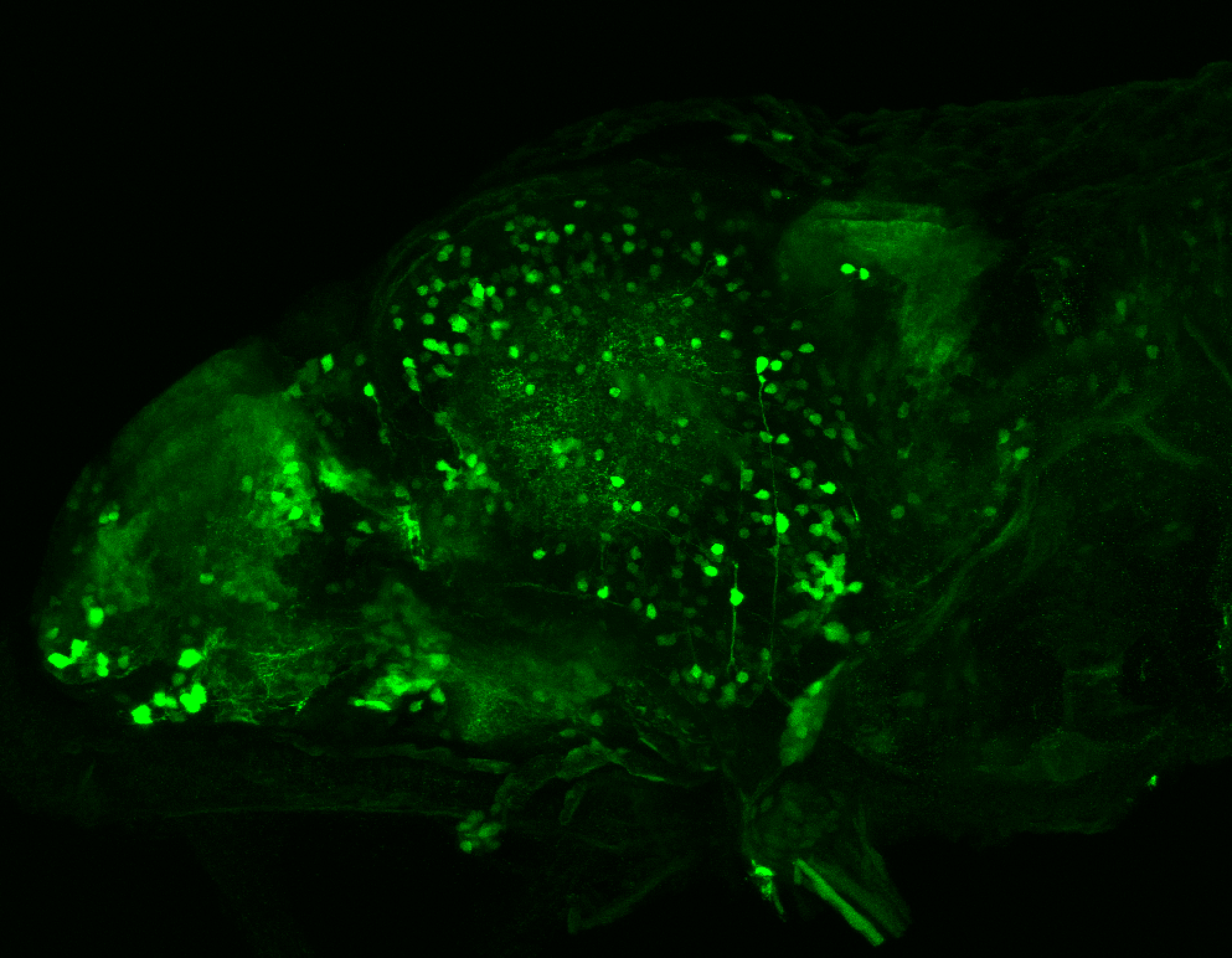
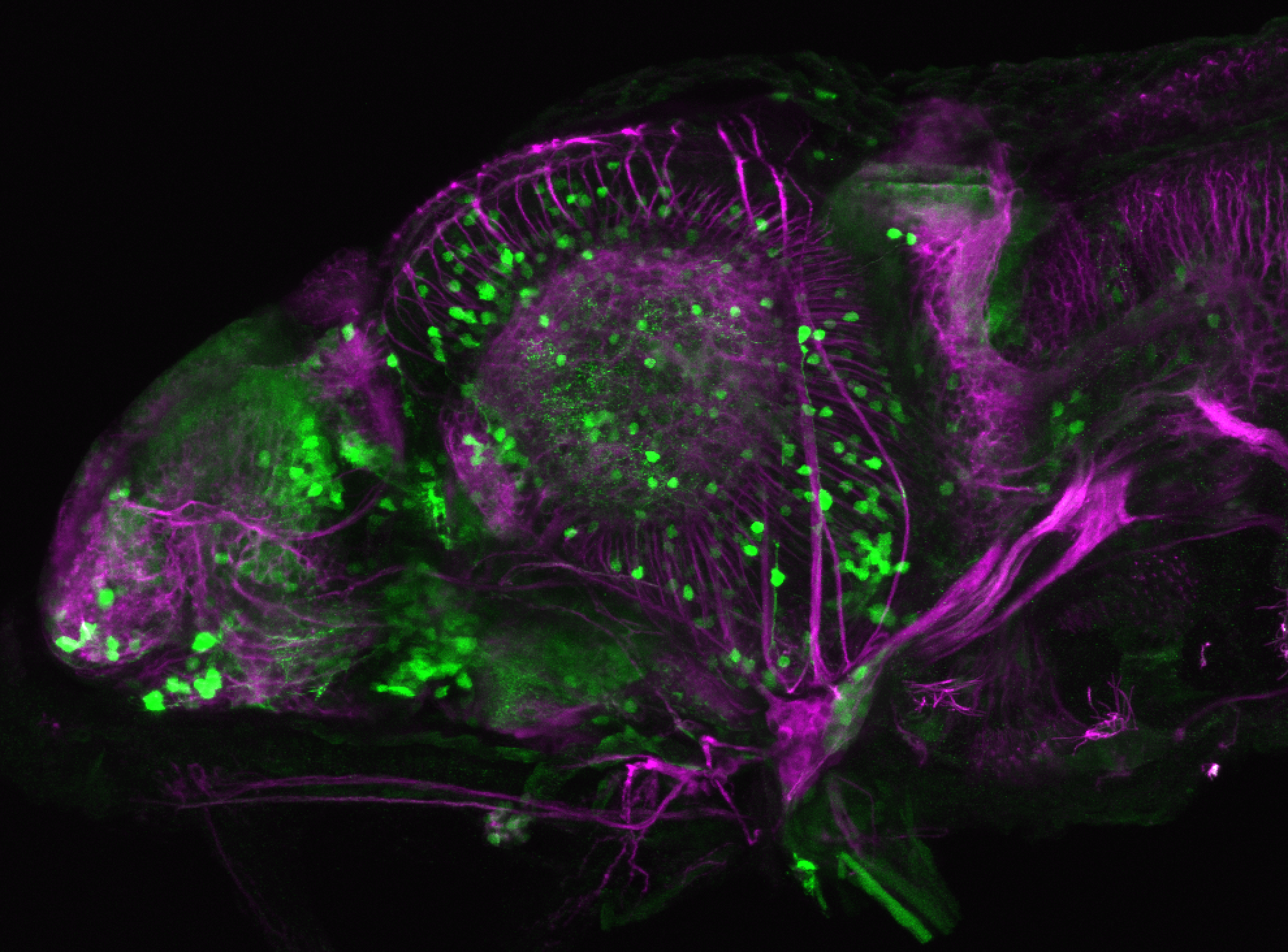
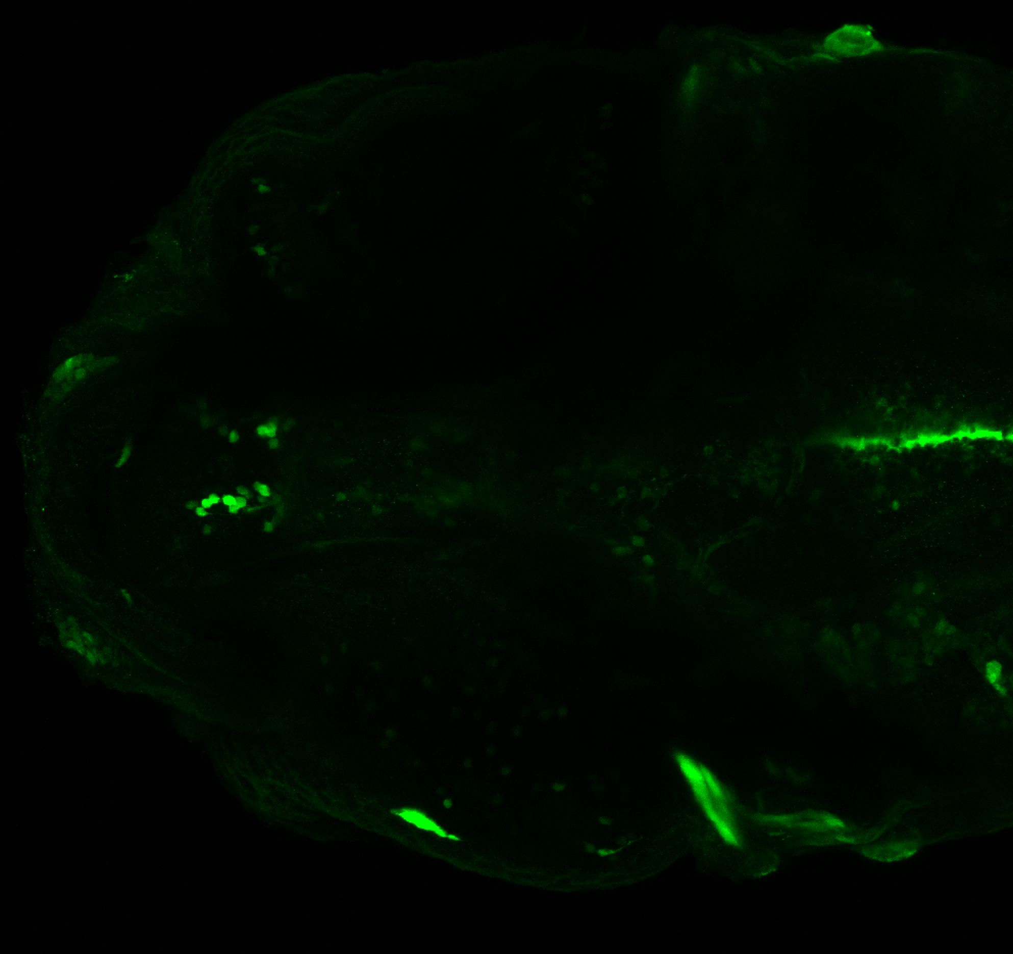
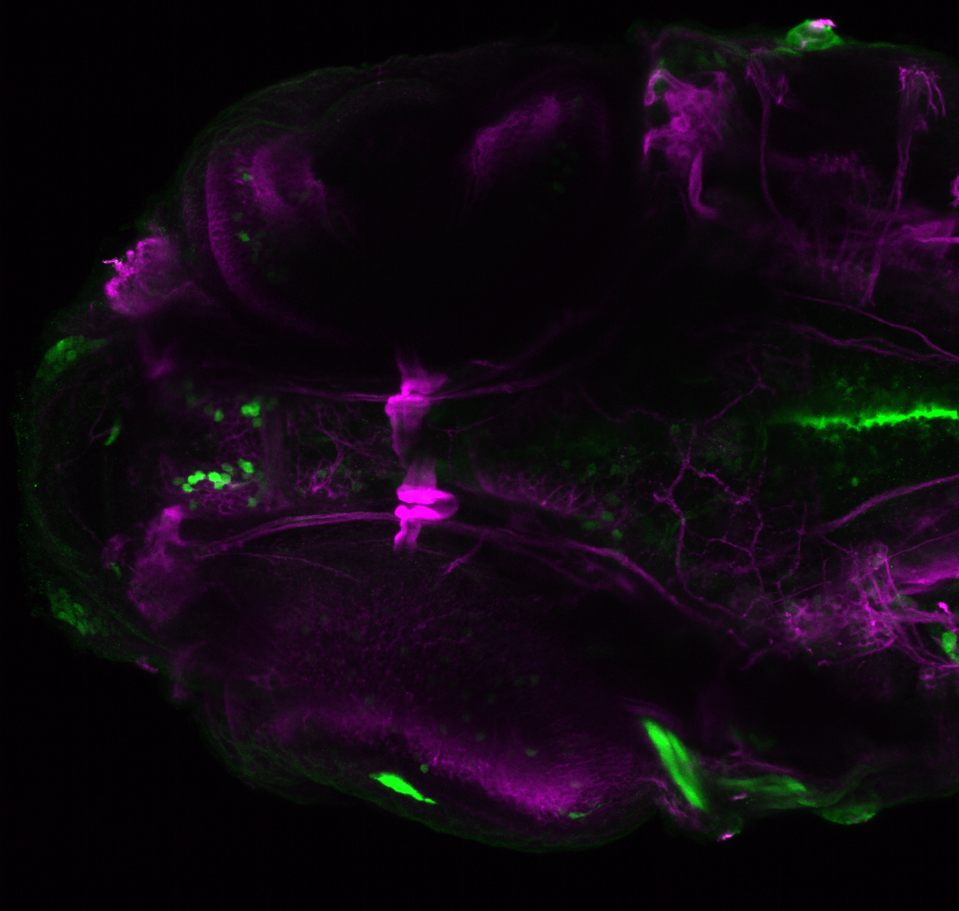
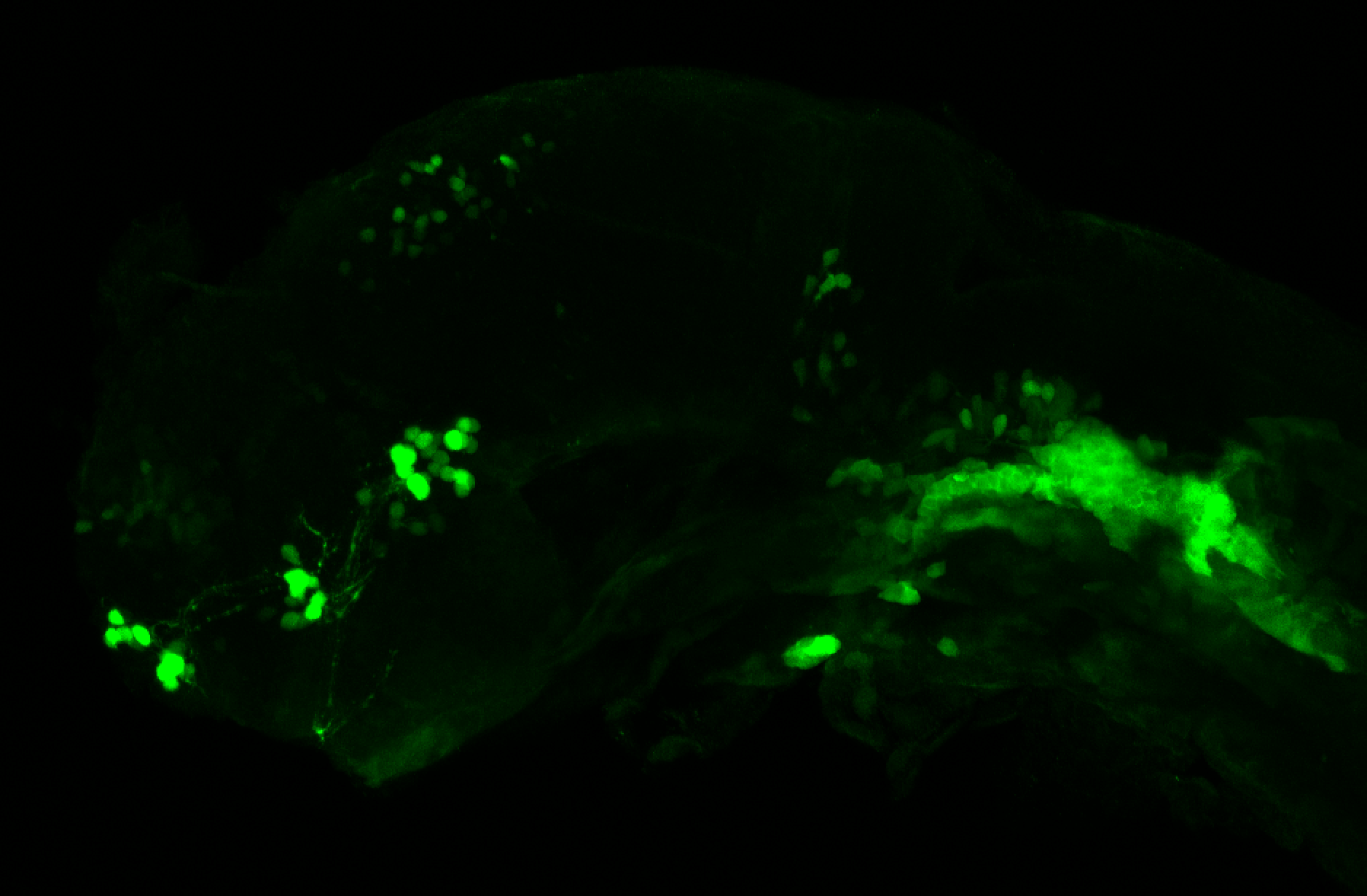
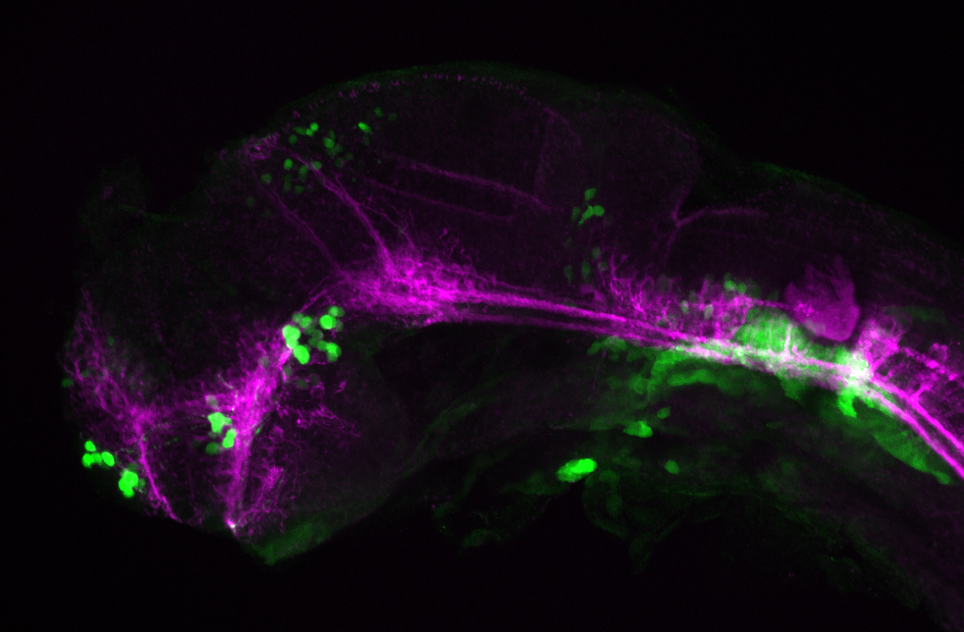
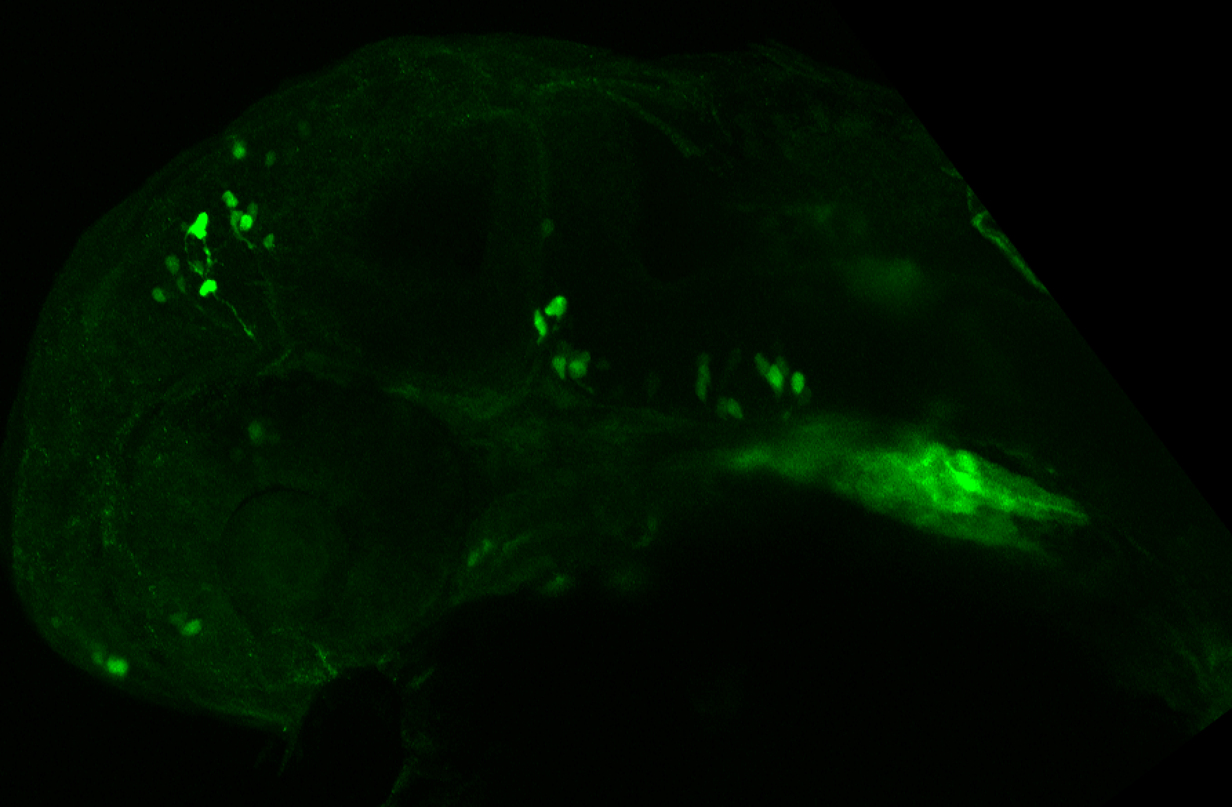
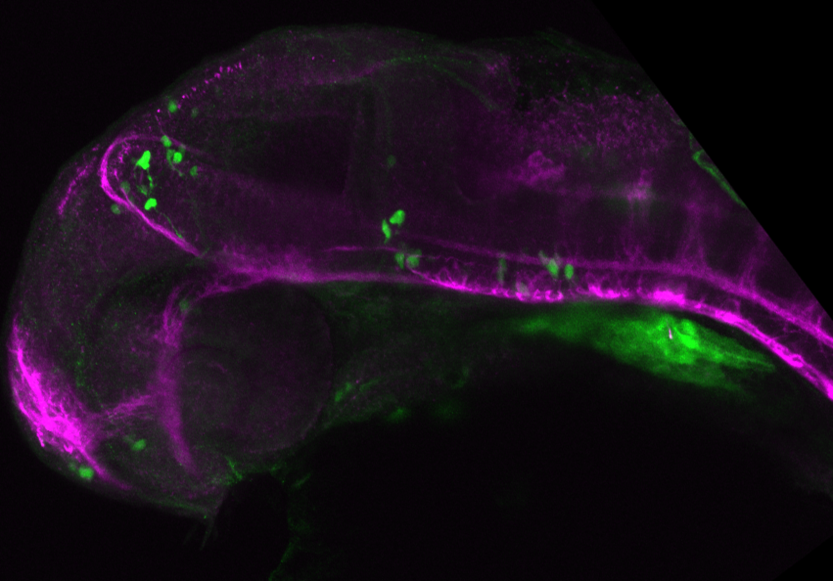
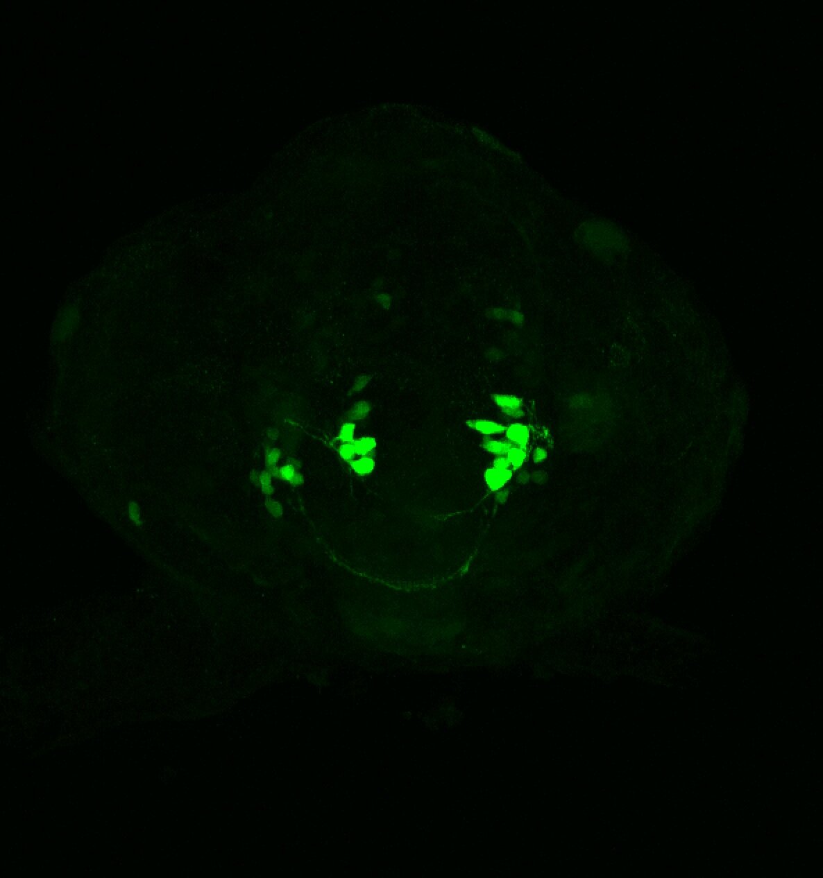
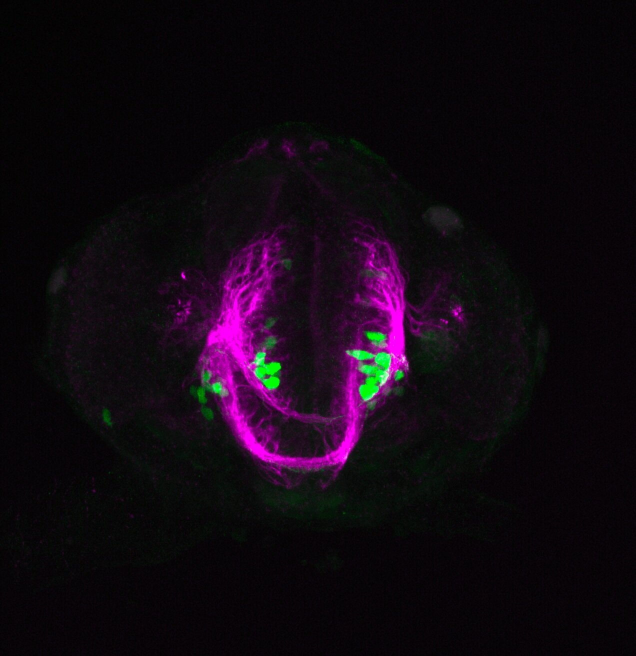
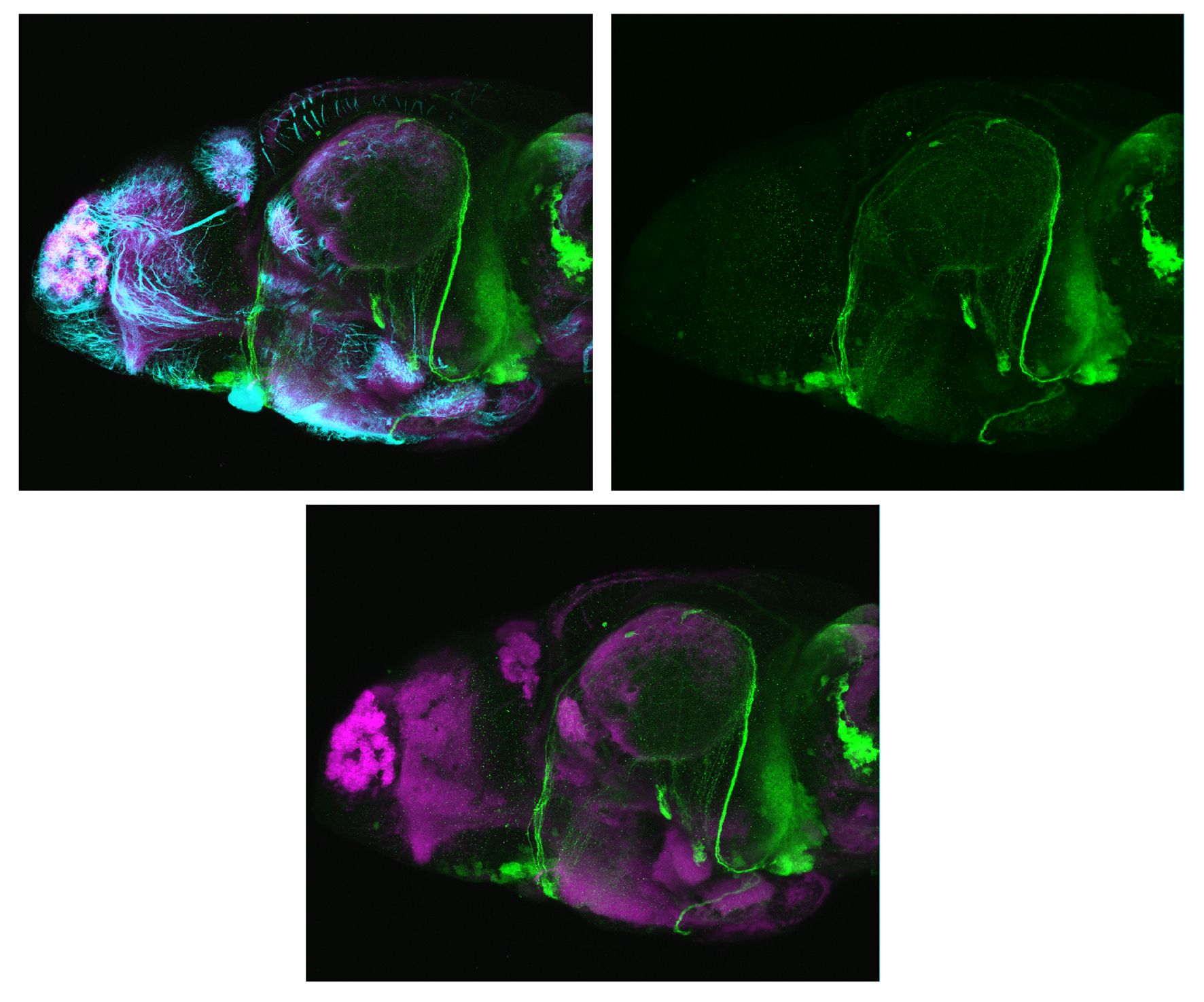
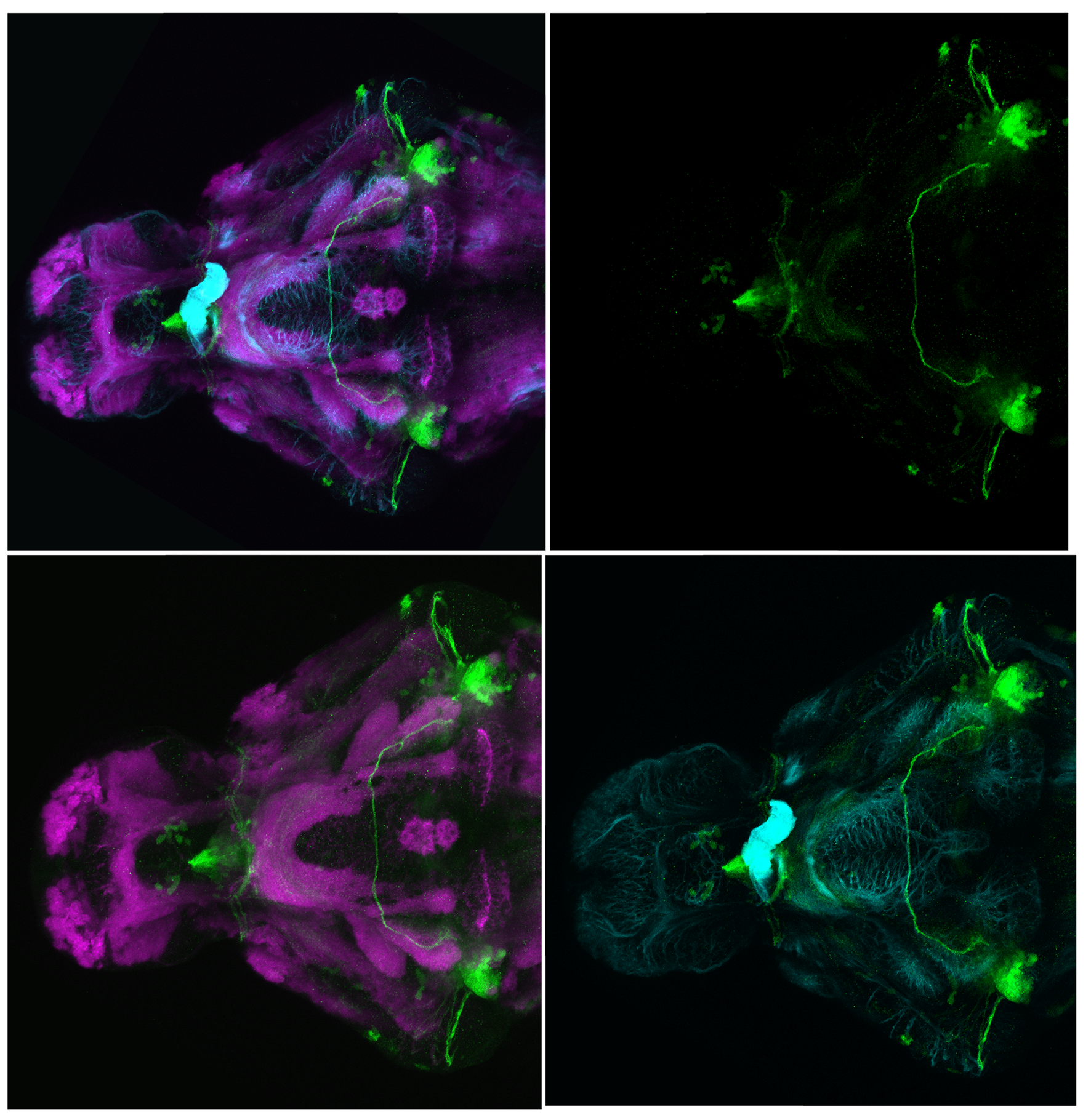
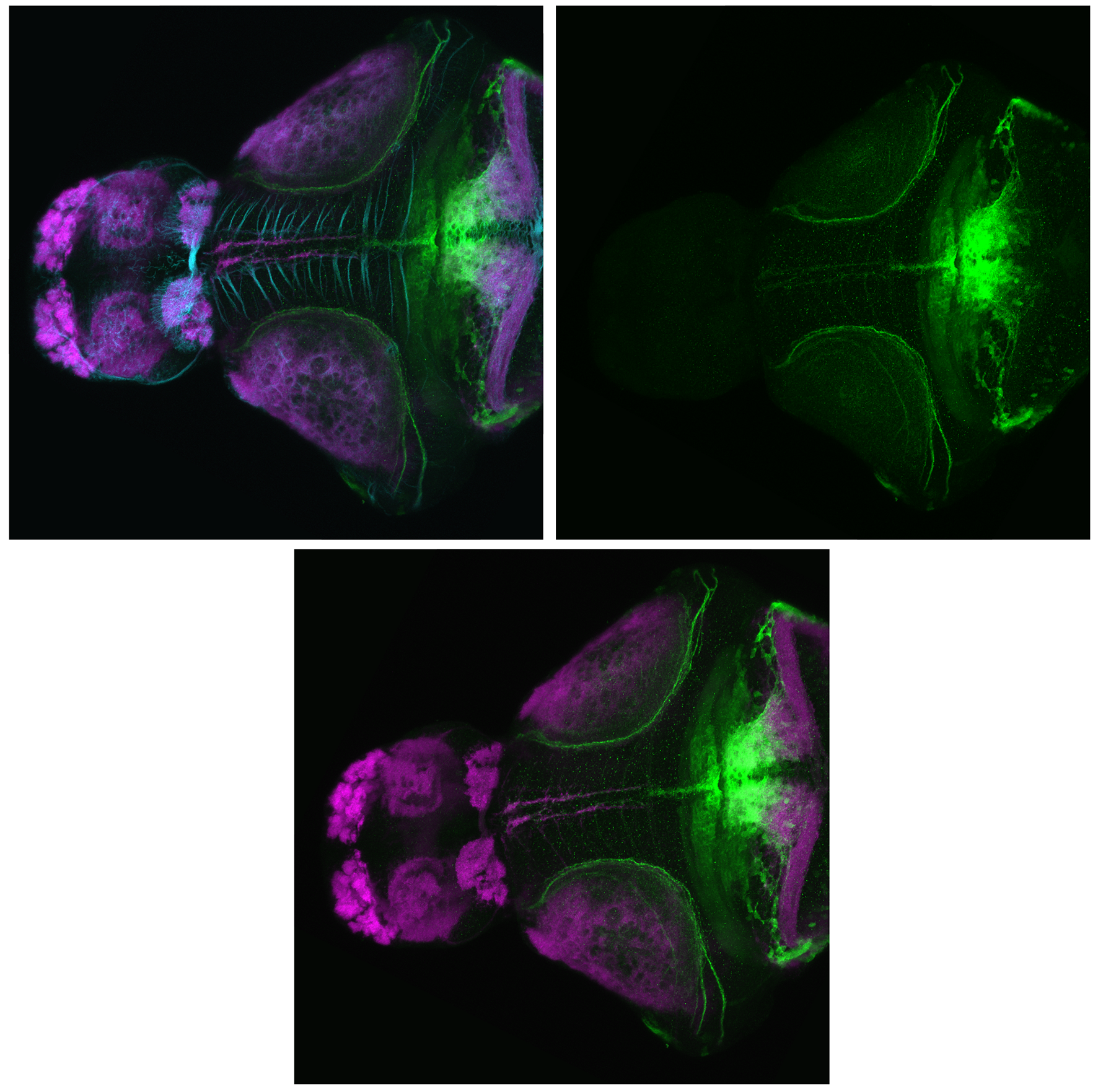
 This transgenic originates from Herwig Baier’s laboratory and is one of many enhancer trap Gal4 lines created by them. Driving Kaede expression in the posterior tuberculum,hypothalamus and cerebellum. There is a very interesting tract labelled that projects between the midbrain tegmentum and posterior tuberculum. The tract skirts the tectal neuropil. The insertion of this transgene is currently unmapped.


External Links:

 ZFIN

Lab or Origin: Baier Lab


Expressed in: 

tectum, posterior tuberculum, tegmentum, hypothalamus, cerebellum. 


Key Publications

Scott, E.K., and Baier, H. (2009) The cellular architecture of the larval zebrafish tectum, as revealed by gal4 enhancer trap lines. Frontiers in neural circuits. 3:13.

Heap, L.A., Goh, C.C., Kassahn, K.S., and Scott, E.K. (2013) Cerebellar output in zebrafish: an analysis of spatial patterns and topography in eurydendroid cell projections. Frontiers in neural circuits. 7:53.

Heap, L.A., Vanwalleghem, G.C., Thompson, A.W., Favre-Bulle, I., Rubinsztein-Dunlop, H., Scott, E.K. (2018) Hypothalamic Projections to the Optic Tectum in Larval Zebrafish. Frontiers in Neuroanatomy. 11:135.

Kani, S., Bae, Y.K., Shimizu, T., Tanabe, K., Satou, C., Parsons, M.J., Scott, E., Higashijima, S.I., and Hibi, M. (2010) Proneural gene-linked neurogenesis in zebrafish cerebellum. Developmental Biology. 343(1-2):1-17. 

	io005Tg/Tg(Xleomes:GFP)io005

io005Tg/Tg(Xleomes:GFP)io005

About

 Synonyms: Tg(Xleomes:GFP)io005, tg(Xeom:GFP)

The tg(Xeom:GFP) transgenic lines were made in the Mione lab and used to study the dorsal to ventral migration of the paraseptal neurons in the subpallium.

“GFP-expressing cells appear around 28 hpf in the telencephalon of tg(Xeom:GFP) transgenic embryos. Paired groups of GFP-expressing cells appear in the lateral region of the telencephalon more or less at the level of the olfactory placode, at the telencephalic/diencephalic border and in the midbrain tegmentum.
We describe the migration of the telencephalic group: these cells originate from the corresponding ventricular zone, they first move towards the lateral side and then proceed rapidly towards the ventral telencephalon. Most of the GFP+ cells will congregate at the level of the septal area just rostral to the anterior commissure The 3 cell groups are interconnected through the anterior commissure and the middle forebrain bundle.” (Mione et al., 2008).

“Telencephalic Xeom:GFP- expressing cells probably correspond to the eomes/tbr1- expressing cells located in the ventral telencephalon of zebrafish [Mione et al., 2001] and of many other vertebrates [Brox et al., 2004; Puelles et al., 2000]. The observation that these cells originate from a dorsal telencephalic position and undergo an extensive migration towards the ventral telencephalon and diencephalon, accompanying the growth of the MOT and MFB, suggests that they may play pioneer roles on the formation of these major axon tracts. “(Mione et al., 2008).


External Links:

 ZFIN

Lab or Origin: Mione Lab


Expressed in: 

 septal region, subpallium, entopeduncular nucleus, thalamic emminence, midbrain tegmentum, optic tectum, cerebellum.


Key Publications

Mione, M., Baldessari, D., Deflorian, G., Nappo, G., and Santoriello, C. (2008)
How neuronal migration contributes to the morphogenesis of the CNS: insights from the zebrafish. Developmental neuroscience. 30(1-3):65-81.

Mione M, Shanmugalingam S, Kimelman D, Griffin K (2001):
Overlapping expression of zebrafish T-brain-1 and eomesodermin during forebrain development.
Mech Dev 100: 93–97.

Puelles L, Rubenstein JL (2003)
Forebrain gene expression domains and the evolving prosomeric model.
Trends Neurosci 26:469–476.

Puelles L, Kuwana E, Puelles E, Bulfone A, Shi- mamura K, Keleher J, Smiga S, Rubenstein JL (2000)
Pallial and subpallial derivatives in the embryonic chick and mouse telencephalon, traced by the expression of the genes Dlx-2, Emx-1, Nkx-2.1, Pax-6, and Tbr-1.
J Comp Neurol 424:409–438.