Viewing entries tagged
thalamic eminence

Et(krt4:EGFP)sqet24

Et(krt4:EGFP)sqet24

About

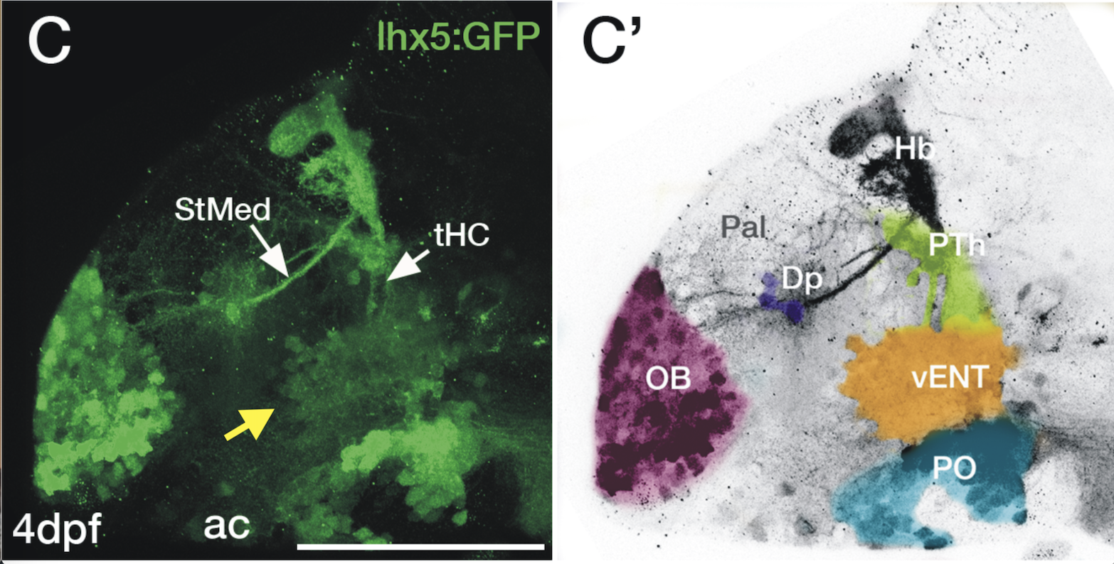
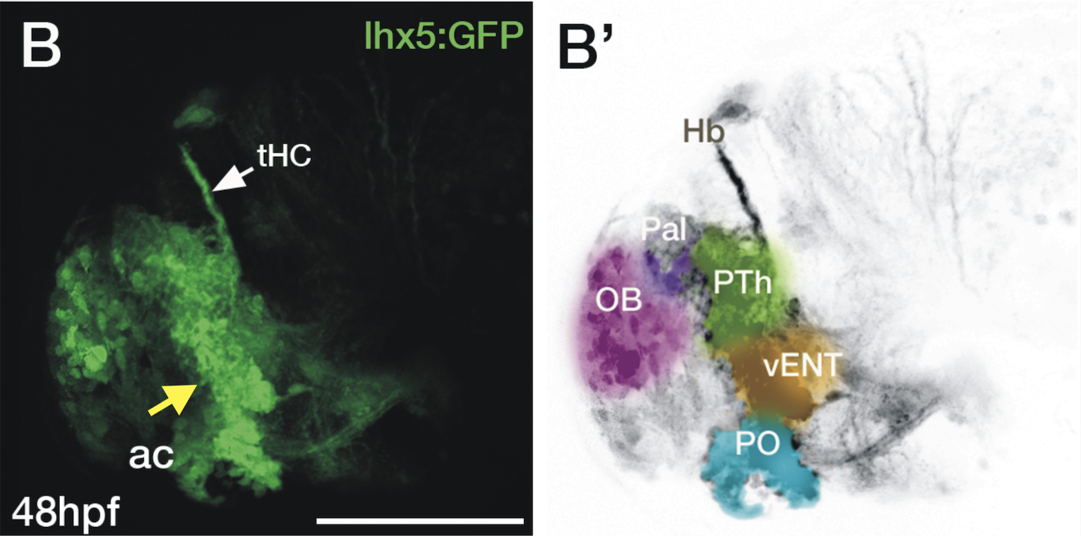
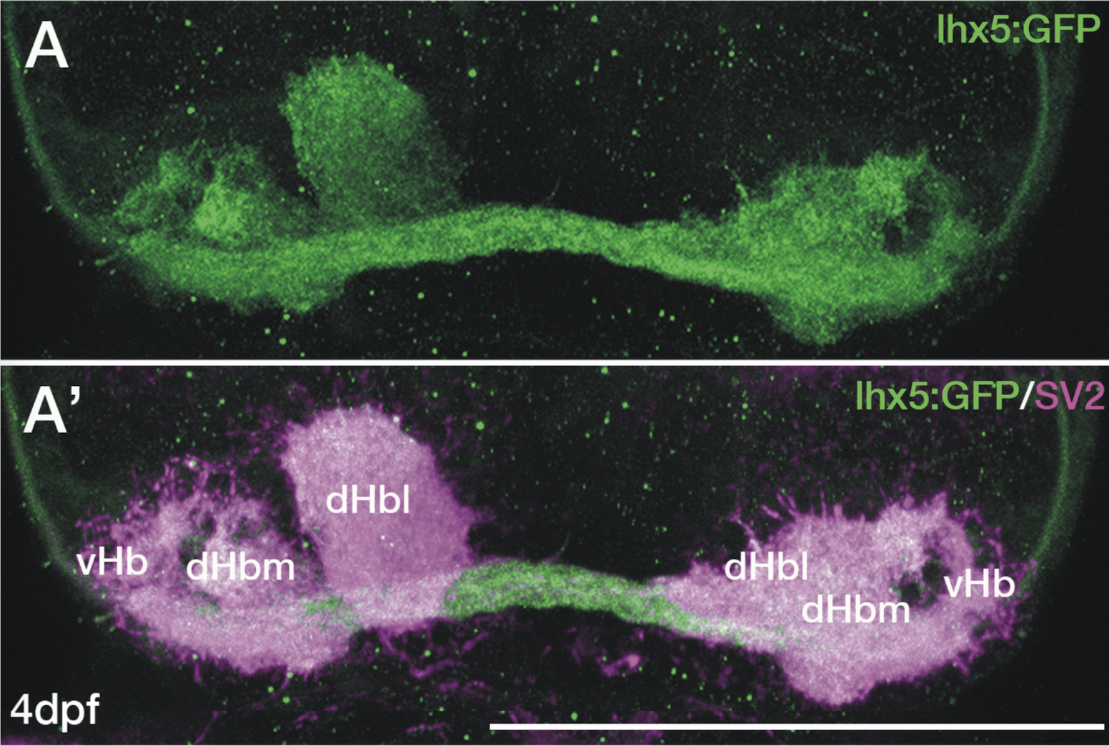
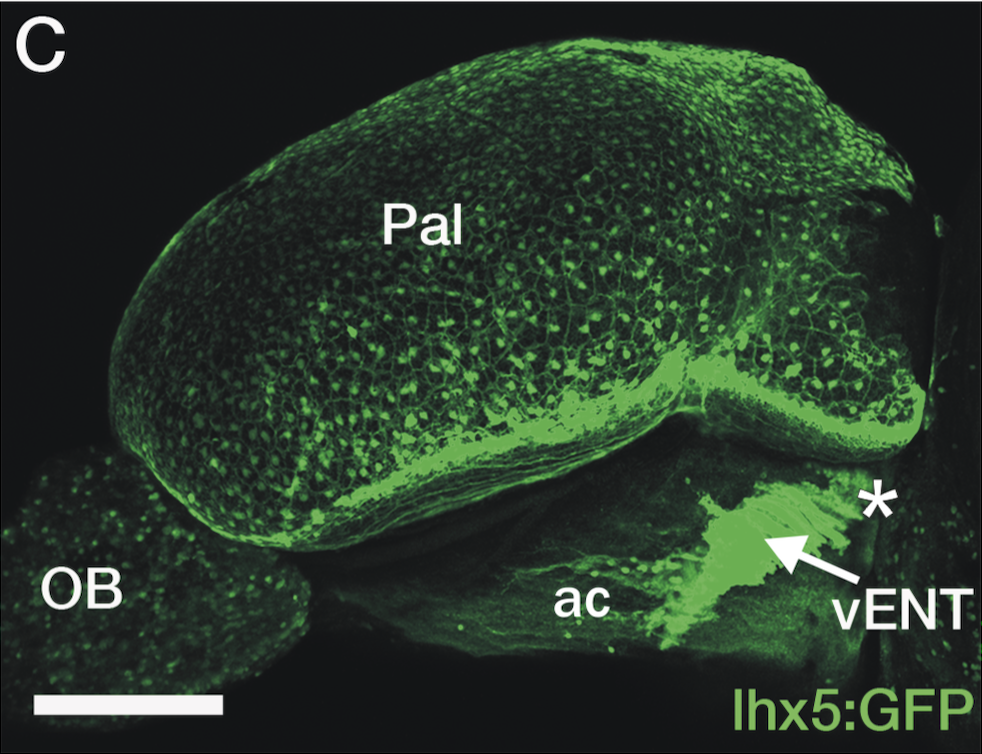
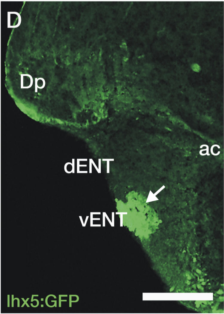
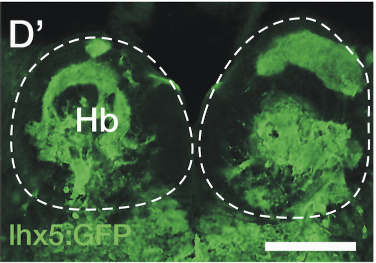
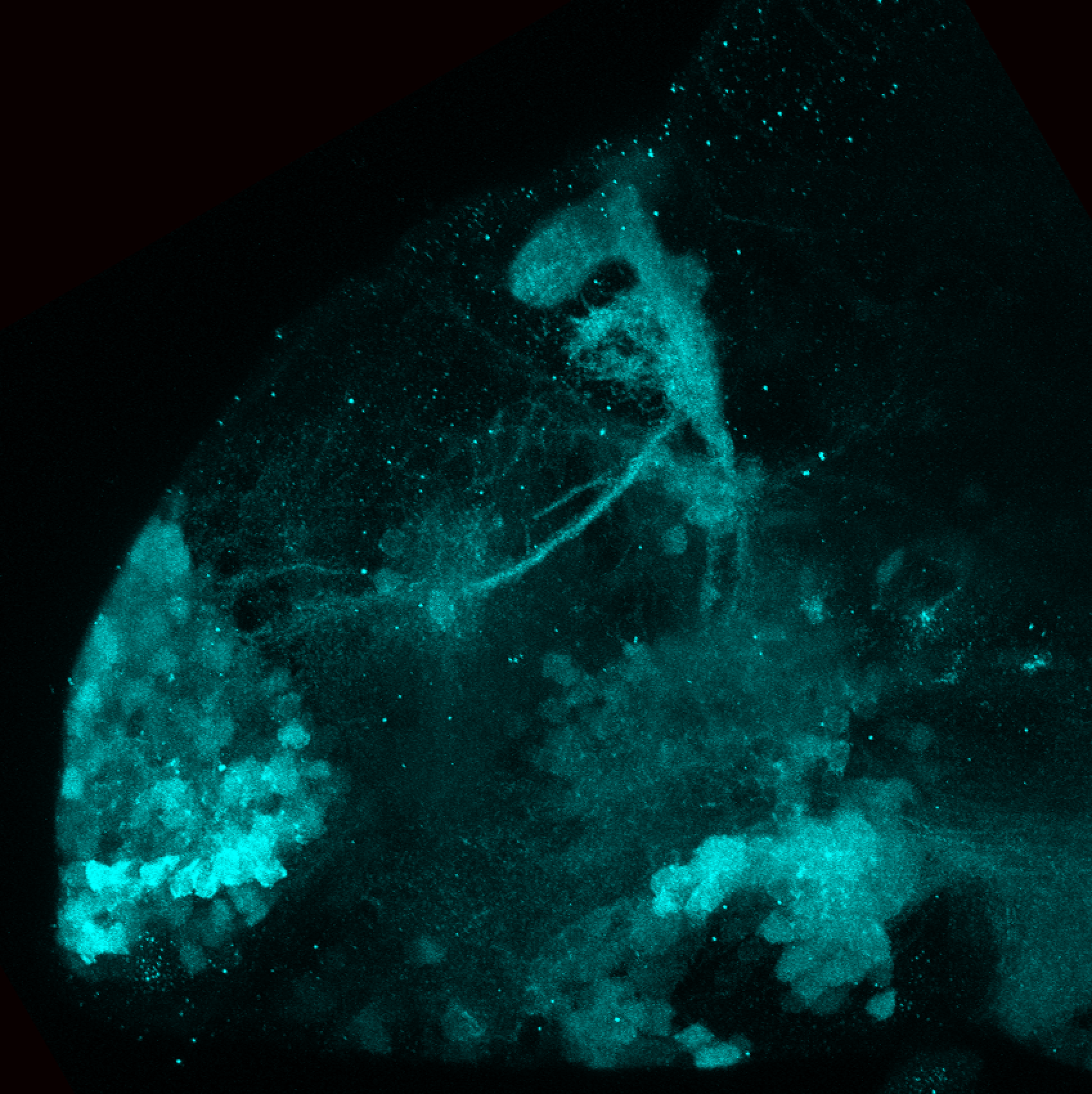
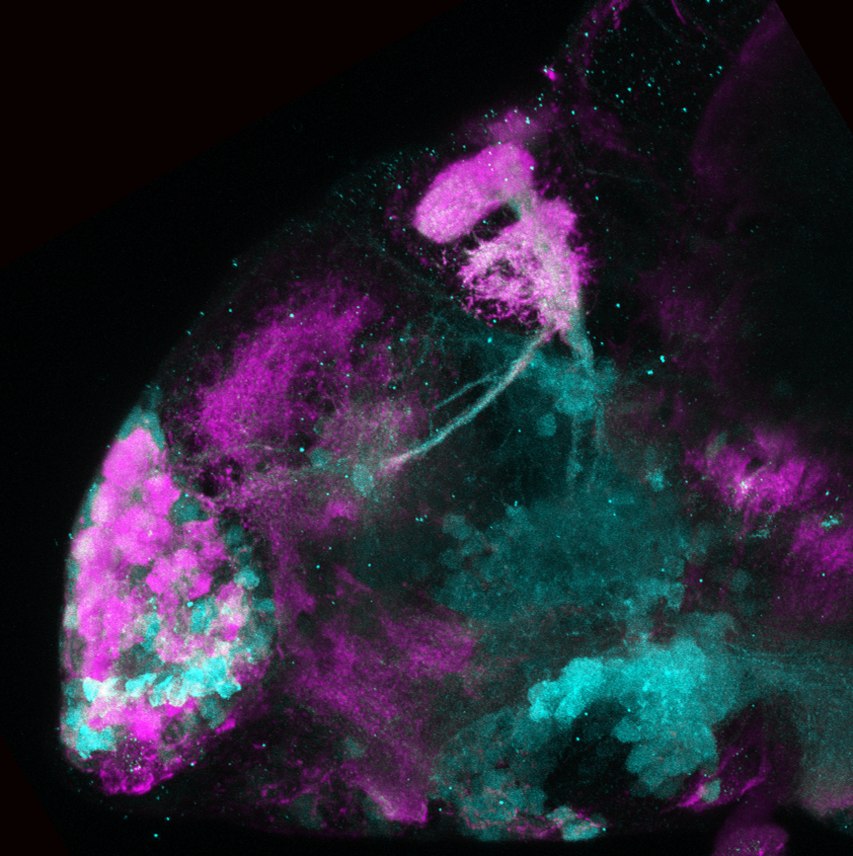
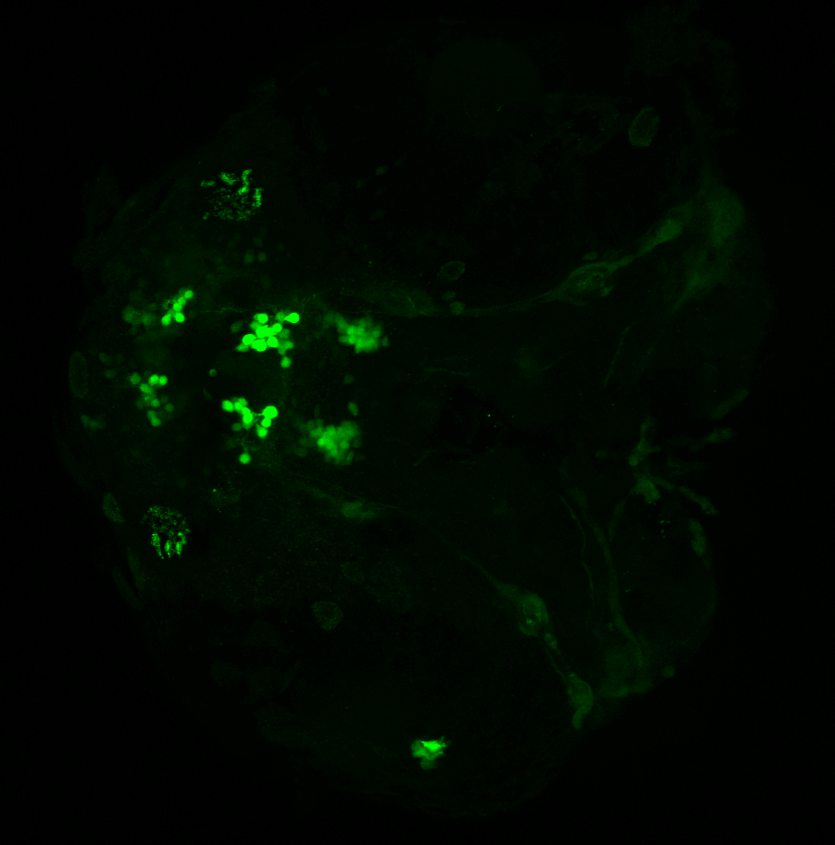
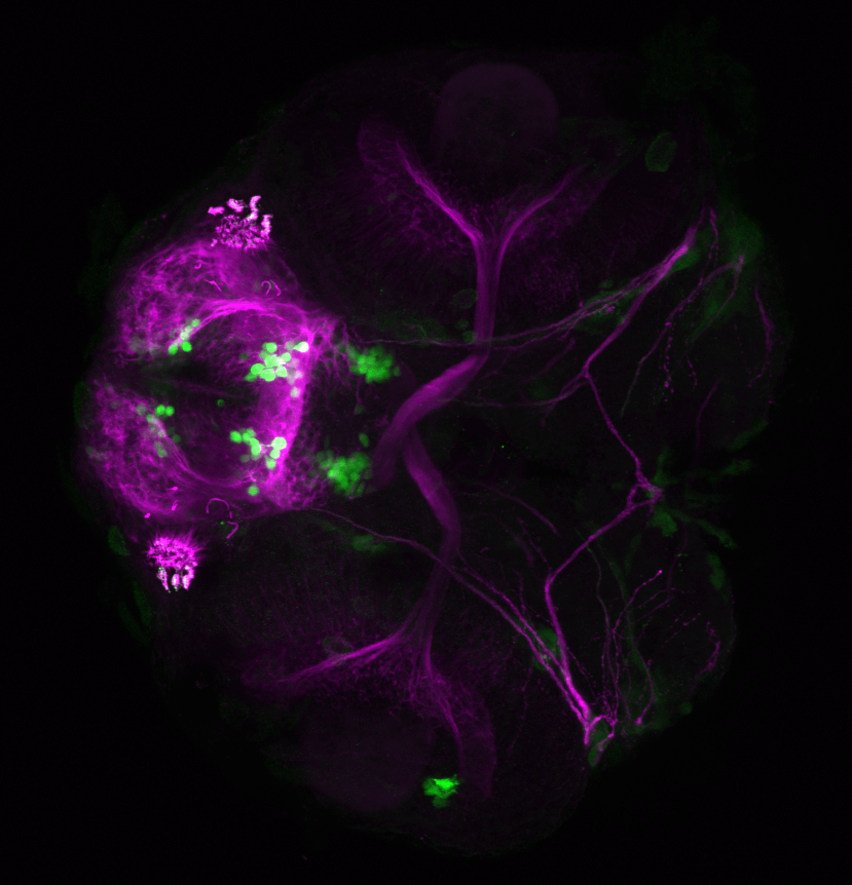
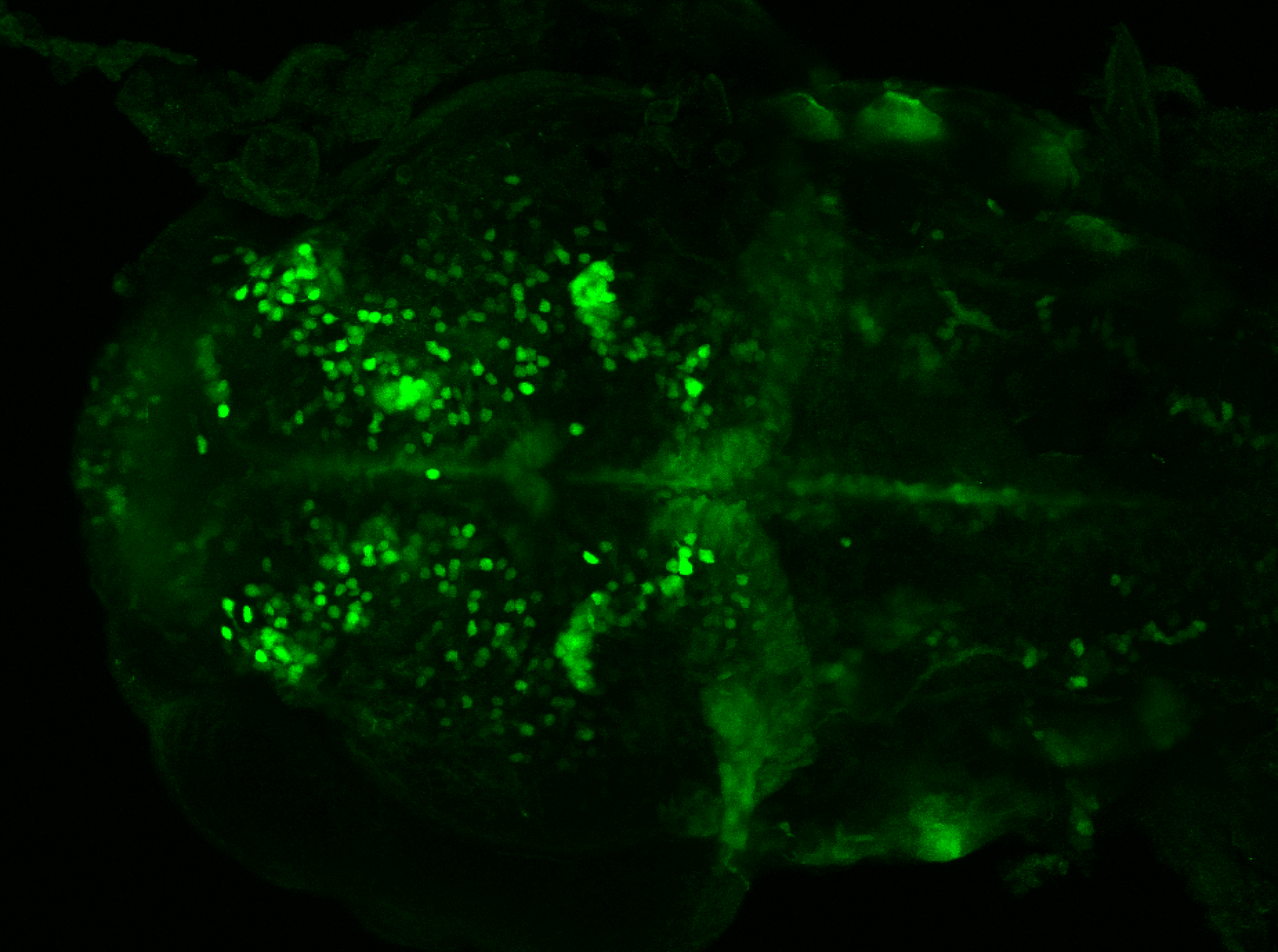
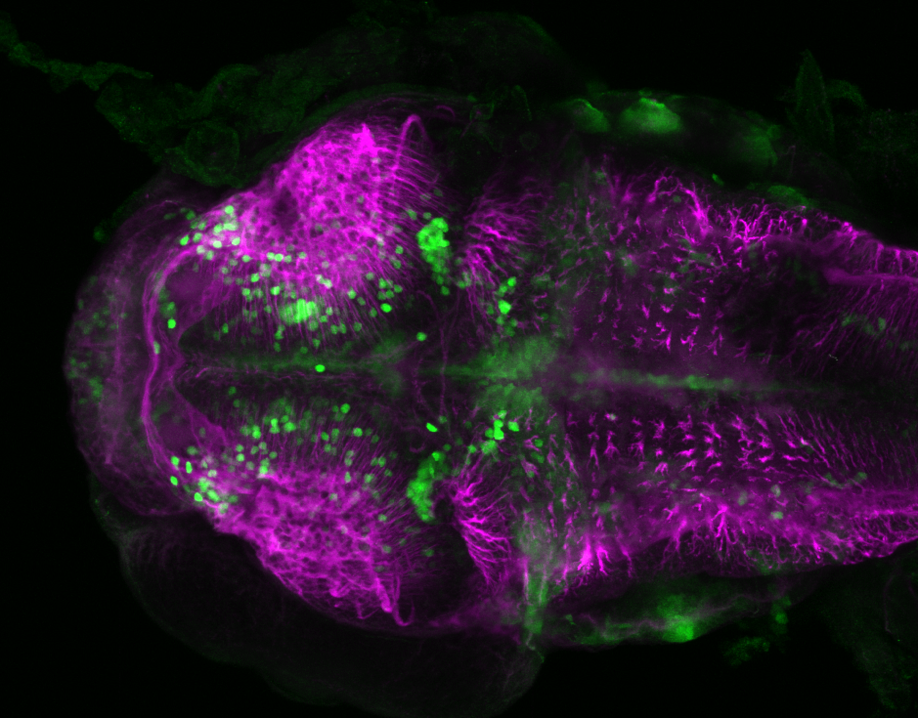
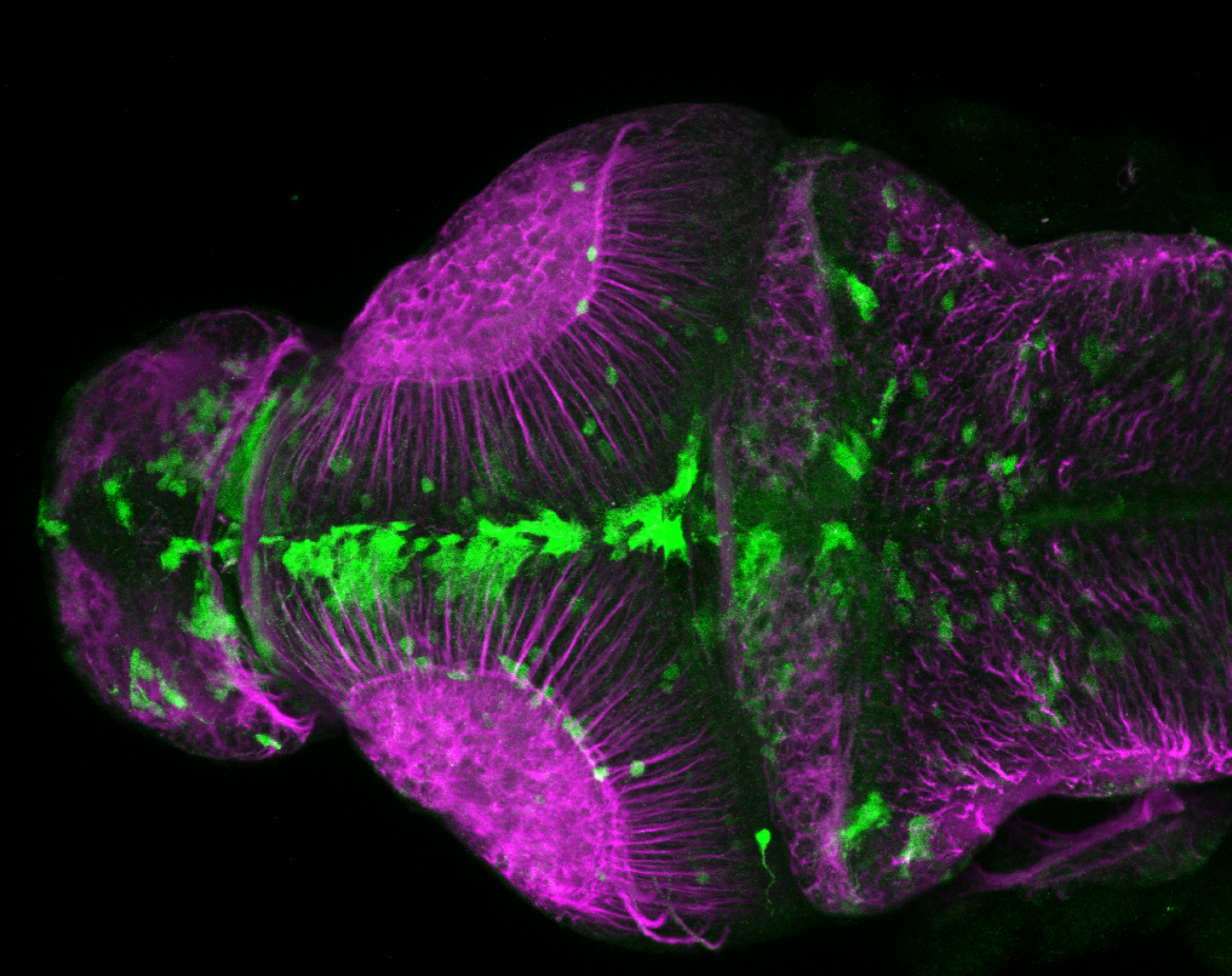
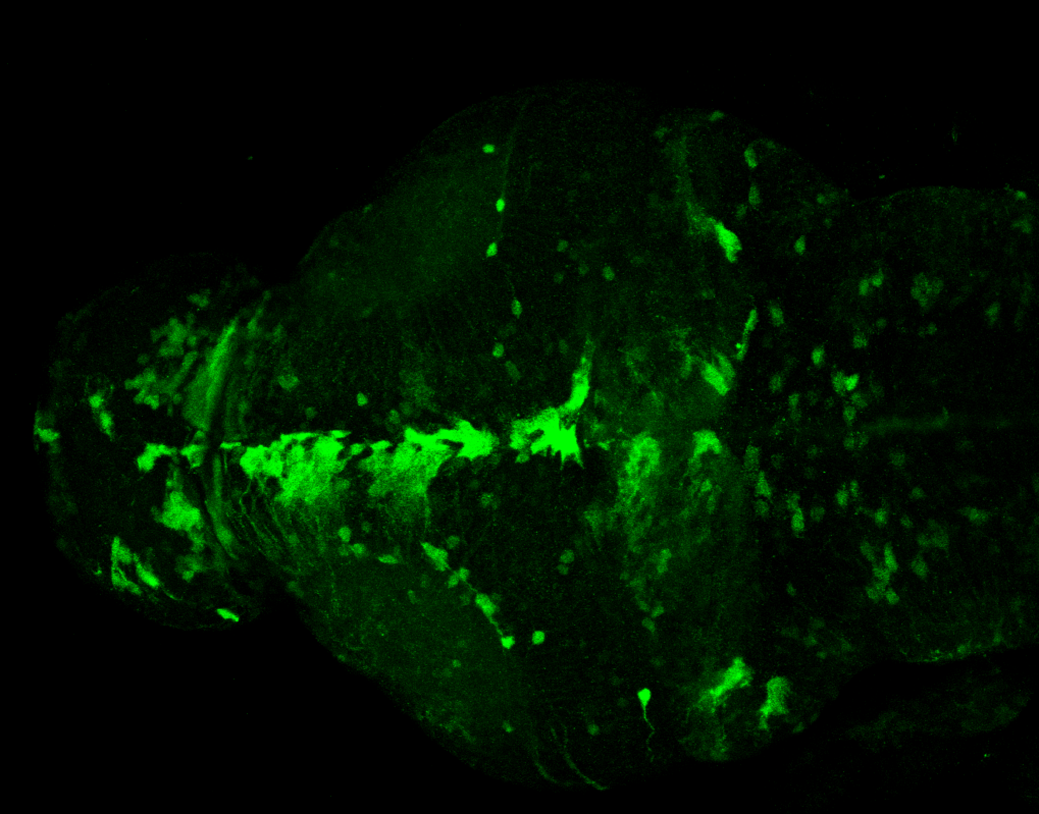
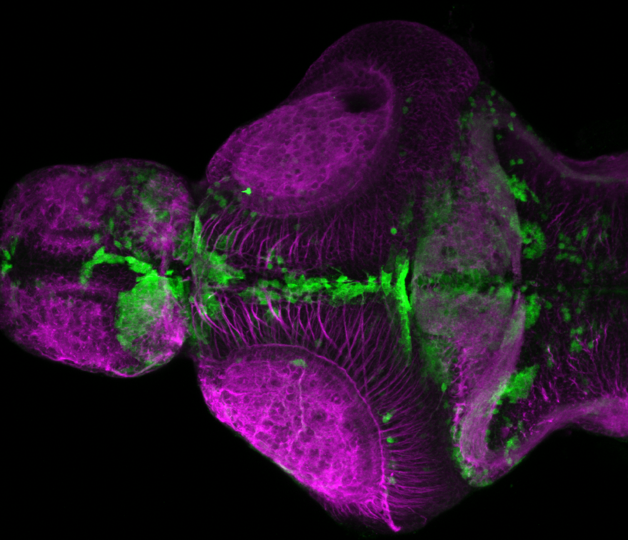
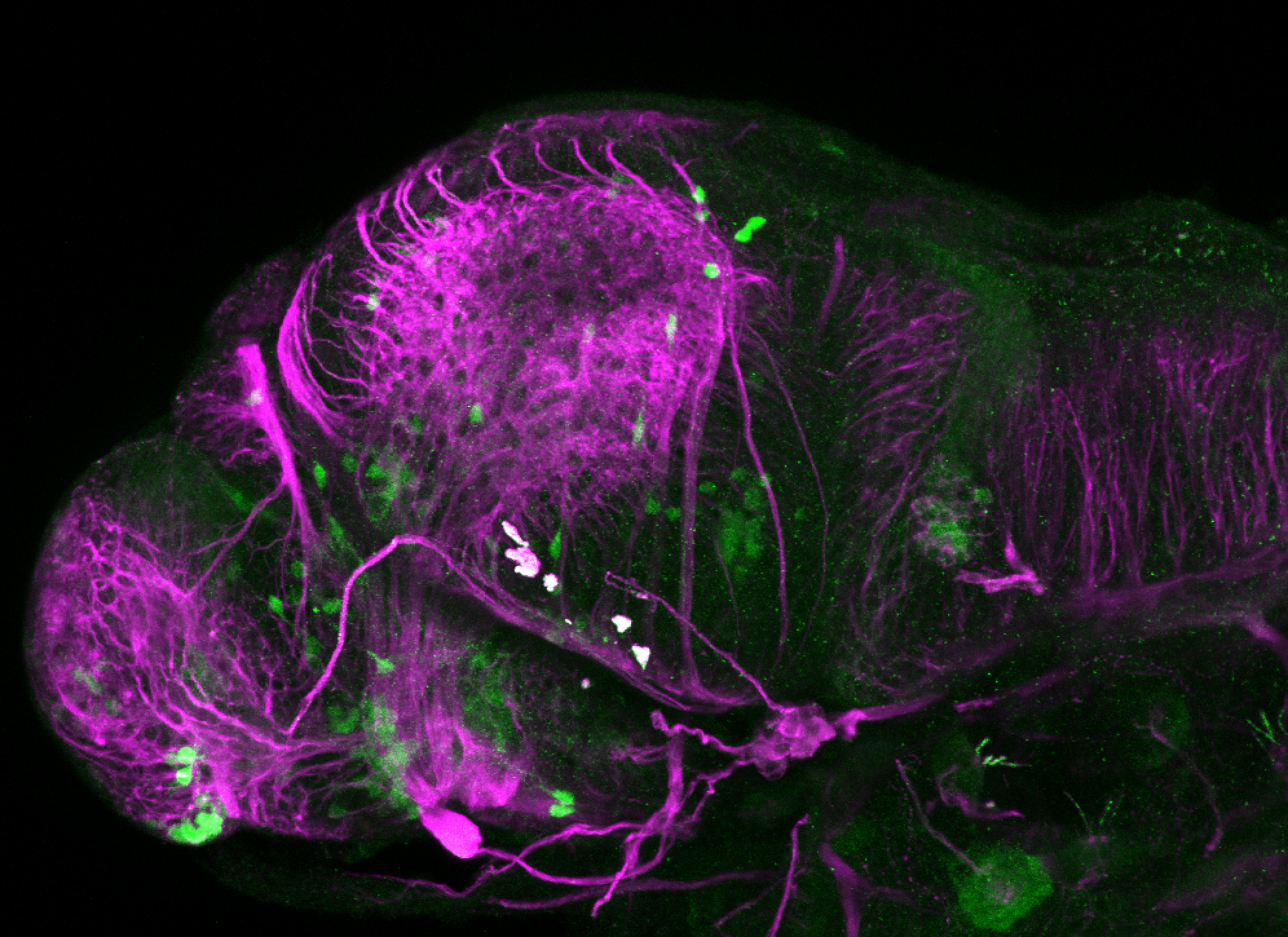
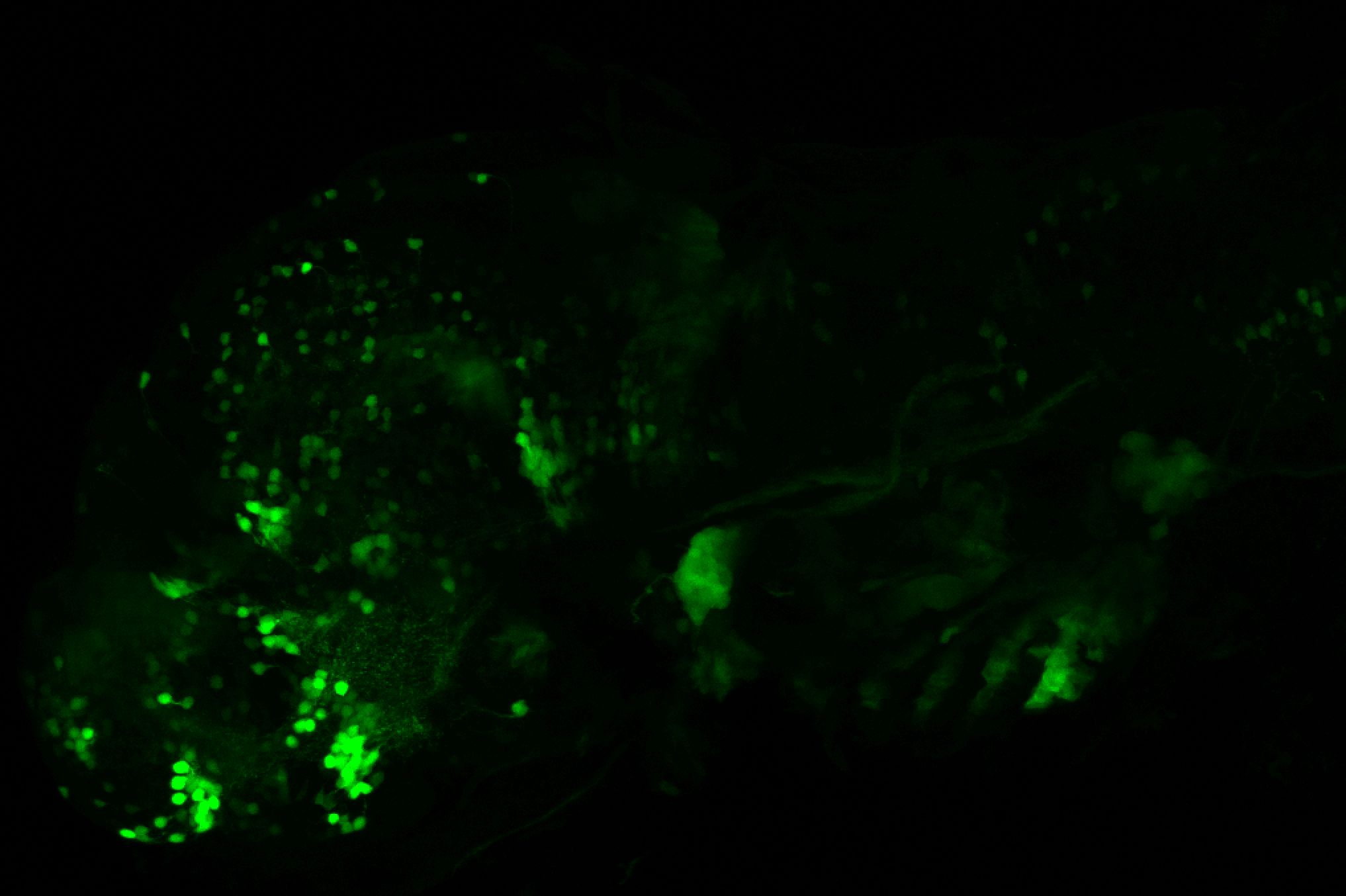
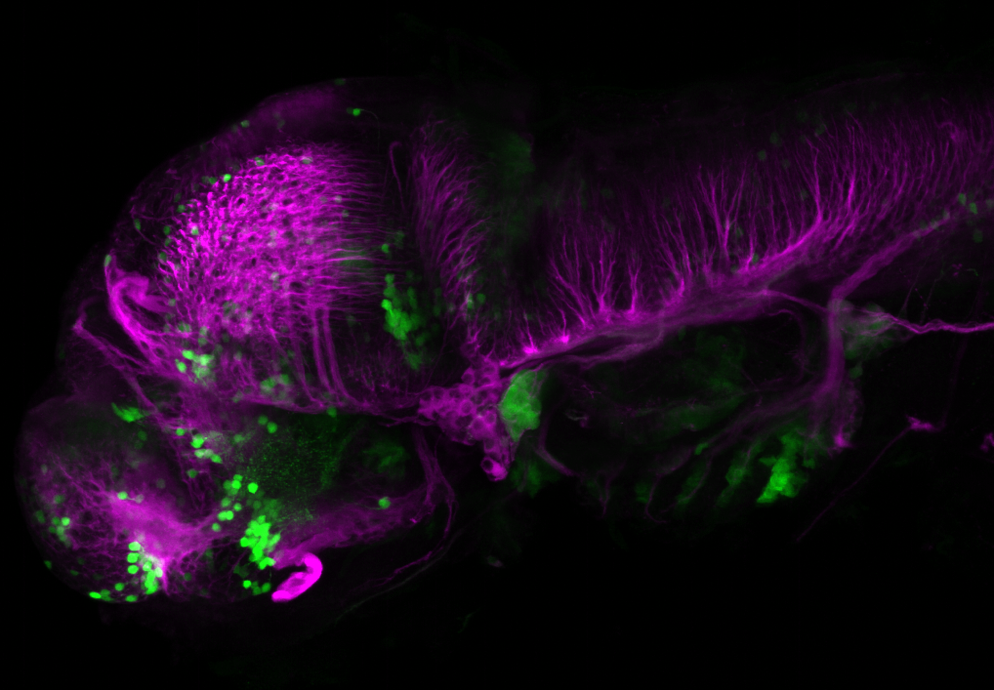
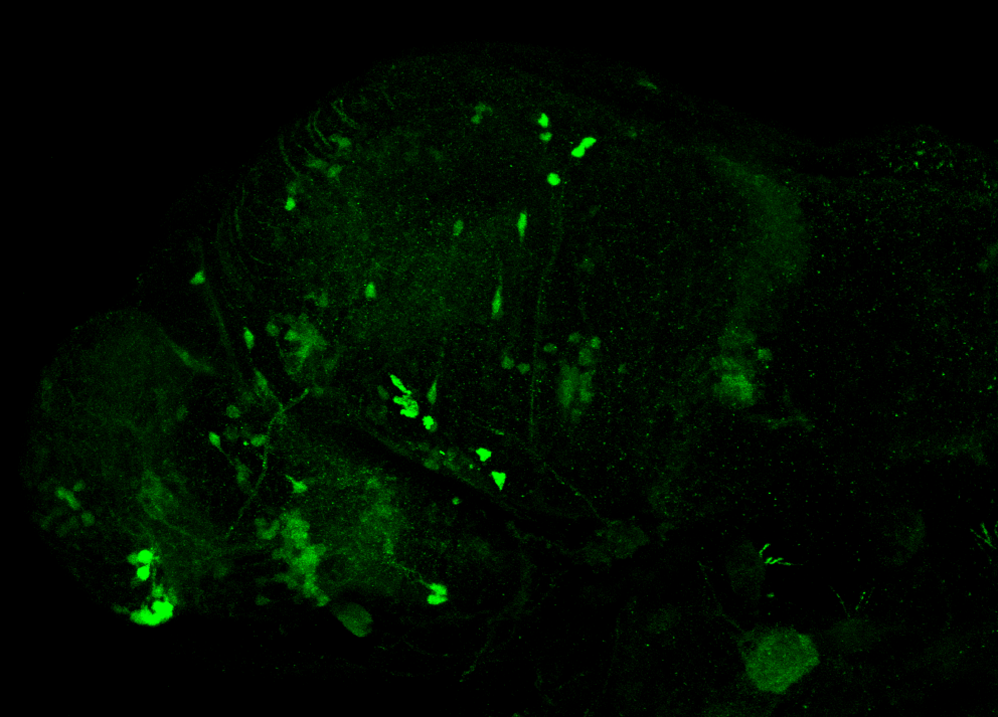
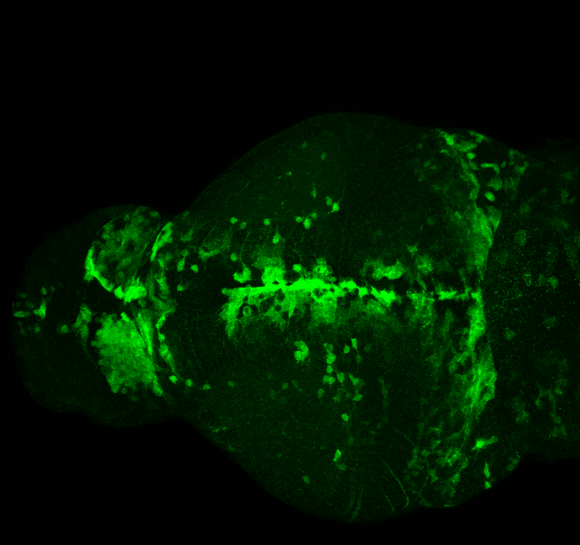
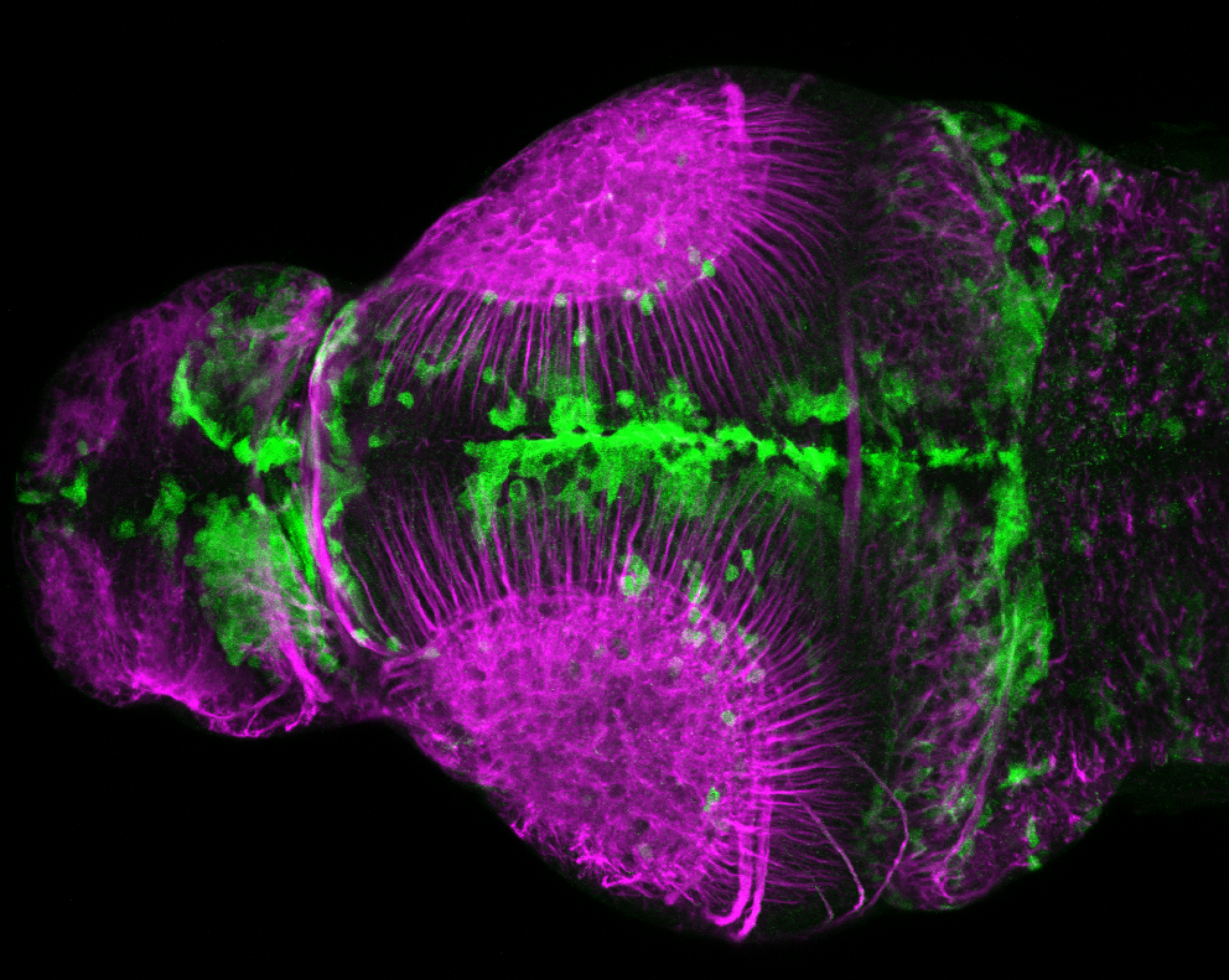
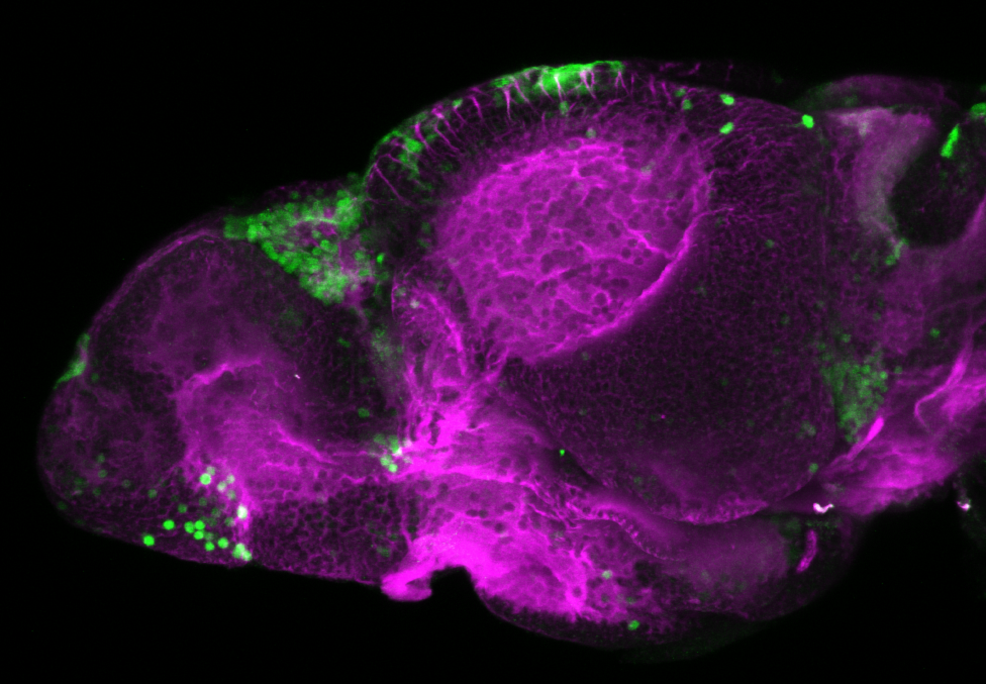
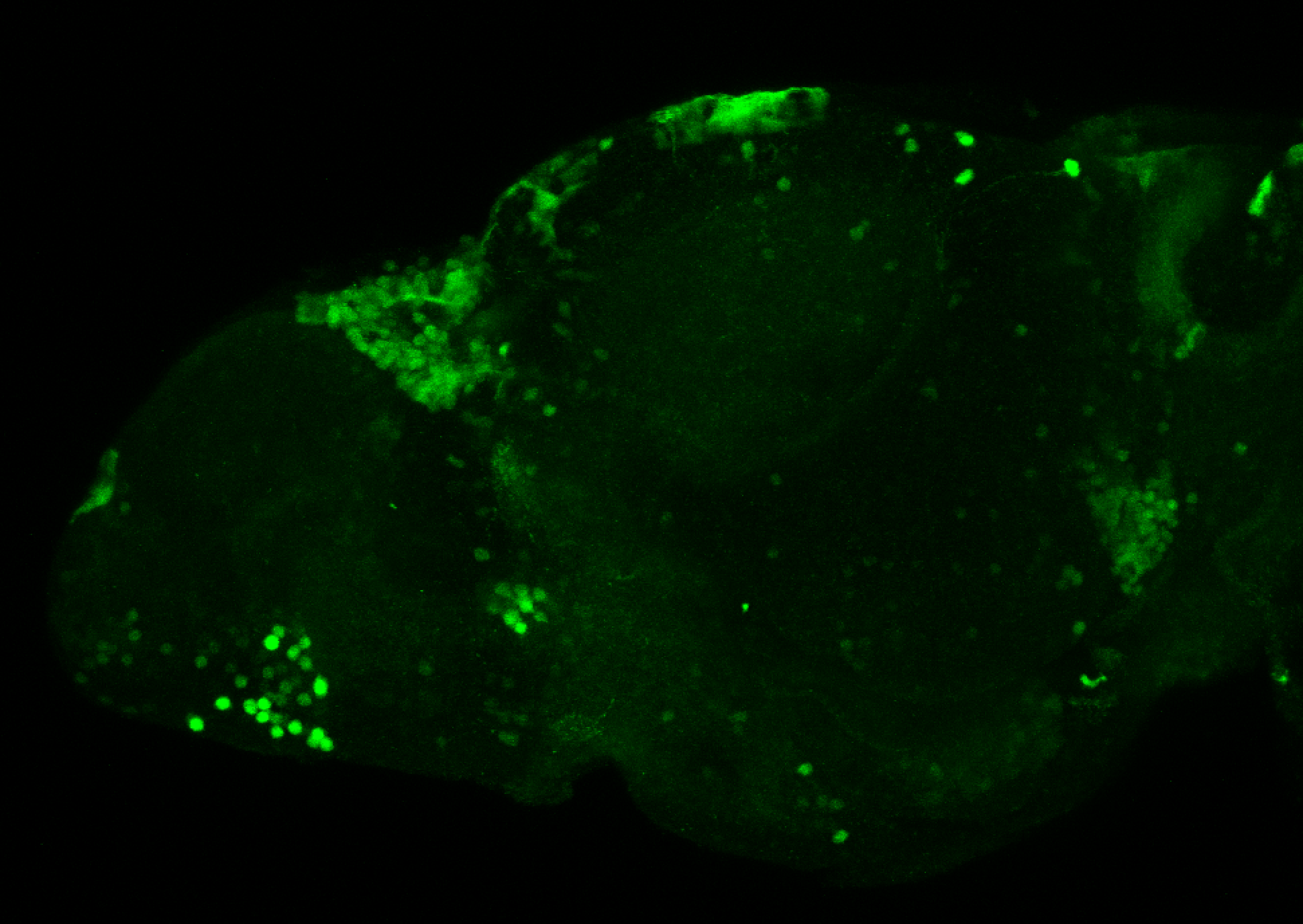
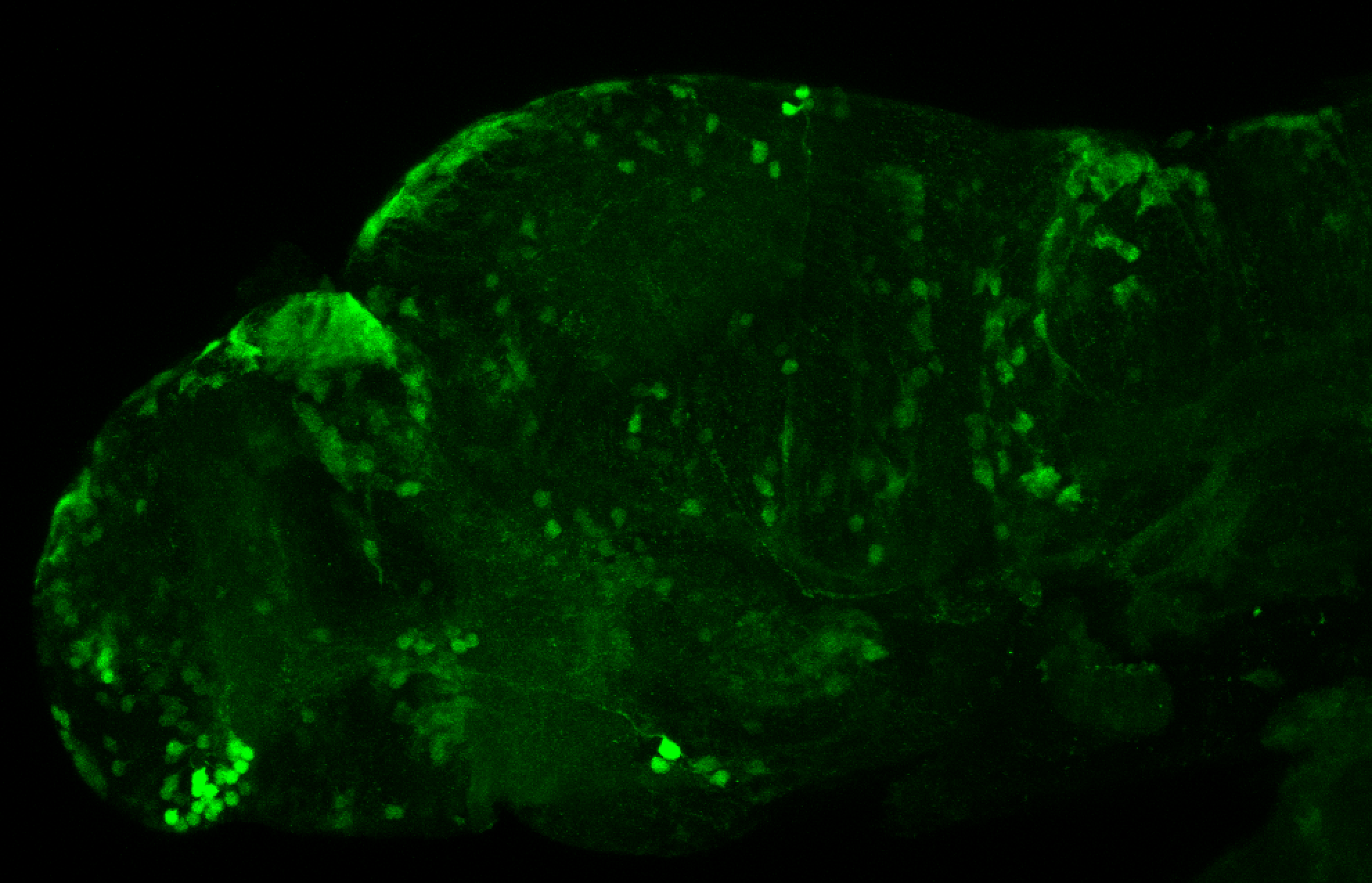
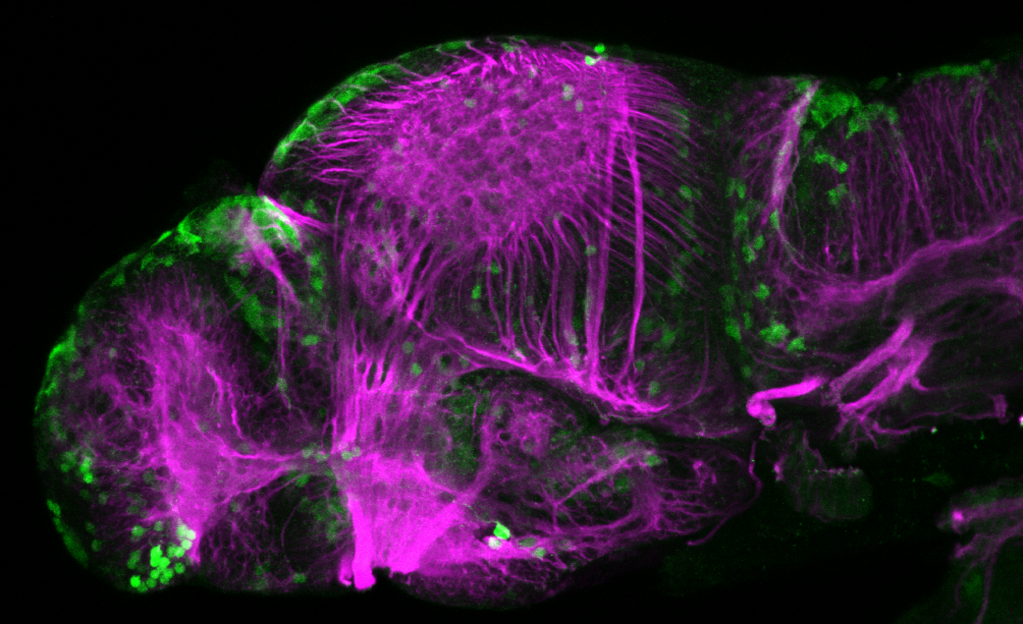
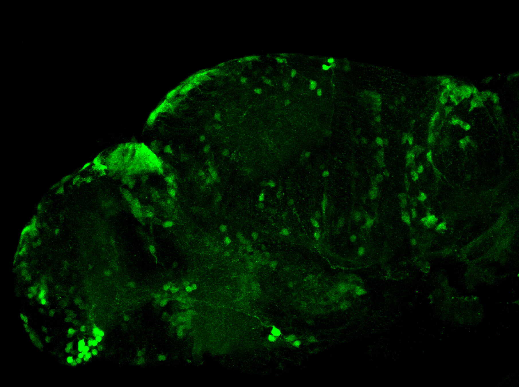
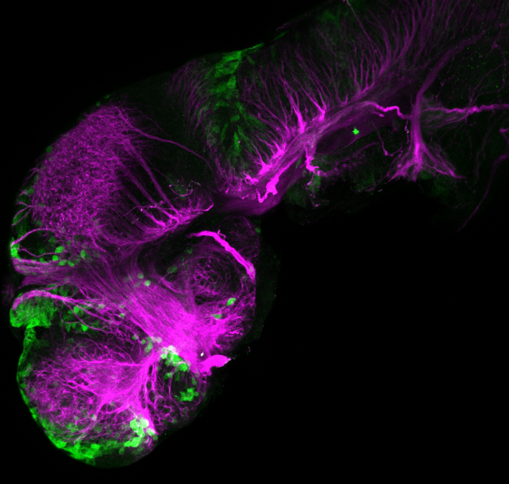
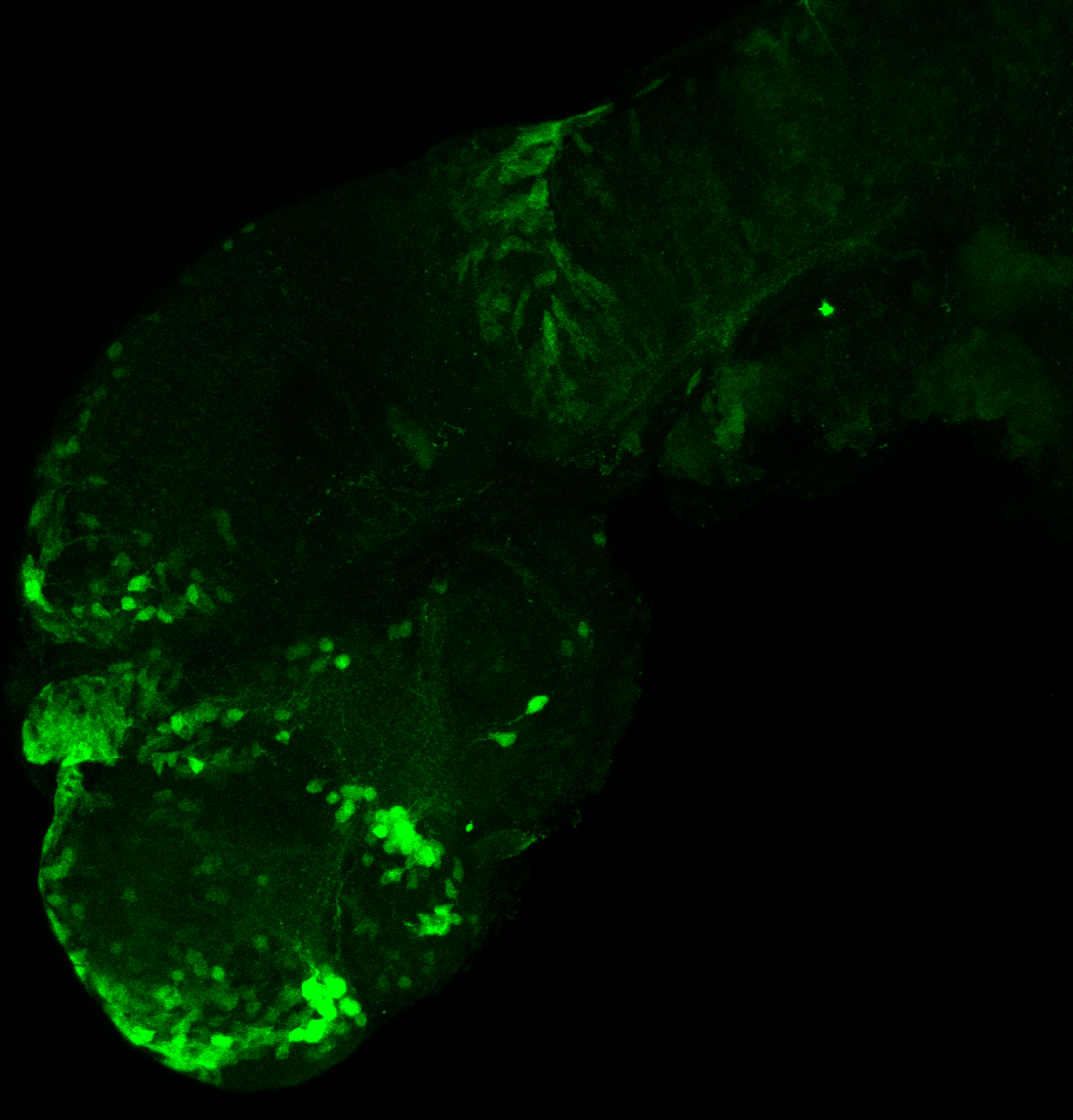
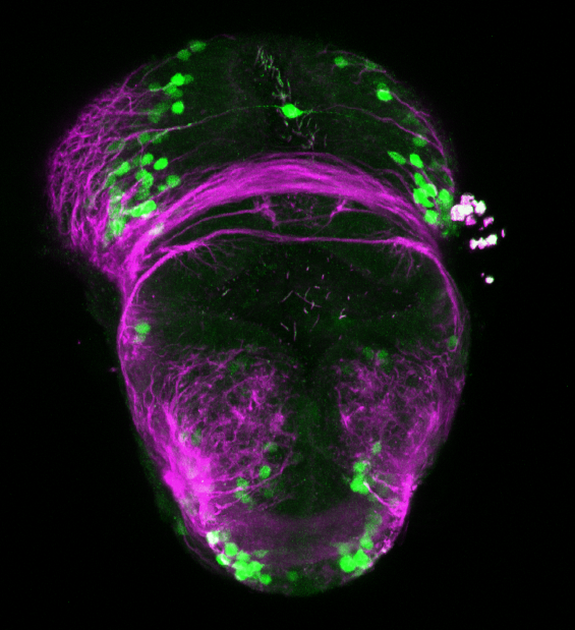
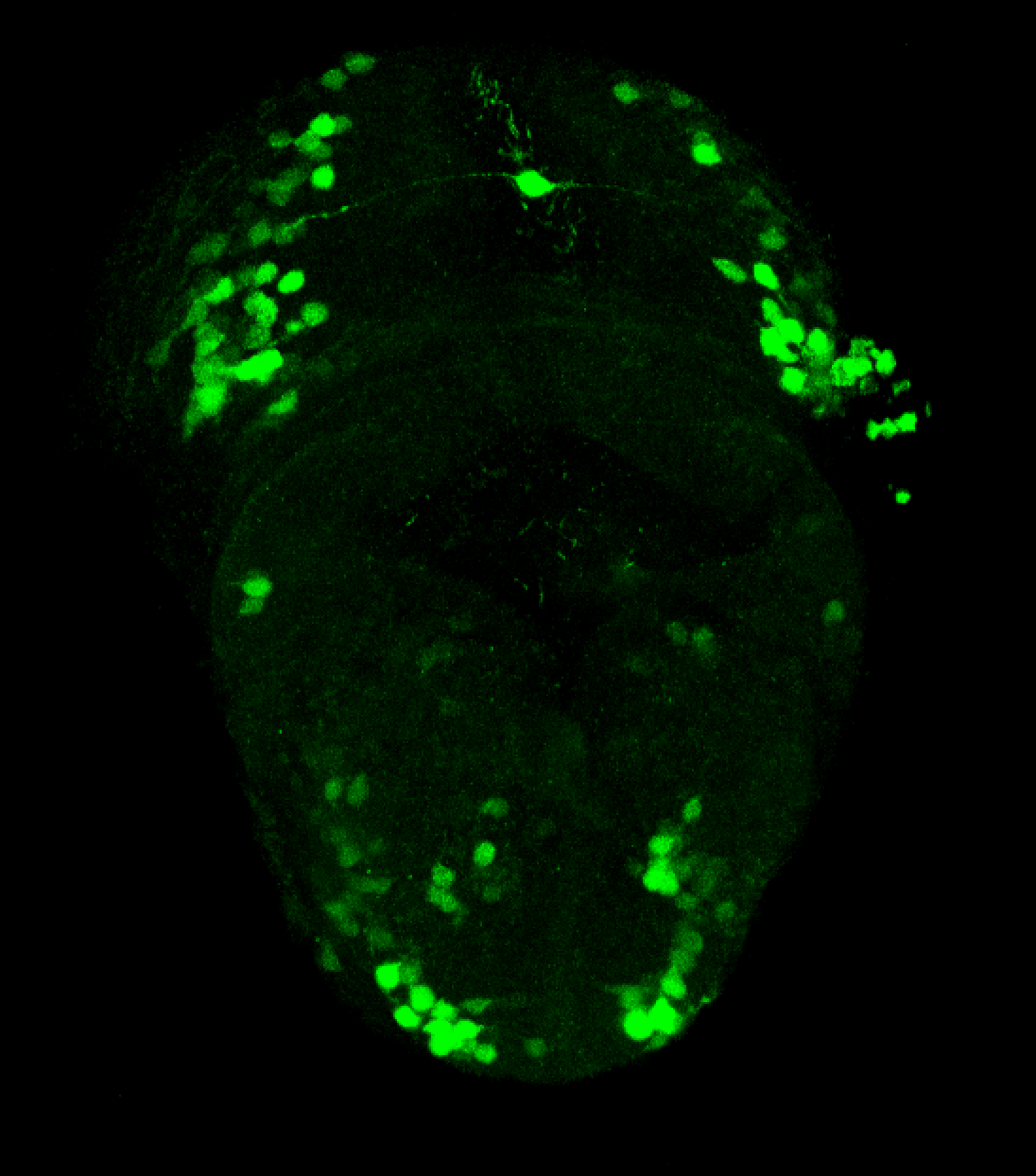
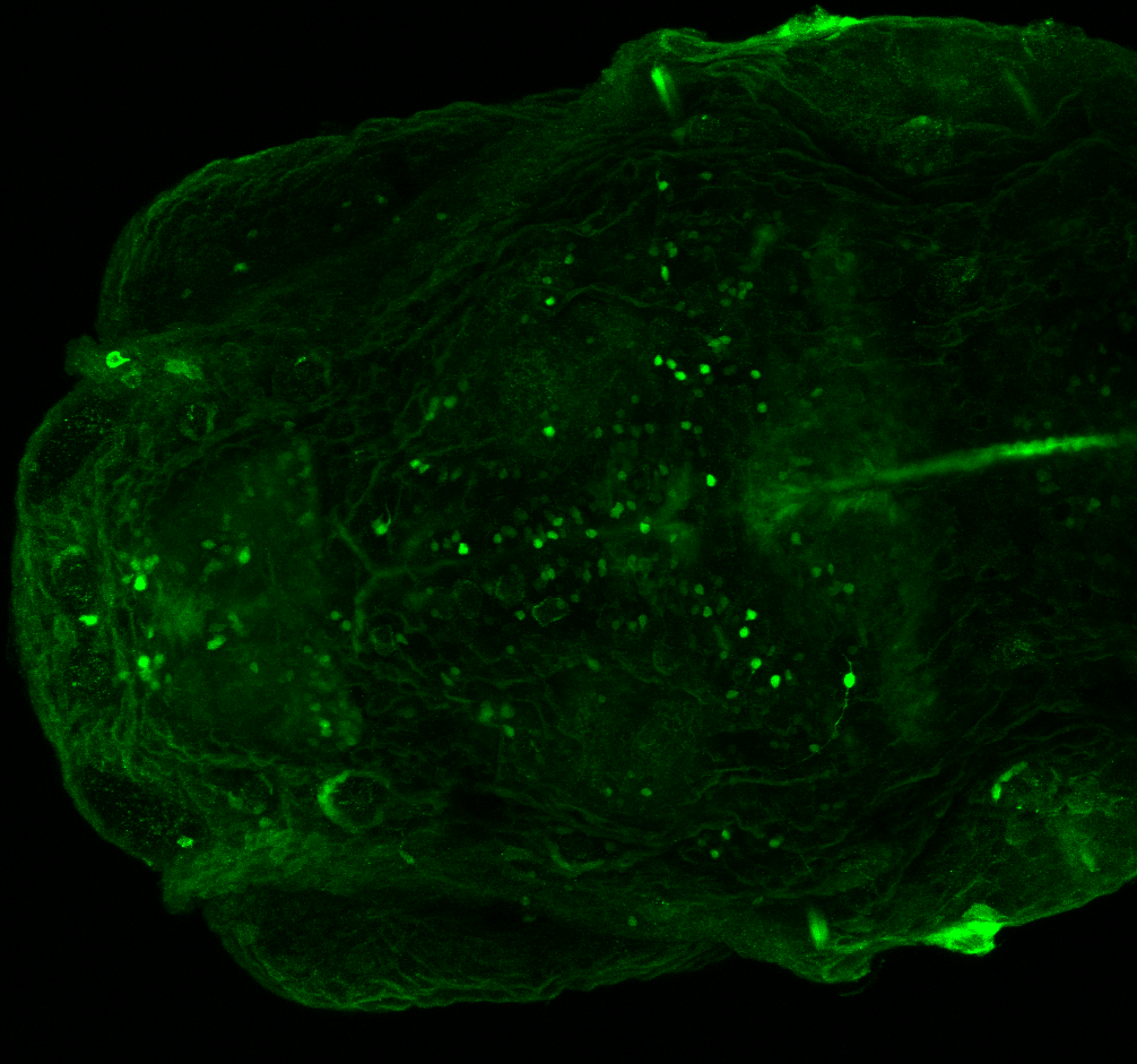
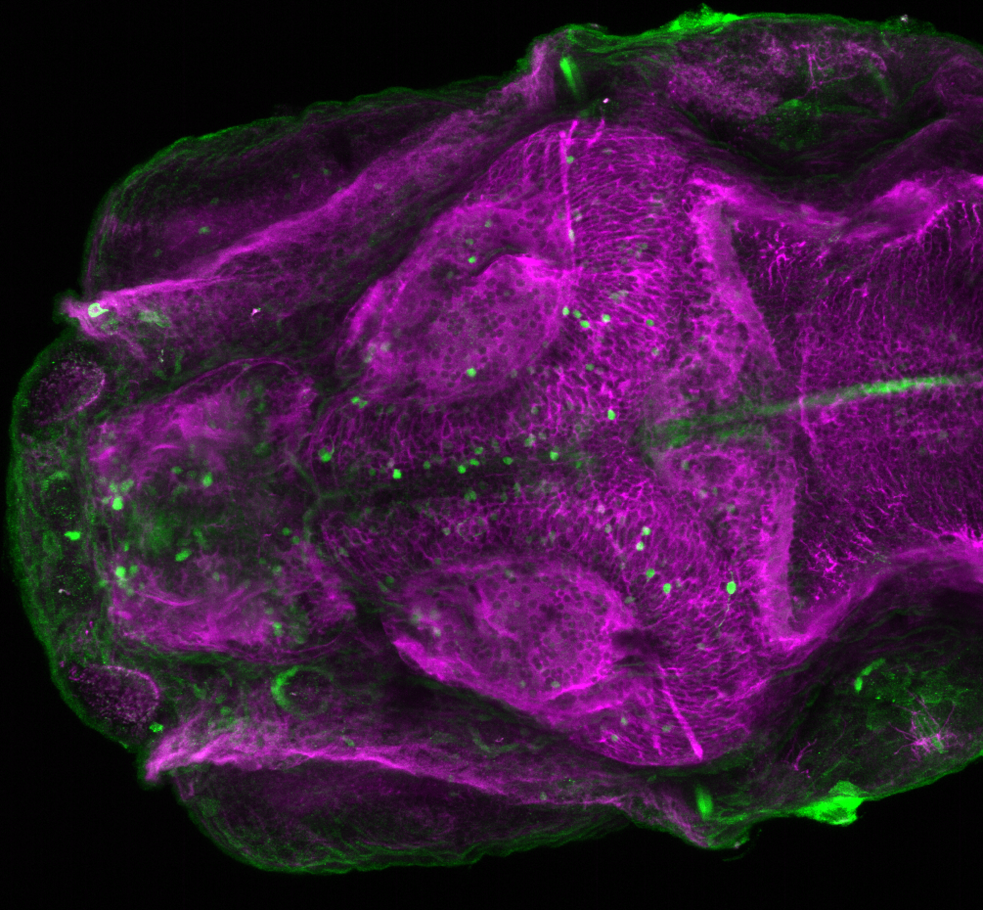
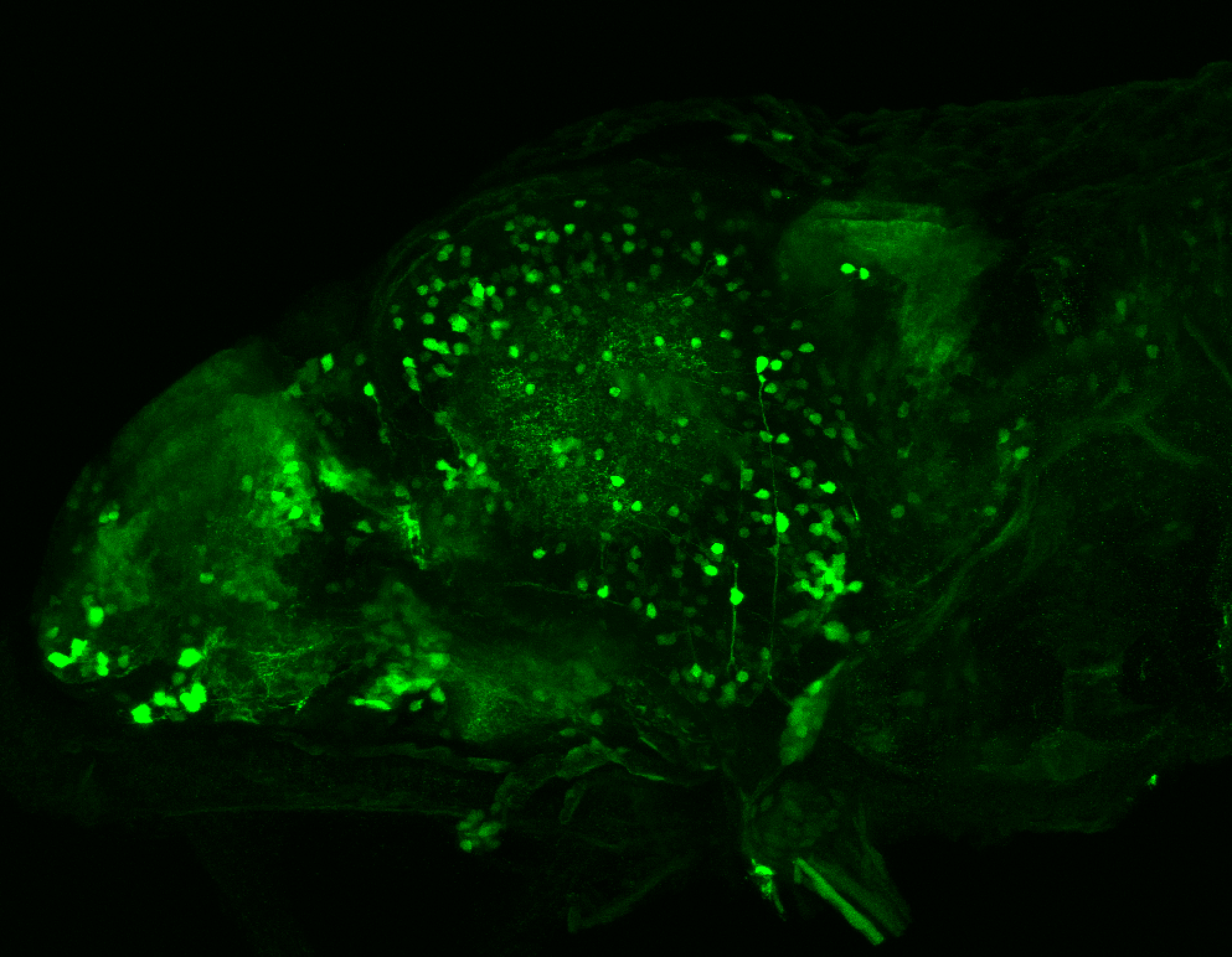
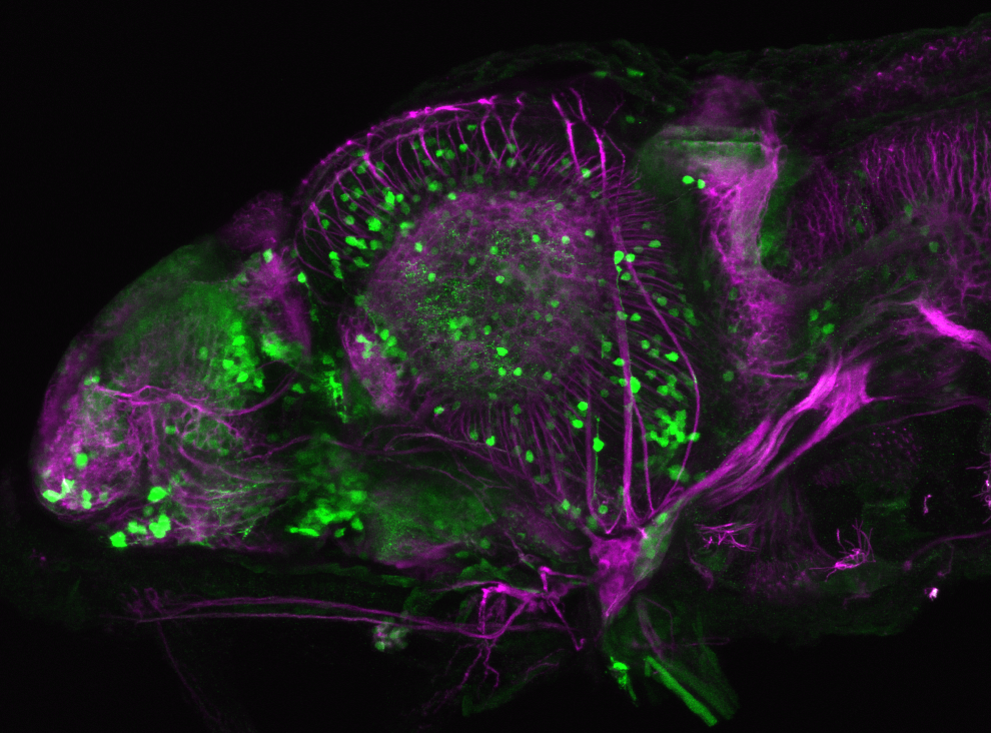
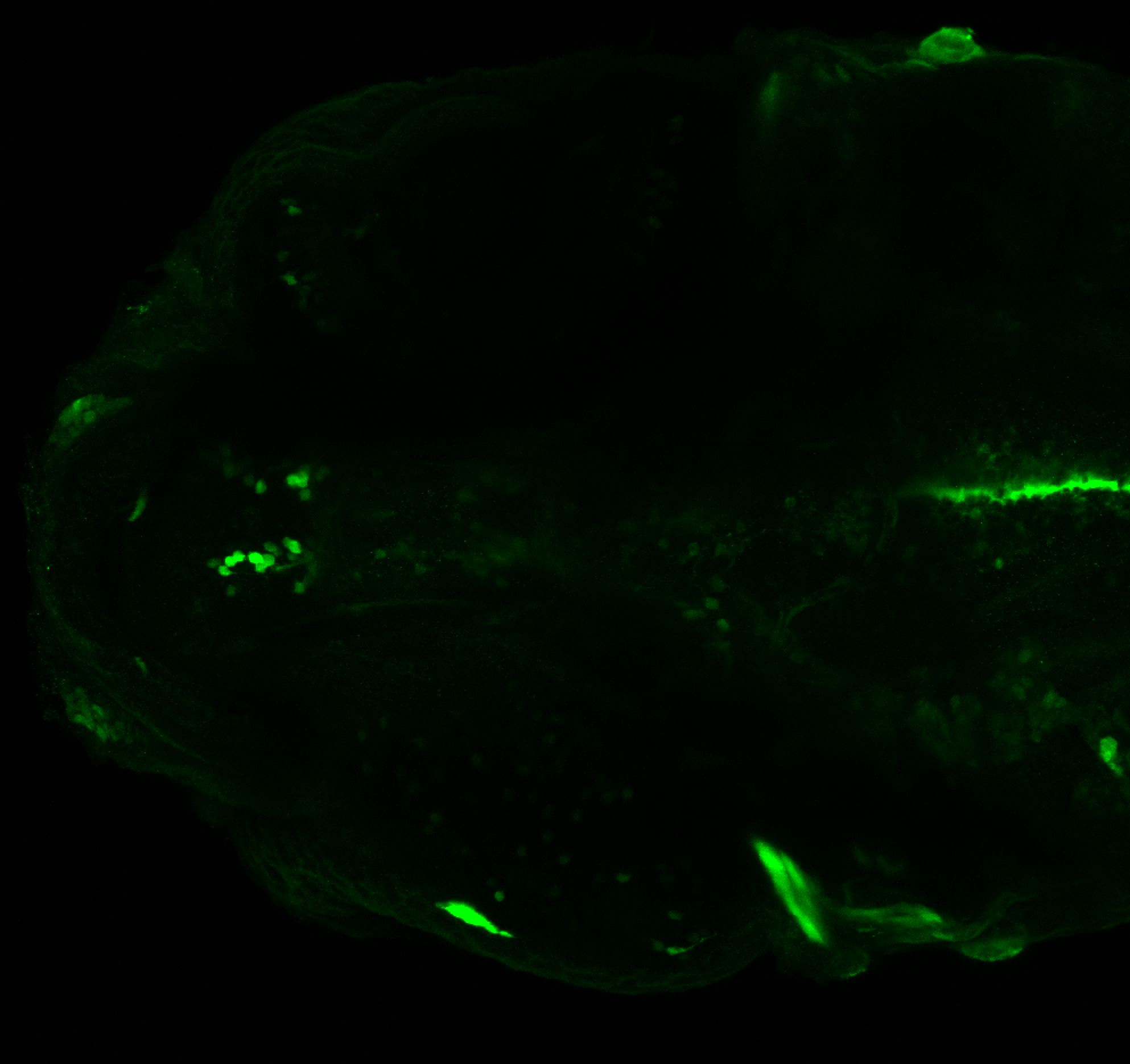
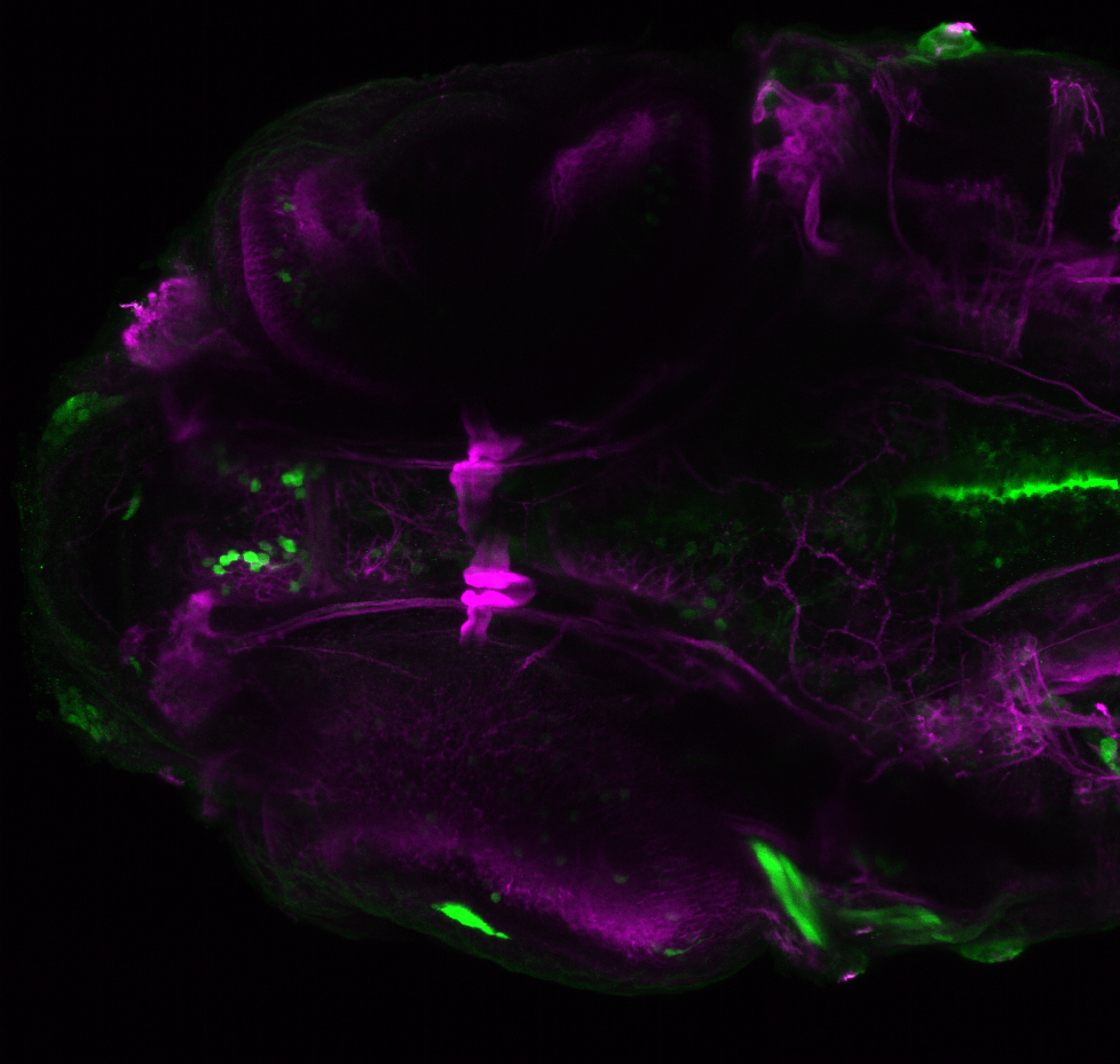
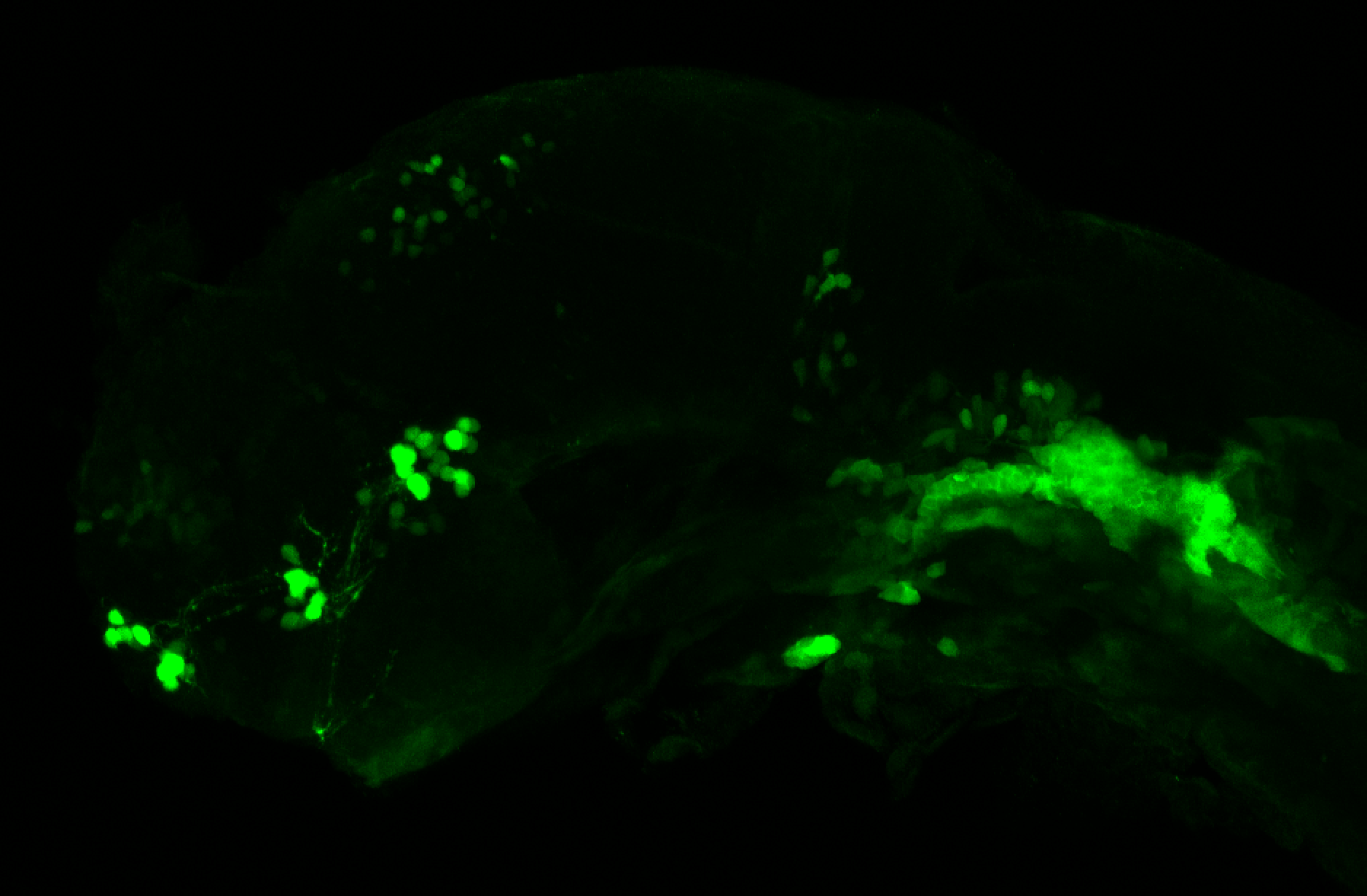
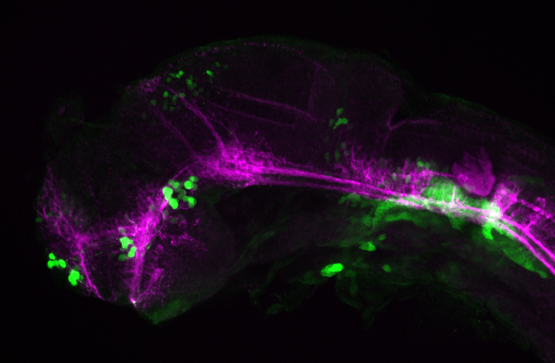
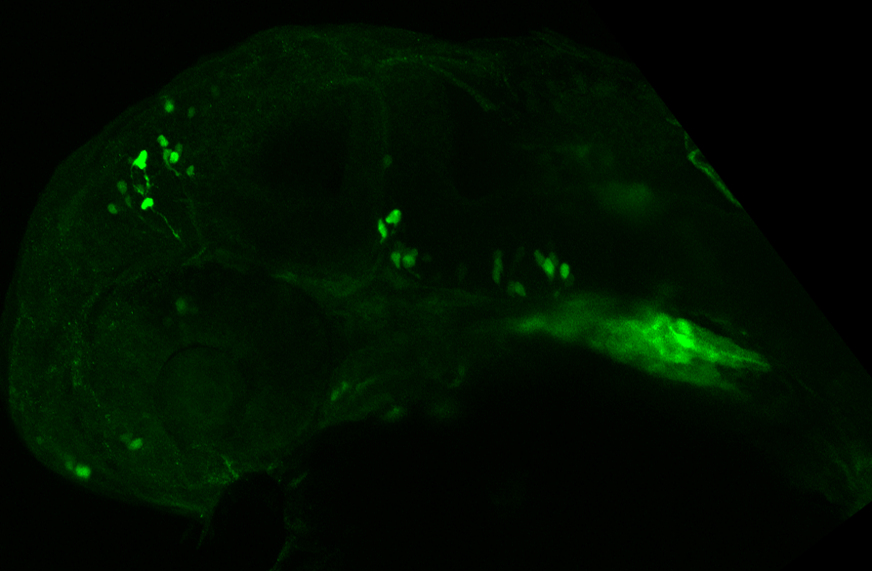
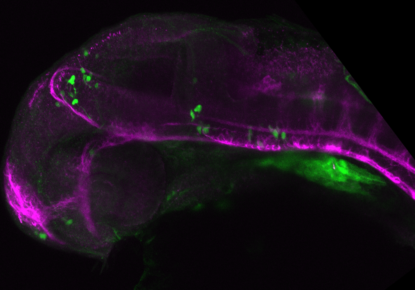
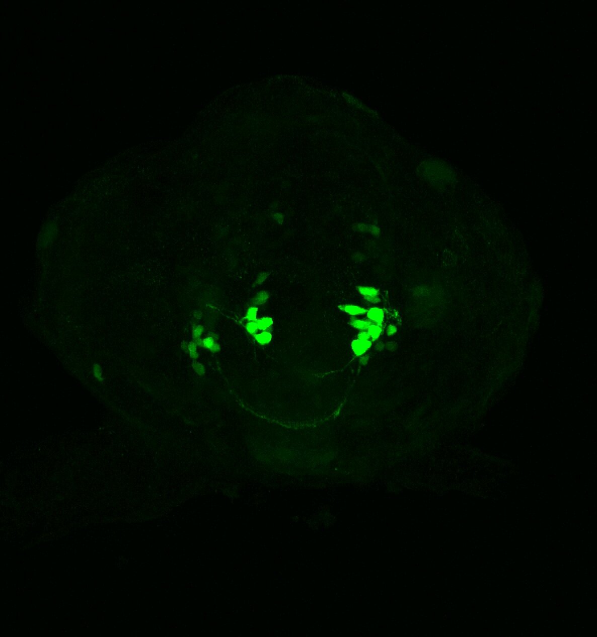
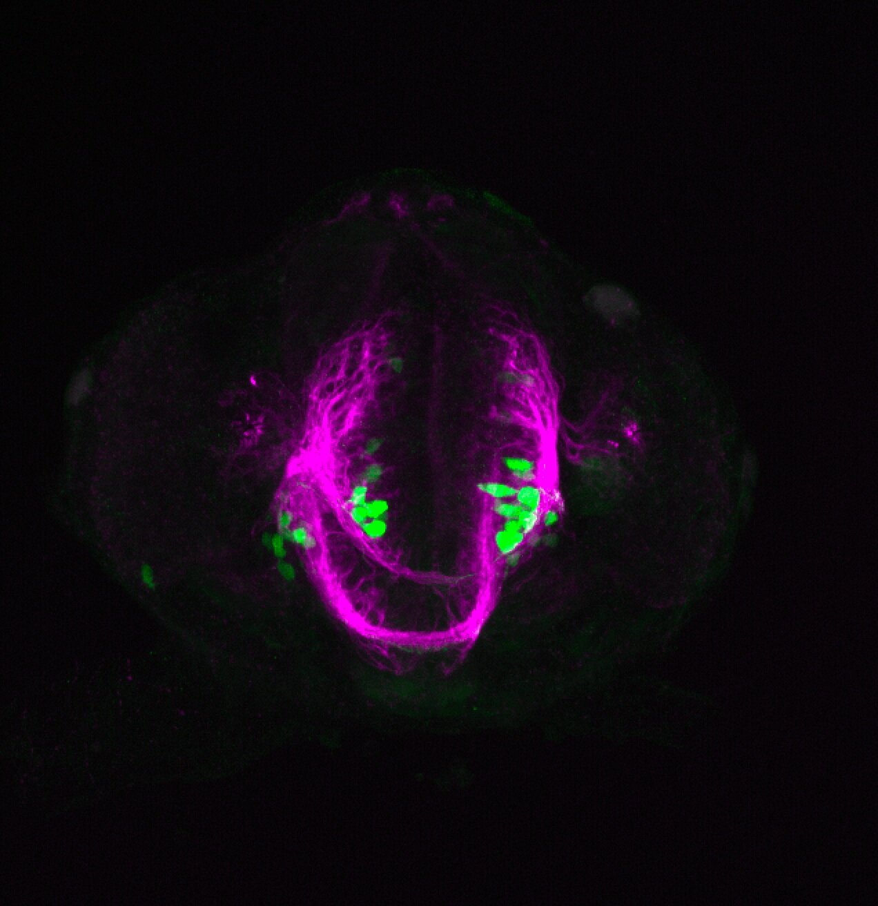
 Enhancer trap line from Vladimir Korzh lab that has EGFP expression in the epithalamus, thalamic eminence, hypothalamic lobes, cerebellum, medulla oblongata.
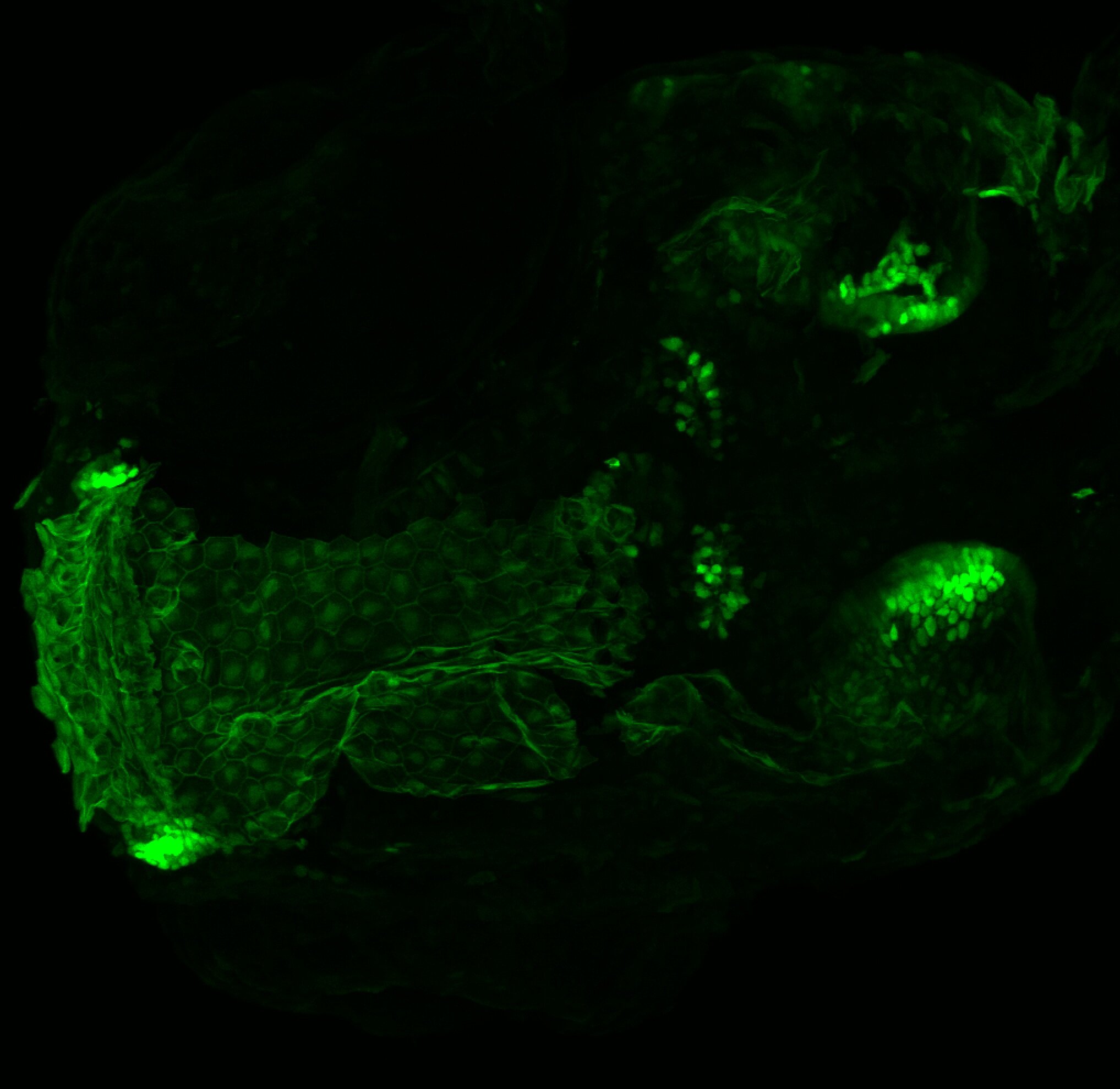
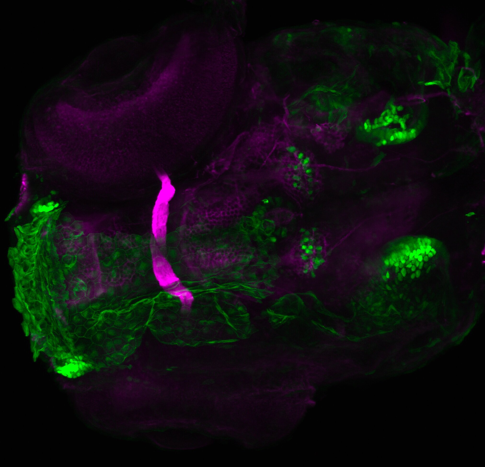
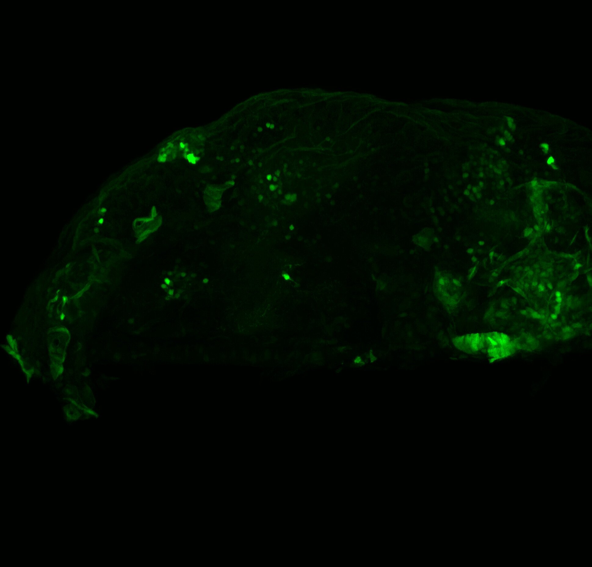
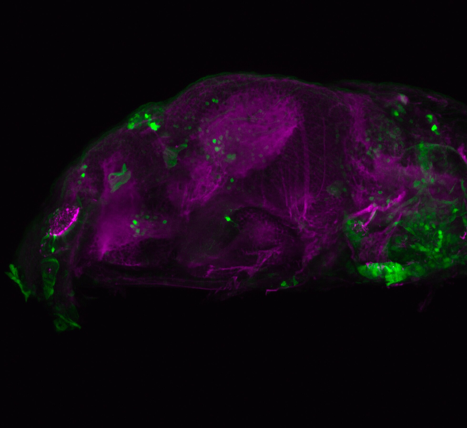
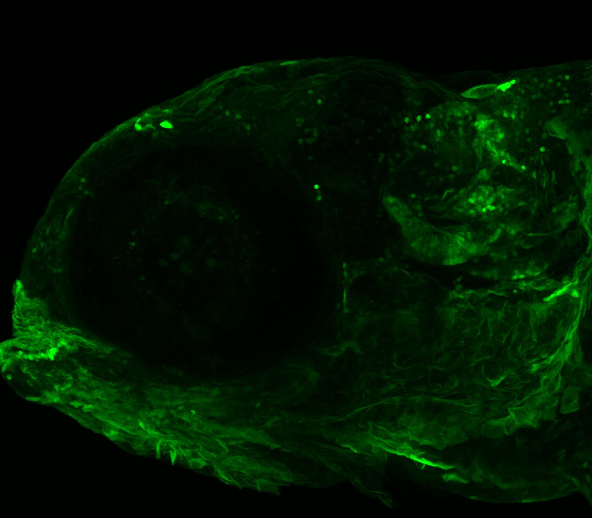
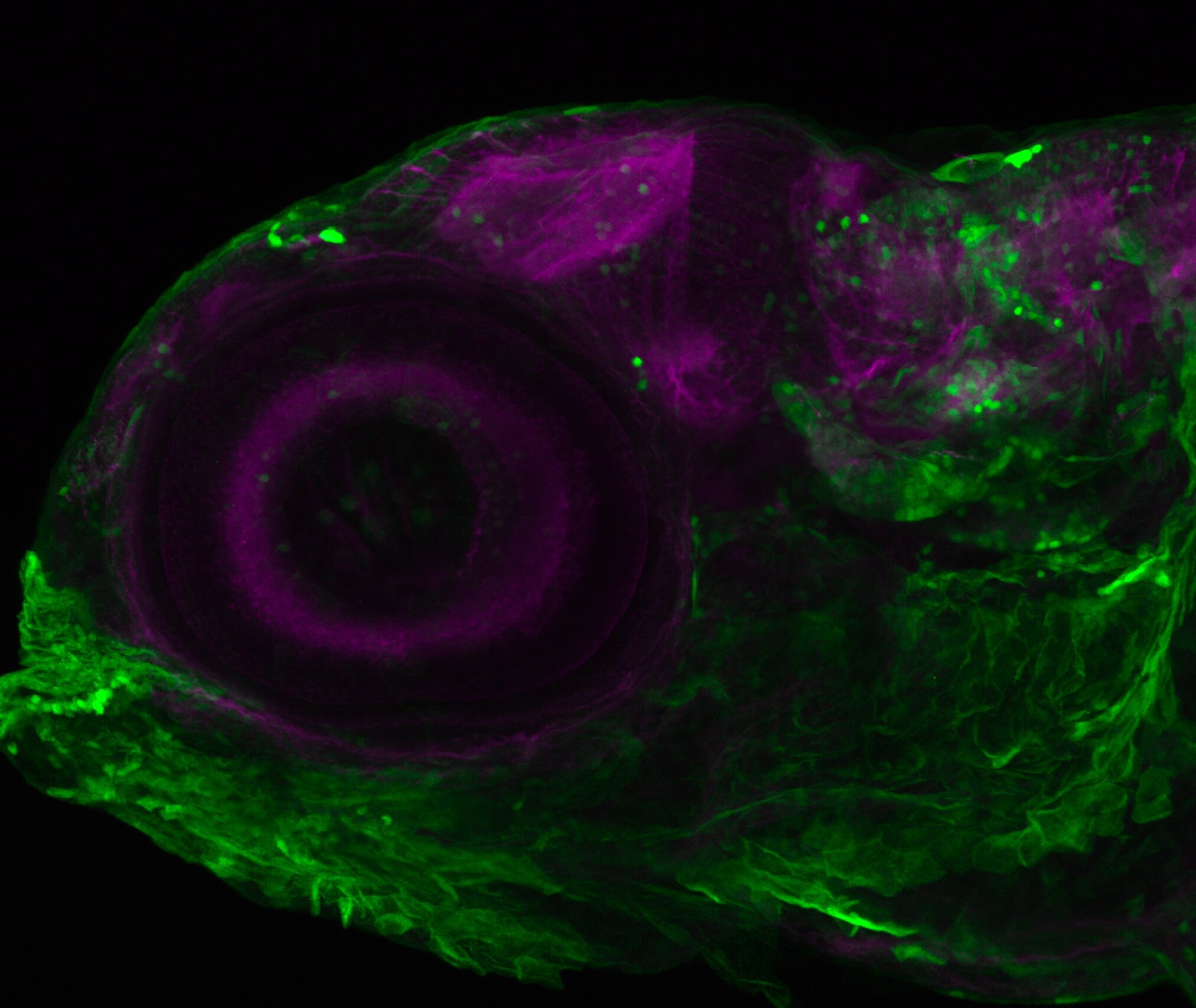

External Links:

 ZFIN

Lab or Origin: Korzh Lab


Expressed in: 

epithalamus, thalamic eminence, hypothalamus, cerebellum, medulla oblongata


Key Publications

Parinov, S., Kondrichin, I., Korzh, V., and Emelyanov, A. (2004)
Tol2 transposon-mediated enhancer trap to identify developmentally regulated zebrafish genes in vivo.
Developmental dynamics : an official publication of the American Association of Anatomists. 231(2):449-459.

Tg(lhx5:GFP)b1205

Tg(lhx5:GFP)b1205

About

 This transgenic line recapitulates the expression of lhx5 transcription factor. Generated by the Westerfield lab using BAC transgenesis. A kaede version of this line is also available. This transgenic has been used to study pathfinding in early telencephalic development (Guo et al., 2012; Zhang et al., 2012, Turner et al., 2019) and also to look at areas afferent to the habenula at later stages of development (Turner et al., 2016).


External Links:

 ZFIN

Lab or Origin: Westerfield Lab


Expressed in: 

 olfactory bulb, pallium, ventral entopeduncular nucleus, preoptic area, prethalamus, stria medularis, tract of the habenula commissure, habenula commisure, habenula neuropil.


Key Publications

Gao, J., Zhang, C., Yang, B., Sun, L., Zhang, C., Westerfield, M., and Peng, G. (2012)
Dcc Regulates Asymmetric Outgrowth of Forebrain Neurons in Zebrafish.
PLoS One. 7(5):e36516.

Zhang, C., Gao, J., Zhang, H., Sun, L., and Peng, G. (2012)
Robo2-Slit and Dcc-Netrin1 Coordinate Neuron Axonal Pathfinding within the Embryonic Axon Tracts.
The Journal of neuroscience : the official journal of the Society for Neuroscience. 32(36):12589-12602.

Turner, K.J., Hawkins, T.A., Yáñez, J., Anadón, R., Wilson, S.W., Folgueira, M. (2016)
Afferent Connectivity of the Zebrafish Habenulae.
Frontiers in neural circuits. 10:30.

Turner, K.J., Hoyle, J., Valdivia, L.E., Cerveny, K.L., Hart, W., Mangoli, M., Geisler, R., Rees, M., Houart, C., Poole, R.J., Wilson, S.W., Gestri, G. (2019)
Abrogation of Stem Loop Binding Protein (Slbp) function leads to a failure of cells to transition from proliferation to differentiation, retinal coloboma and midline axon guidance deficits.
PLoS One. 14:e0211073.

	io005Tg/Tg(Xleomes:GFP)io005

io005Tg/Tg(Xleomes:GFP)io005

About

 Synonyms: Tg(Xleomes:GFP)io005, tg(Xeom:GFP)

The tg(Xeom:GFP) transgenic lines were made in the Mione lab and used to study the dorsal to ventral migration of the paraseptal neurons in the subpallium.

“GFP-expressing cells appear around 28 hpf in the telencephalon of tg(Xeom:GFP) transgenic embryos. Paired groups of GFP-expressing cells appear in the lateral region of the telencephalon more or less at the level of the olfactory placode, at the telencephalic/diencephalic border and in the midbrain tegmentum.
We describe the migration of the telencephalic group: these cells originate from the corresponding ventricular zone, they first move towards the lateral side and then proceed rapidly towards the ventral telencephalon. Most of the GFP+ cells will congregate at the level of the septal area just rostral to the anterior commissure The 3 cell groups are interconnected through the anterior commissure and the middle forebrain bundle.” (Mione et al., 2008).

“Telencephalic Xeom:GFP- expressing cells probably correspond to the eomes/tbr1- expressing cells located in the ventral telencephalon of zebrafish [Mione et al., 2001] and of many other vertebrates [Brox et al., 2004; Puelles et al., 2000]. The observation that these cells originate from a dorsal telencephalic position and undergo an extensive migration towards the ventral telencephalon and diencephalon, accompanying the growth of the MOT and MFB, suggests that they may play pioneer roles on the formation of these major axon tracts. “(Mione et al., 2008).


External Links:

 ZFIN

Lab or Origin: Mione Lab


Expressed in: 

 septal region, subpallium, entopeduncular nucleus, thalamic emminence, midbrain tegmentum, optic tectum, cerebellum.


Key Publications

Mione, M., Baldessari, D., Deflorian, G., Nappo, G., and Santoriello, C. (2008)
How neuronal migration contributes to the morphogenesis of the CNS: insights from the zebrafish. Developmental neuroscience. 30(1-3):65-81.

Mione M, Shanmugalingam S, Kimelman D, Griffin K (2001):
Overlapping expression of zebrafish T-brain-1 and eomesodermin during forebrain development.
Mech Dev 100: 93–97.

Puelles L, Rubenstein JL (2003)
Forebrain gene expression domains and the evolving prosomeric model.
Trends Neurosci 26:469–476.

Puelles L, Kuwana E, Puelles E, Bulfone A, Shi- mamura K, Keleher J, Smiga S, Rubenstein JL (2000)
Pallial and subpallial derivatives in the embryonic chick and mouse telencephalon, traced by the expression of the genes Dlx-2, Emx-1, Nkx-2.1, Pax-6, and Tbr-1.
J Comp Neurol 424:409–438.