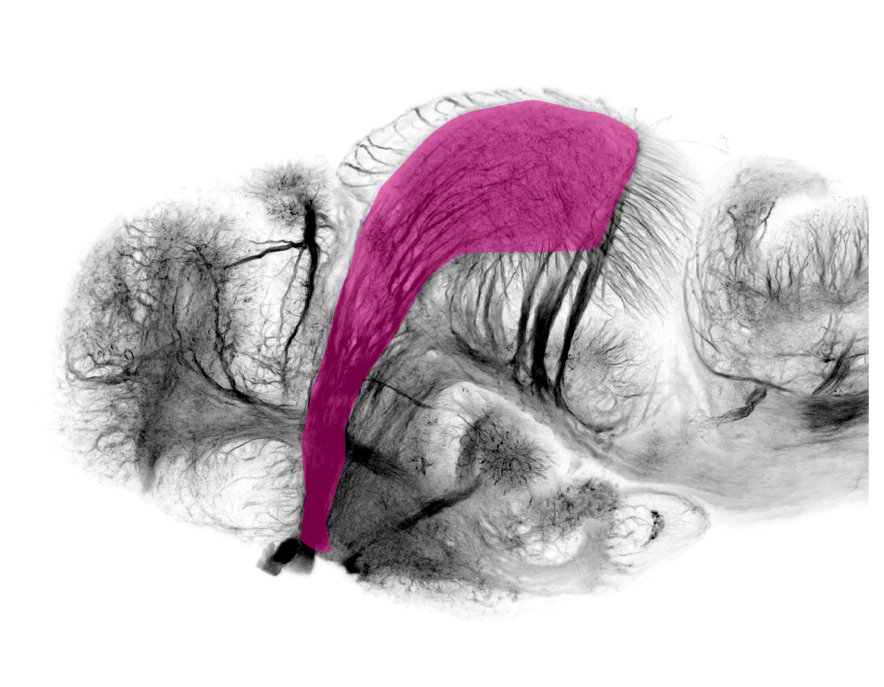
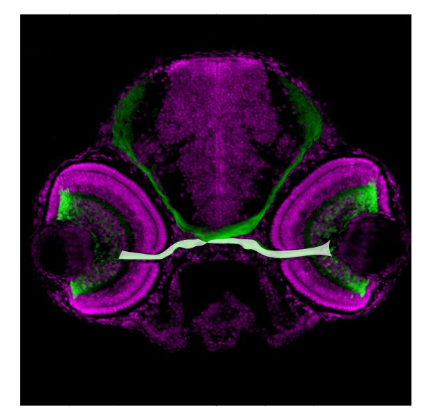
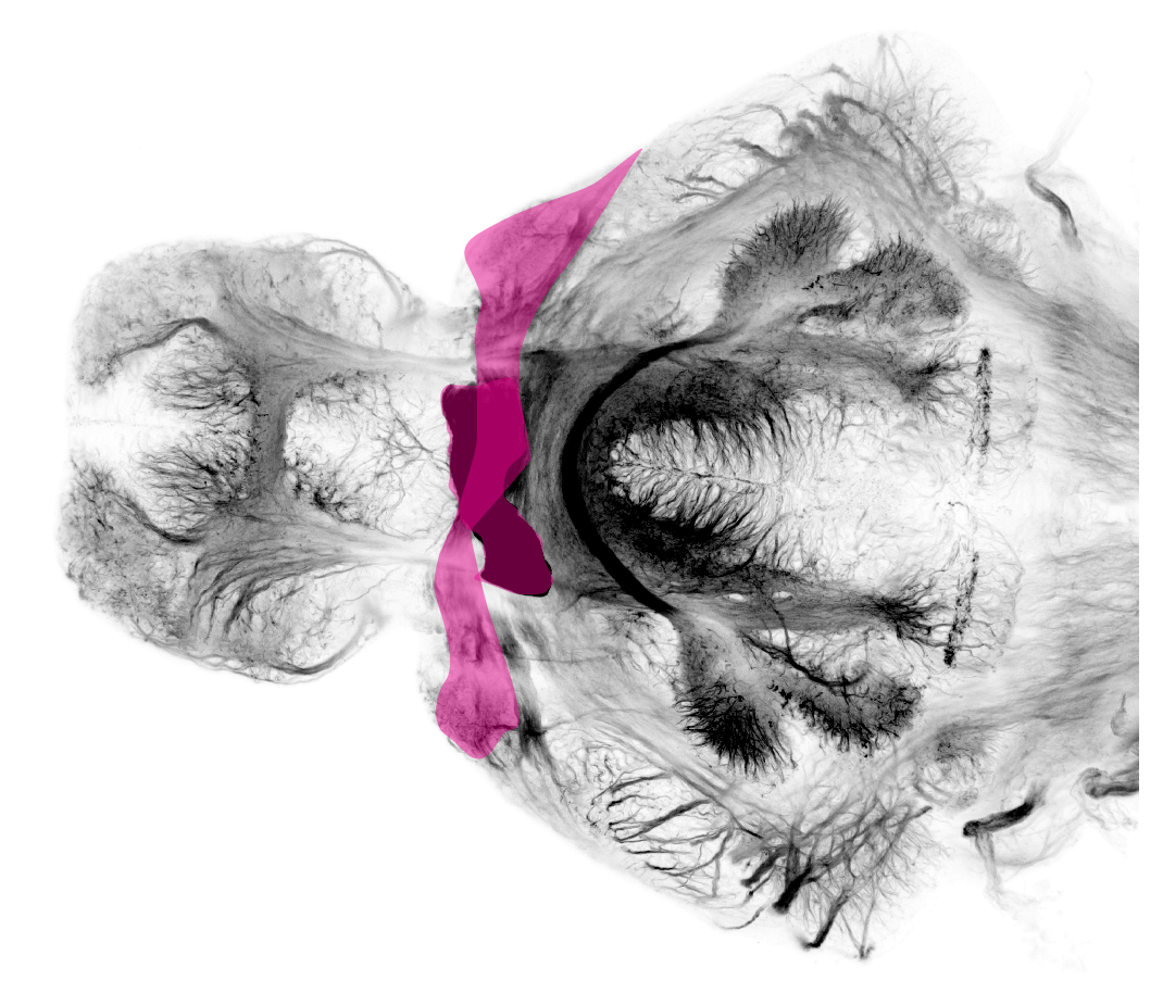
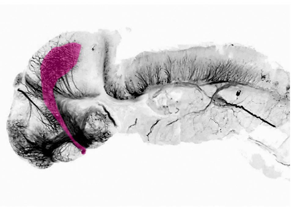
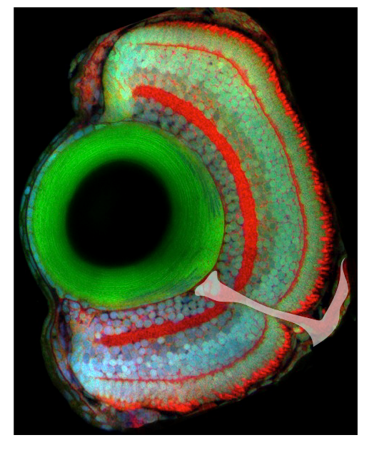
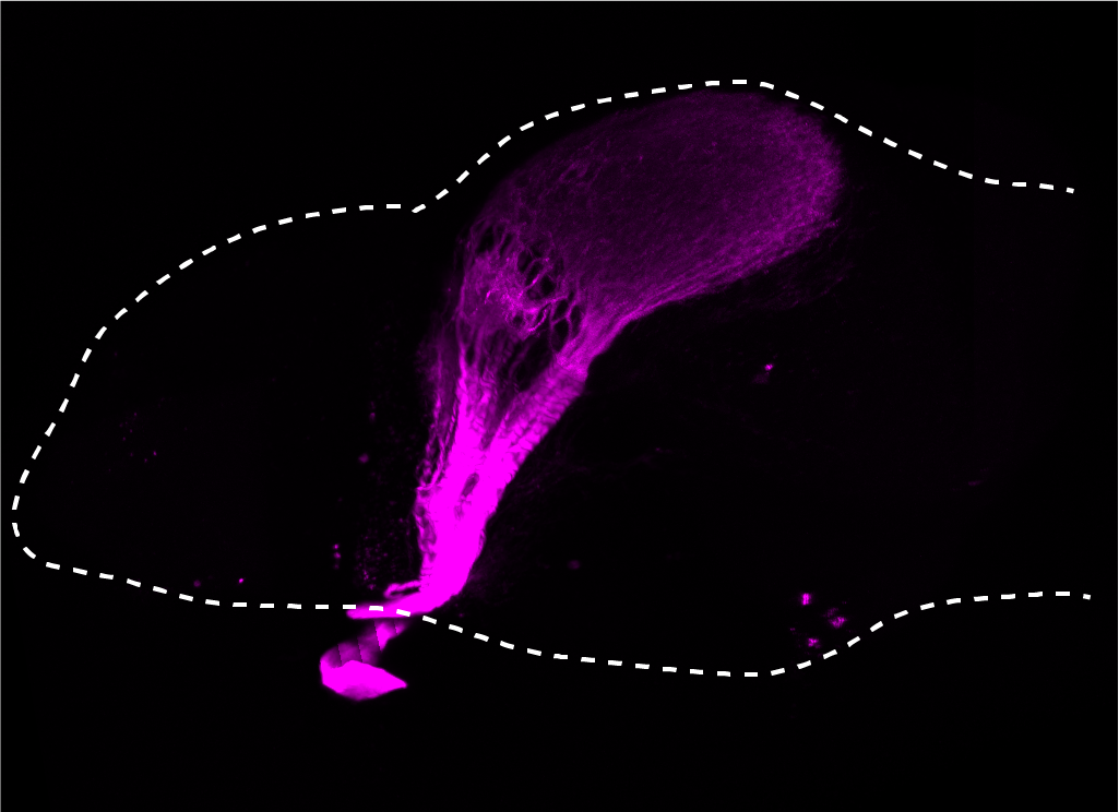
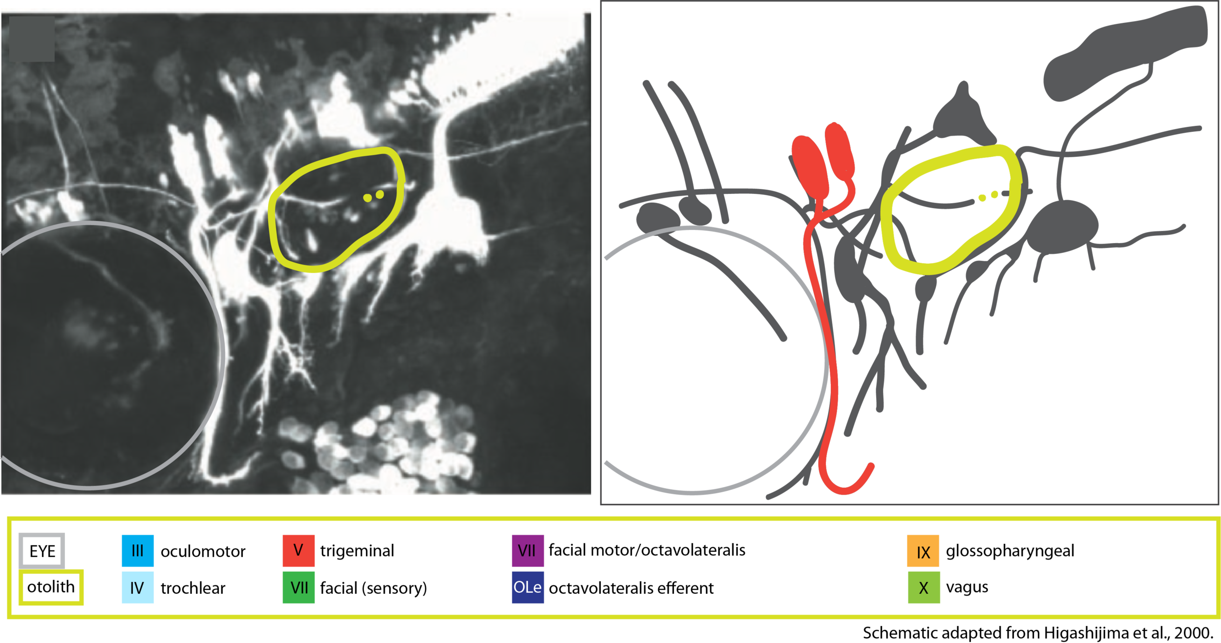
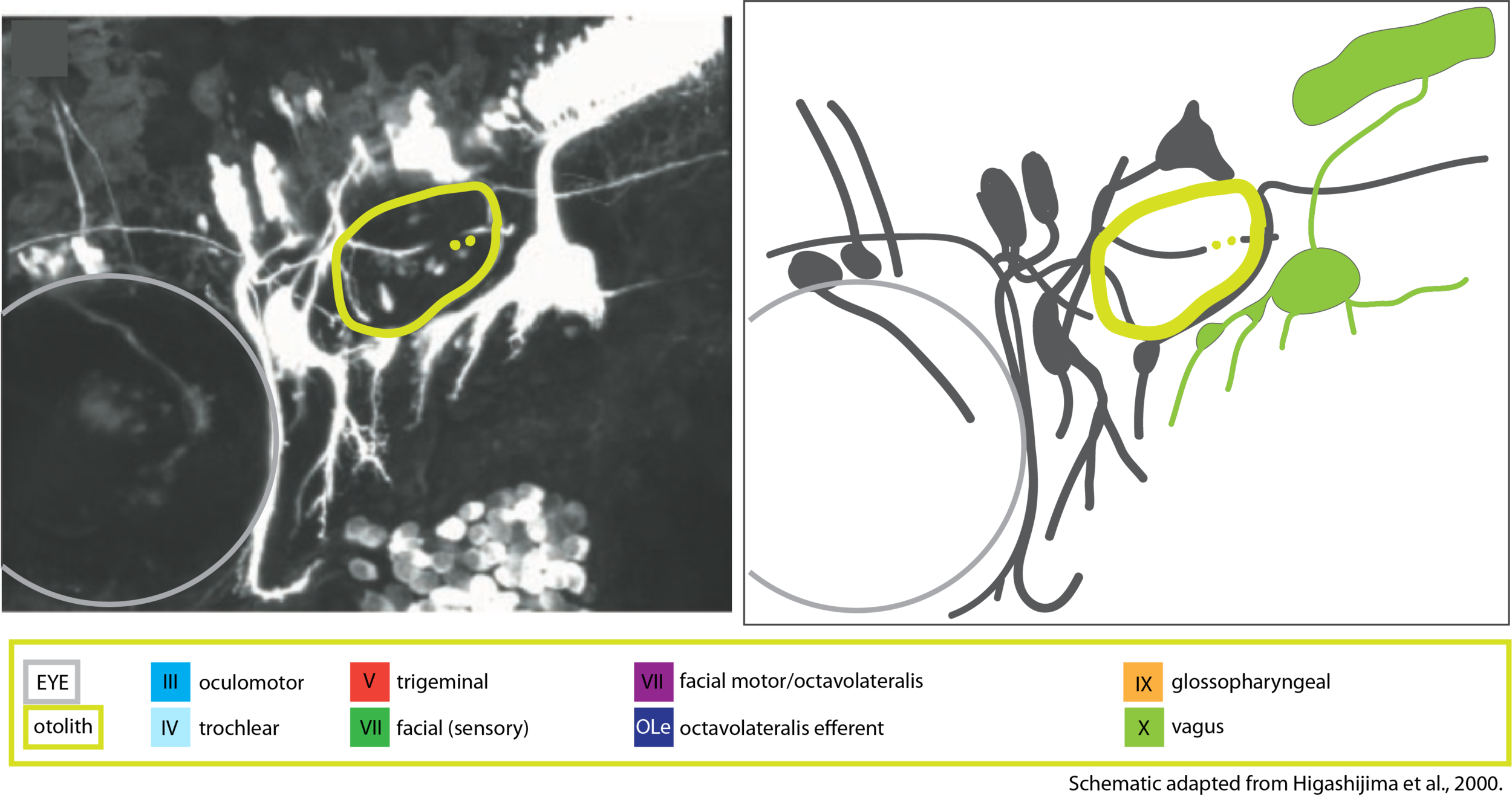
“The mechanosensory lateral line detects water movement via clusters of hair cells positioned over the surface of the head and body in specialized structures called neuromasts (Coombs et al., 1989). The hair cells of the lateral line are virtually identical to those found in the inner ear, and they sense movement by deflection of stereocilia. Two lateral line nerves are found anterior to the otic vesicle: the anterodorsal nerve innervates neuromasts of the supraorbital, infraorbital, and otic lines, whereas the anteroventral nerve innervates the mandibular and opercular lines. “(Raible & Kruse, 2000)
Key publications
DAVID W. RAIBLE AND GREGORY J. KRUSE
Organization of the Lateral Line System in Embryonic Zebrafish
THE JOURNAL OF COMPARATIVE NEUROLOGY 421:189–198 (2000)
Valdivia, L.E., Young, R.M., Hawkins, T.A., Stickney, H.L., Cavodeassi, F., Schwarz, Q., Pullin, L.M., Villegas, R., Moro, E., Argenton, F., Allende, M.L., and Wilson, S.W. (2011)
Lef1-dependent Wnt/β-catenin signalling drives the proliferative engine that maintains tissue homeostasis during lateral line development.
Development. 138(18):3931-3941.
McGraw, H.F., Drerup, C.M., Culbertson, M.D., Linbo, T., Raible, D.W., and Nechiporuk, A.V. (2011)
Lef1 is required for progenitor cell identity in the zebrafish lateral line primordium.
Development. 138(18):3921-3930.
Ghysen, A., and Dambly-Chaudière, C. (2007)
The lateral line microcosmos.
Genes and Development. 21(17):2118-2130.
Gompel, N., Cubedo, N., Thisse, C., Thisse, B., Dambly-Chaudière, C., and Ghysen, A. 2001.
Pattern formation in the lateral line of zebrafish.
Mech. Dev. 105: 69–77.
Gilmour, D., Knaut, H., Maischein, H.M., and Nusslein-Vol- hard, C. 2004.
Towing of sensory axons by their migrating target cells in vivo.
Nat. Neurosci. 7: 491–492.
Haas, P. and Gilmour, D. 2006.
Chemokine signaling mediates self-organizing tissue migration in the zebrafish lateral line.
Dev. Cell 10: 673–680.
Metcalfe, W.K., Kimmel, C.B., and Schabtach, E. 1985.
Anatomy of the posterior lateral line system in young larvae of the zebrafish.
J. Comp. Neurol. 233: 377–389.
Jacqueline F. Webb and Jonathan E. Shirey
Postembryonic Development of the Cranial Lateral Line Canals and Neuromasts in Zebrafish
DEVELOPMENTAL DYNAMICS 228:370–385, 2003
Melanie Haehnel-Taguchi, António M. Fernandes, Margit Böhler, Ina Schmitt, LenaTittel1 and WolfgangDriever
Projections of the Diencephalospinal Dopaminergic System to Peripheral Sense Organs in Larval Zebrafish (Danio rerio)
Front. Neuroanat., 19 March 2018 | https://doi.org/10.3389/fnana.2018.00020
Manuel, R., Iglesias Gonzalez, A.B., Habicher, J., Koning, H.K., Boije, H. (2021)
Characterization of Individual Projections Reveal That Neuromasts of the Zebrafish Lateral Line are Innervated by Multiple Inhibitory Efferent Cells.
Frontiers in Neuroanatomy. 15:666109.
Bricaud, O., Chaar, V., Dambly-Chaudiere, C., and Ghysen, A. (2001)
Early efferent innervation of the zebrafish lateral line.
The Journal of comparative neurology. 434(3):253-261.