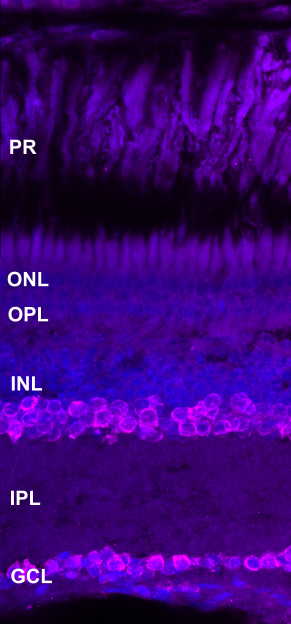
Viewing entries tagged
Amacrine cells

Calbindin

Calbindin

ABOUT THIS ANTIBODY

Rabbit Polyclonal anti-Calbindin (Swant, Cat#1CB38, dilution 1:200)

image

by Ryan MacDonald


labels these retinal cell types

Amacrine and bipolar cells


HuC/D

HuC/D

HuC/D

ABOUT THIS ANTIBODY

Anti-HuC/D labels amacrine and ganglion cells in the retina

HuC/D antibody which has been found to label HuC/D neuronal proteins which are expressed in neurons leaving the mitotic cycle. It labels amacrine cells in the inner nuclear layer and ganglion cells in the ganglion cell layer in the zebrafish (Gorsuch et al., 2017). In the killifish it labelled cells in the inner nuclear layer and cells in the ganglion cell layer.

Mouse monoclonal anti-HuC/D (Invitrogen, Cat#A21271, dilution 1:200)

imageS

Section of 2 mpf male killifish sodium citrate antigen retrieval anti-HuC/D (magenta) and DAPI (blue)

PS = photoreceptors, ONL = outer nuclear layer, OPL = outer plexiform layer, INL = inner nuclear layer, IPL = inner plexiform layer, GCL = ganglion cell layer


labels these retinal cell types

Amacrine cells in the inner nuclear layer and ganglion cells in the ganglion cell layer


key publications

Gorsuch RA, Lahne M, Yarka CE, Petravick ME, Li J and Hyde DR. 2017. Sox2 regulates Müller glia reprogramming and proliferation in the regenerating zebrafish retina via Lin28 and Ascl1a. Experimental Eye Research. 161:174-192.

Parvalbumin (PV)

Parvalbumin (PV)

ABOUT THIS ANTIBODY

Anti-PV labels amacrine cells

Parvalbumin (PV) antibody binding to parvalbumin which is a regulator of neuronal intracellular calcium dynamics and found to stain amacrine cells in zebrafish (Ahmed, Kojima and Masai, 2022). In the killifish it stained cells in the inner nuclear and ganglion cell layer; and linear speckled staining in the inner nuclear, plexiform and ganglion cell layer.

Mouse Monoclonal anti-PV (Sigma, Cat#P3088, dilution 1:200)

imageS


labels these retinal cell types

Amacrine cells


key publications

Ahmed M, Kojima Y and Masai I. 2022. Strip1 regulates retinal ganglion cell survival by suppressing Jun-mediated apoptosis to promote retinal neural circuit formation. eLife. 11:1-34.

Paired Box 6 protein (Pax6)

Paired Box 6 protein (Pax6)

ABOUT THIS ANTIBODY

Rabbit Polyclonal anti-Pax6 (Proteintech, Cat#12323-1-AP, dilution 1:100)

image

by Ryan MacDonald


labels these retinal cell types

Amacrine cells and ganglion cells