Viewing entries in
antibody

Tumor protein P53

Tumor protein P53

Tumor protein P53

ABOUT THIS ANTIBODY

Anti-P53 labels apoptosis

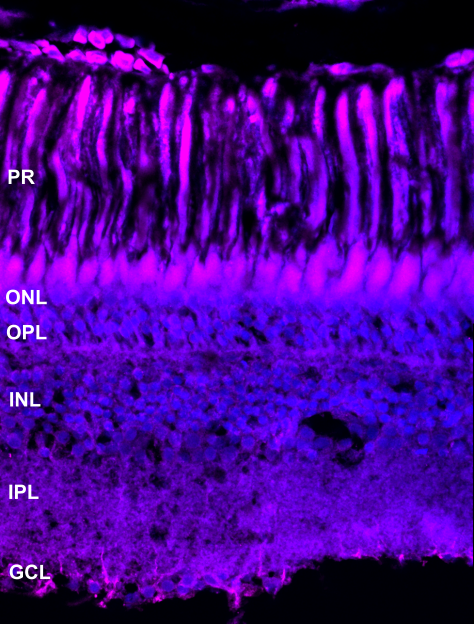
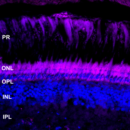
Tumor protein P53 is a sequence specific transcription factor which is being activated when cells are under stress. P53 antibody stains cell death in mice (Dong et al., 2019). In killifish it labelled one cell in the outer nuclear layer (white arrow).

Rabbit Polyclonal anti-P53 (Proteintech, Cat#21891-1-AP, dilution 1:100)

image

by Eva-Maria Breitenbach

Section of 4 mpf female killifish sodium citrate antigen retrieval anti-P53 (green) and DAPI (blue)

PS = photoreceptors, ONL = outer nuclear layer, OPL = outer plexiform layer, INL = inner nuclear layer, IPL = inner plexiform layer, GCL = ganglion cell layer


labels these retinal cell types

Apoptosis


key publications

Dong S, Ji J, Hu L and Wang H. 2019. Dihydromyricetin alleviates acetaminophen-induced liver injury via the regulation of transformation, lipid homeostasis, cell death and regeneration. Life Sciences. 227:20-29.

Vimentin

Vimentin

ABOUT THIS ANTIBODY

Mouse monoclonal anti-Vimentin (Proteintech, Cat#60330-1-IG, dilution 1:100)

image

by Ryan MacDonald


labels these retinal cell types

Müller glia


Yes associated protein (YAP)

Yes associated protein (YAP)

Yes associated protein (YAP)

ABOUT THIS ANTIBODY

Anti-YAP labels Müller glia proliferation

Yes associated protein (YAP) is part of the hippo pathway and its antibody has been found to mark proliferating Müller glia in rats (Zhang and Kong, 2020). In the killifish it stained some cells in the ganglion cell layer, possibly Müller glia cells.

Mouse Monoclonal anti-YAP (Santa Cruz, Cat#sc-101199, dilution 1:200)

imageS


labels these retinal cell types

Müller glia proliferation


key publications

Zhang W and Kong Y. 2020. YAP is essential for TGF‐β‐induced retinal fibrosis in diabetic rats via promoting the fibrogenic activity of Müller cells. Journal of Cellular and Molecular Medicine. 24:12390-12400.

Zpr-1

Zpr-1

ABOUT THIS ANTIBODY

Anti-Zpr-1 labels double cone photoreceptors

Zpr-1 antibody detects an unknown antigen specific to double cones in zebrafish (Yin et al., 2012). Great photoreceptors staining was found in the killifish.

Mouse Monoclonal anti-Zpr-1 (ZIRC, Cat#AB_10013803, dilution 1:1000)

imageS


labels these retinal cell types

Double cone photoreceptors


key publications

Yin J, Brocher J, Linder B, Hirmer A, Sundaramurthi H, Fischer U and Winkler C. 2012. The 1D4 antibody labels outer segments of long double cone but not rod photoreceptors in zebrafish. Investigative Ophthalmology & Visual Science, 53:4943-4951.