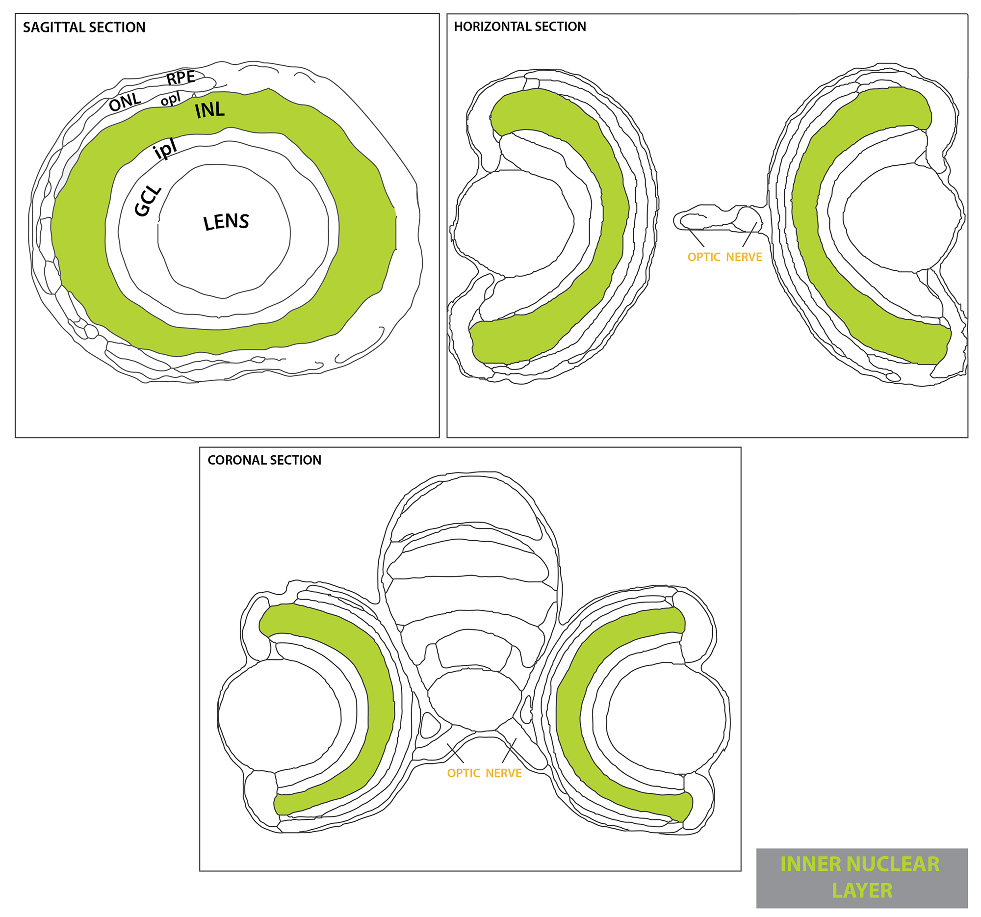
• Home
  • Groups
    • Alexandre Lab
    • Bianco Lab
    • Dreosti Lab
    • Frankel Lab
    • Tada Lab
    • RIhel Lab
    • MacDonald Lab
    • Mongera Lab
    • Payne Lab
    • Tuschl Lab
    • Wilson Lab
    • Wong Lab
    • Young Lab
    • Zebrafish Facility
  • ZebrafishBrain
  • UCL

Zebrafish UCL

  • Home
  • Groups
    • Alexandre Lab
    • Bianco Lab
    • Dreosti Lab
    • Frankel Lab
    • Tada Lab
    • RIhel Lab
    • MacDonald Lab
    • Mongera Lab
    • Payne Lab
    • Tuschl Lab
    • Wilson Lab
    • Wong Lab
    • Young Lab
    • Zebrafish Facility
  • ZebrafishBrain
  • UCL
Retina inner nuclear layer

Retina inner nuclear layer


July 4, 2018
by Kate Turner

INL-03.png

Description

Layer of the neural retina that contains the interneurons, the horizontal, bipolar and amacrine cells.

Retinal Cell types with cell body in the INL

Featured
Horizontal Cells
Horizontal Cells
Bipolar Cells
Bipolar Cells
Amacrine Cells
Amacrine Cells
muller glia
muller glia
Microglia
Microglia

Ontology
 

is part of: neural retina

has parts: horizontal cell, bipolar cell, amacrine cell.

INL L.png Inner Nuclear Layer

ZFIN

OLS Tree Diagram


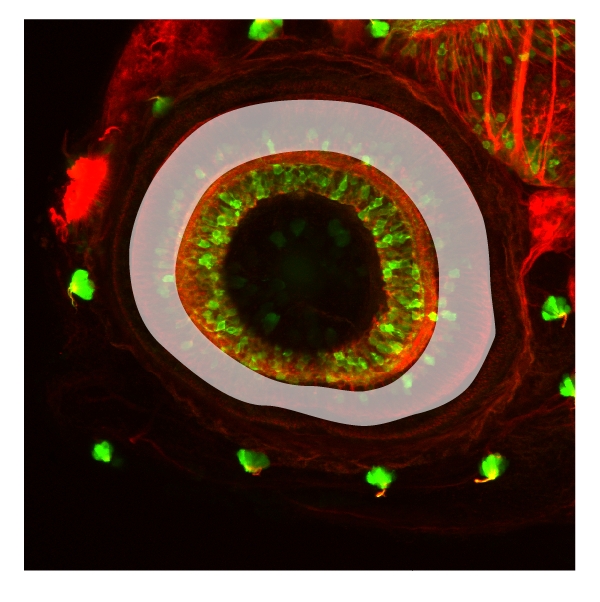
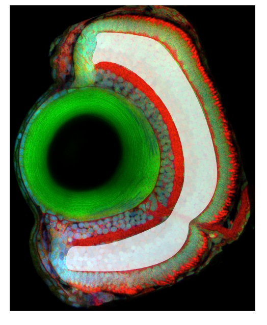
Transgenic Lines/Antibodies that label this brain region


Key Publications

tagged with retina, retinal outer nuclear layer T
Newer
Older
Top
  • Alexandre Lab
  • Bianco Lab
  • Dreosti Lab
  • Frankel Lab
  • MacDonald Lab
  • Mongera Lab
  • Payne Lab
  • Rihel Lab
  • Tada lab
  • Tuschl Lab
  • Wilson Lab
  • Wong Lab
  • Young Lab
  • Zebrafish Facility


Contact us

1st Floor, Anatomy Building, University College London
Gower St, London, WC1E 6BT
tel 020 3549 5642

Copyright © 2017 UCL Disclaimer