Glycine

Glycine

Glycine is an inhibitory neurotransmitter. Glycinergic transmission is found particularly in the retina, brainstem and spinal cord. Following activation of the glycinergic receptor, negatively charged chloride anions enter the postsynaptic neuron leading to an inhibitory postsynaptic potential. Glycine also acts as a co-agonist for glutamatergic signalling through NMDA receptors.

Antibodies that label glycinergic neurons

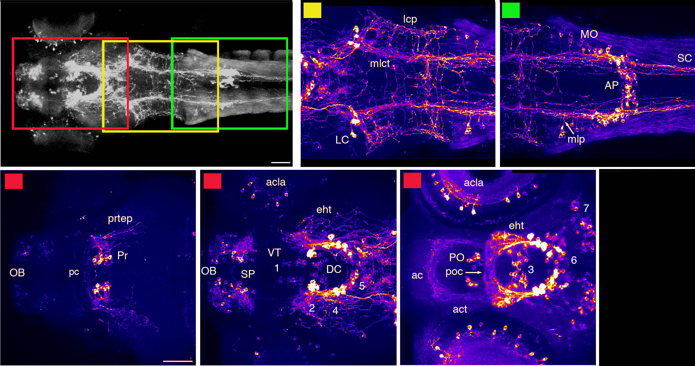
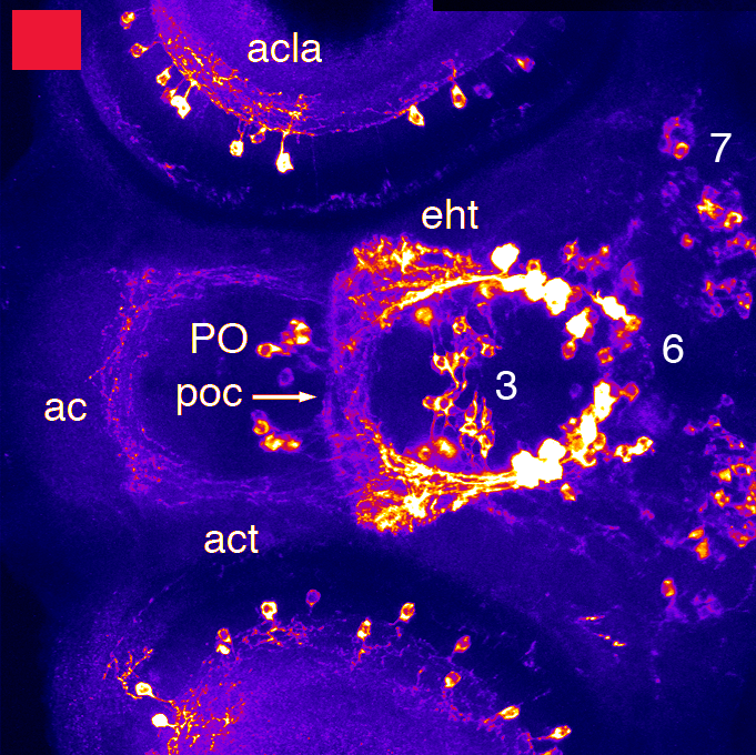
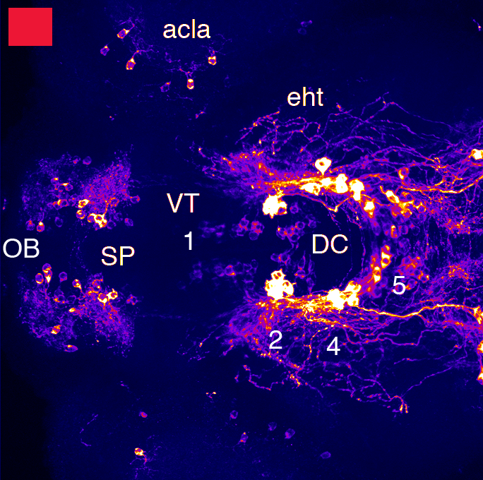
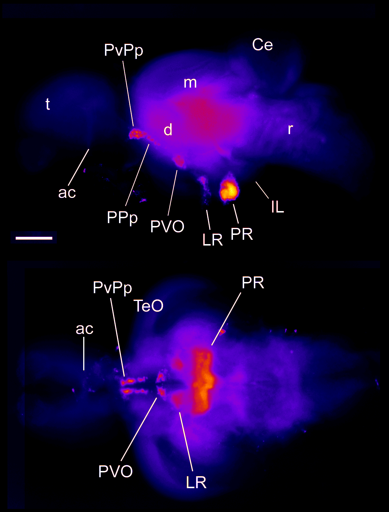
Transgenic lines that label glycinergic neurons

Tg(glyt2:GFP) or Tg(slc6a5:GFP) can also be viewed on ZBB at ZBB>Aliases>Neurotransmitters>Glycinergic


Probes that label glycinergic neurons


Key Publications

Jusuf, P.R., Almeida, A.D., Randlett, O., Joubin, K., Poggi, L., and Harris, W.A. (2011)
Origin and determination of inhibitory cell lineages in the vertebrate retina.
The Journal of neuroscience : the official journal of the Society for Neuroscience. 31(7):2549-2562.

Barreiro-Iglesias, A., Mysiak, K.S., Adrio, F., Rodicio, M.C., Becker, C.G., Becker, T., and Anadón, R. (2013) Distribution of glycinergic neurons in the brain of glycine transporter-2 Tg(glyt2:gfp) transgenic adult zebrafish: Relation with brain-spinal descending systems.
The Journal of comparative neurology. 521(2):389-425.

Acetylcholine

Acetylcholine

Acetylcholine is the neurotransmitter used by motor neurons at the neuro-muscular junction. Other “cholinergic” neurons in the brain also use acetylcholine as a neurotransmitter and neuromodulator. Ach is also used as a neurotransmitter in the autonomic nervous system. There are two types of Ach receptors in the brain:

  • nicotinic receptors: Ligand-gated ion channels permeable to sodium, calcium and potassium ions.

  • muscarinic receptors: G-protien coupled receptors that exert their longer term effects through second messenger systems. The effect of acetylcholine binding to muscarinic acetylcholine receptors M1, M3 and M5 is usually excitatory on the post-synaptic cell. Ach plays a role in plasticity, arousal and reward in the brain.

Antibodies that label cholinergic neurons

Transgenic lines that label cholinergic neurons

Probes that label cholinergic neurons

Key Publications

Mueller, T., Vernier, P., and Wullimann, M.F. (2004)
The adult central nervous cholinergic system of a neurogenetic model animal, the zebrafish Danio rerio.
Brain research. 1011(2):156-169.

Edwards, J.G., Greig, A., Sakata, Y., Elkin, D., and Michel, W.C. (2007)
Cholinergic innervation of the zebrafish olfactory bulb.
The Journal of comparative neurology. 504(6):631-645.

Hong, E., Santhakumar, K., Akitake, C.A., Ahn, S.J., Thisse, C., Thisse, B., Wyart, C., Mangin, J.M., and Halpern, M.E. (2013)
Cholinergic left-right asymmetry in the habenulo-interpeduncular pathway.
Proc. Natl. Acad. Sci. USA. 110(52):21171-21176.

Bertuzzi, M., Ampatzis, K. (2018)
Spinal cholinergic interneurons differentially control motoneuron excitability and alter the locomotor network operational range.
Scientific Reports. 8:1988.

Arenzana, F.J., Clemente, D., Sanchez-Gonzalez, R., Porteros, A., Aijon, J., and Arevalo, R. (2005)
Development of the cholinergic system in the brain and retina of the zebrafish.
Brain research bulletin. 66(4-6):421-425.

Kaslin, J., Nystedt, J.M., Ostergard, M., Peitsaro, N., and Panula, P. (2004)
The orexin/hypocretin system in zebrafish is connected to the aminergic and cholinergic systems.
The Journal of neuroscience : the official journal of the Society for Neuroscience. 24(11):2678-2689.

Glutamate

Glutamate

Glutamate is the anion of glutamic acid(an amino acid). It is the most abundant excitatory neurotransmitter in the nervous system of the zebrafish and other vertebrates. Post-synaptic transmission of glutamate is mediated by four types of glutamate receptors:

  • NMDA receptors: ionotropic transmembrane receptor that requires in addition to the binding of glutamate, glycine to also bind as a co-agonist to open the ion channel. The ion channel associated with the NMDAR is cation NMDAR current flow is directly related to membrane depolarization, the opening probability of the channel increases with depolarization allowing voltage dependent flow of sodium and calcium ions into the post-synaptic neuron and potassium ions out of the cell.

  • AMPA receptors: ionotropic transmembrane receptor permeable to cations including calcium, sodium and potassium ions. AMPA receptors open and close very quickly and are responsible for most of the fast excitatory transmission in the nervous system. They also play a role in synaptic plasticity and are required for long-term potentiation LTP.

  • metabotropic glutamate receptors: These receptors are indirectly linked with ion channels and mediate their responses through second messengers such as G-proteins, they can initiate a variety of cellular effects, such as ion channel opening or other cellular effects through triggering signal transduction cascades. Ion channel opening via metabotropic receptors can last for seconds to minutes, as opposed to milliseconds in the case of ionotropic receptors. This means the effects of metabotropic glutamtergic signalling can last much longer and can have more widespread effects throughout the cell.

  • kainate receptors: the rarest type of glutamate receptor in the nervous system, kainate receptors can be both pre and post synaptic. Kainate receptors are ionotropic receptors whose ion channels are permeable to sodium and potassium ions when activated by glutamate leading to depolarisation of the post-synaptic neuron.

Antibodies that label glutamatergic neurons


Transgenic lines that label glutamatergic neurons

Tg(slc17a6b:DsRed) or vglut:DsRed can also be viewed on ZBB at ZBB>Aliases>Neurotransmitters>Glutamatergic


Probes that label glutamatergic neurons


Key Publications


Higashijima, S.I., Mandel, G., and Fetcho, J.R. (2004)
Distribution of prospective glutamatergic, glycinergic, and GABAergic neurons in embryonic and larval zebrafish.
The Journal of comparative neurology. 480(1):1-18.

Filippi, A., Mueller, T., and Driever, W. (2014)
Vglut2 and gad expression reveal distinct patterns of dual GABAergic versus glutamatergic cotransmitter phenotypes of dopaminergic and noradrenergic neurons in the zebrafish brain.
The Journal of comparative neurology. 522(9):2019-37.

 GABA

GABA

gamma-Aminobutyric acid or GABA is the principal inhibitory neurotransmitter in the brain. The role of GABA is to reduce excitability of neurons. Two types of GABA receptor exist in the nervous system GABAA and GABAB receptors. GABAA receptors are ligand activated ion channels and GABAB receptors are G-protein coupled receptors that open and close channels via intermediary G proteins. The flow of negatively charged Chloride ions into a neuron or positively charged potassium ions out of a neuron leads to hyperpolarozation of the cell.


Antibodies that label GABA-ergic neurons


Transgenic lines that label GABA-ergic neurons

Tg(gad1b:GFP) (Satou et al., 2013) can be viewed at ZBB>aliases>Neurotransmitters>GABAergic.


Probes that label GABA-ergic neurons


Key Publications

Satou, C., Kimura, Y., Hirata, H., Suster, M.L., Kawakami, K., and Higashijima, S. (2013)
Transgenic tools to characterize neuronal properties of discrete populations of zebrafish neurons. Development (Cambridge, England). 140(18):3927-3931.

Higashijima, S.I., Mandel, G., and Fetcho, J.R. (2004)
Distribution of prospective glutamatergic, glycinergic, and GABAergic neurons in embryonic and larval zebrafish.
The Journal of comparative neurology. 480(1):1-18.

Mueller, T., Wullimann, M.F., and Guo, S. (2008)
Early teleostean basal ganglia development visualized by Zebrafish Dlx2a, Lhx6, Lhx7, Tbr2 (eomesa), and GAD67 gene expression.
The Journal of comparative neurology. 507(2):1245-1257.

Filippi, A., Mueller, T., and Driever, W. (2014)
Vglut2 and gad expression reveal distinct patterns of dual GABAergic versus glutamatergic cotransmitter phenotypes of dopaminergic and noradrenergic neurons in the zebrafish brain.
The Journal of comparative neurology. 522(9):2019-37.

Mueller, T., and Guo, S. (2009)
The distribution of GAD67-mRNA in the adult zebrafish (teleost) forebrain reveals a prosomeric pattern and suggests previously unidentified homologies to tetrapods.
The Journal of comparative neurology. 516(6):553-568.

Solek, C.M., Feng, S., Perin, S., Weinschutz Mendes, H.C., Ekker, M. (2017)
Lineage tracing of dlx1a/2a and dlx5a/6a expressing cells in the developing zebrafish brain.
Developmental Biology. 427(1):131-147.

Jusuf, P.R., Almeida, A.D., Randlett, O., Joubin, K., Poggi, L., and Harris, W.A. (2011)
Origin and determination of inhibitory cell lineages in the vertebrate retina.
The Journal of neuroscience : the official journal of the Society for Neuroscience. 31(7):2549-2562.

Monesson-Olson, B., McClain, J.J., Case, A.E., Dorman, H.E., Turkewitz, D.R., Steiner, A.B., Downes, G.B. (2018) Expression of the eight GABAA receptor α subunits in the developing zebrafish central nervous system.
PLoS One. 13:e0196083.

Tabor, R., Yaksi, E., and Friedrich, R.W. (2008)
Multiple functions of GABA(A) and GABA(B) receptors during pattern processing in the zebrafish olfactory bulb.
The European journal of neuroscience. 28(1):117-127.

Noradrenaline

Noradrenaline

Key Publications

Ma P. M. (1994).
Catecholaminergic systems in the zebrafish. II. Projection pathways and pattern of termination of the locus coeruleus.
J. Comp. Neurol. 344, 256–269. doi: 10.1002/cne.903440207

Kastenhuber E., Kratochwil C. F., Ryu S., Schweitzer J., Driever W. (2010).
Genetic dissection of dopaminergic and noradrenergic contributions to catecholaminergic tracts in early larval zebrafish.
J. Comp. Neurol. 518, 439–458. doi: 10.1002/cne.22214

Farrar, M.J., Kolkman, K.E., Fetcho, J.R. (2018)
Features of the structure, development and activity of the Zebrafish Noradrenergic System explored in new CRISPR transgenic lines.
The Journal of comparative neurology. 526(15):2493-2508.

Schweitzer, J., Löhr, H., Filippi, A., & Driever, W. (2012).
Dopaminergic and noradrenergic circuit development in zebrafish.
Developmental Neurobiology, 72(3), 256–268.

Tay, T. L., Ronneberger, O., Ryu, S., Nitschke, R., & Driever, W. (2011).
Comprehensive catecholaminergic projectome analysis reveals single-neuron integration of zebrafish ascending and descending dopaminergic systems.
Nature Communications, 2(1), 171–112.

McLean, D. L., & Fetcho, J. R. (2004).
Ontogeny and innervation patterns of dopaminergic, noradrenergic, and serotonergic neurons in larval zeb- rafish.
The Journal of Comparative Neurology, 480(1), 38–56.

Wen, L., Wei, W., Gu, W., Huang, P., Ren, X., Zhang, Z., Zhu, Z., Lin, S., and Zhang, B. (2008)
Visualization of monoaminergic neurons and neurotoxicity of MPTP in live transgenic zebrafish. Developmental Biology. 314(1):84-92.

Histamine

Histamine

Key Publications

Kaslin J., Panula P. (2001).
Comparative anatomy of the histaminergic and other aminergic systems in zebrafish (Danio rerio).
J. Comp. Neurol. 440, 342–377. doi: 10.1002/cne.1390

Serotonin

Serotonin

Zebrafish Serotinergic System

Serotonergic schematic

A simple schematic representing a lateral view of the larval zebrafish brain at around 3dpf (anterior to the left). The approximate locations of serotonergic nuclei. The principle regions of 5HT expression are the raphe, the periventricular region of the caudal hypothalamus (Hc) the posterior tegmentum (PT), the pretectum (Pretect) and the pineal (Pin). Tect = tectum, Tel = telencephalon, OB = olfactory bulb and AC is anterior commissure Based on Lillesaar 2011

The serotonergic system describes the system of neurons in the brain that employ serotonin (5-hydroxytryptamine;5HT) as a neurotransmitter.  The system has been fairly well characterised in the adult (see Lillesaar review ), there have also been many efforts to characterise the system in the embryo and larva.  The system is a current focus of multiple laboratories because of its clinical importance in several psychiatric (eg affective disorders) and neurological (eg migraine) pathologies.

Anatomically there are four main groups of serotonergic neurons in the brain: 1. the pretectal population, the hypothalamic and posterior tuberculum populations and the raphe populations, there are also 5HT-positive neurons in the epiphysis and area postrema.

Serotonin belongs to the tryptamine class of neurotransmitters as they are synthesised from tryptophan.  The tryptamines in turn are part of the monoaminergic class of neurotransmitters which additionally includes the catecholamines (including dopamine, synthesised from tyrosine), Histamine and other amines.

Transgenics and molecular markers

Antibodies raised against 5HT can be used in immunohistochemical interrogation of zebrafish brain preparations, examples are shown in images associated with this tutorial.  In addition to this, the Tg(pet1:GFP)(Lillesaar et al., 2012), Tg(pet1:KALTA4) and Tg(VMAT:GFP) transgenic line marks some of the populations of serotonergic neurons.

Antibodies that label serotinergic neurons

Transgenic lines that label serotinergic neurons

Key Publications

Lillesaar.
The serotonergic system in fish.
J Chem Neuroanat (2011) vol. 41 (4) pp. 294-308

Gaspar & Lillesaar.
Probing the diversity of serotonin neurons.
Philos Trans R Soc Lond B Biol Sci. 2012 Sep 5;367(1601):2382-94. doi: 10.1098/rstb.2011.0378.

Lillesaar et al.
Axonal projections originating from raphe serotonergic neurons in the developing and adult zebrafish, Danio rerio, using transgenics to visualize raphe-specific pet1 expression.
J Comp Neurol. 2009 Jan 10;512(2):158-82. doi: 10.1002/cne.21887.

Panula et al.
Modulatory neurotransmitter systems and behavior: towards zebrafish models of neurodegenerative diseases.
Zebrafish (2006) vol. 3 (2) pp. 235-47

McLean and Fetcho.
Ontogeny and innervation patterns of dopaminergic, noradrenergic, and serotonergic neurons in larval zebrafish.
J. Comp. Neurol. (2004) vol. 480 (1) pp. 38-56

Wen et al.
Visualization of monoaminergic neurons and neurotoxicity of MPTP in live transgenic zebrafish. Developmental Biology (2008) vol. 314 (1) pp. 84-92


Dopamine

Dopamine

Zebrafish dopaminergic system

Dopamine is a monoamine neurotransmitter, part of the catecholamine family of monoamines, which also includes noradrenaline and adrenaline.

Dopaminergic (DA) neurons are found throughout the forebrain in distinct clusters, each with characteristic local and/or long-range projections.   There are no DA neurons in the midbrain or hindbrain of zebrafish.  The catecholaminergic cells in the hindbrain are noradrenergic (NA) neurons located in the locus coeruleus and the medulla oblongata.  DA and NA neurons make up some of the major long-range projection systems in the vertebrate brain (Tay et al., 2011).

 In addition to its role as a neurotransmitter, dopamine also functions as a hormone released from the hypophysis (pituitary gland).  Through its neurotransmitter and hormonal functions dopamine controls a wide range of behaviours in the brain of both vertebrates and invertebrates.  These functions include cognition, voluntary movement, motivation, reward pathways, mood, attention and learning (Schweitzer et al., 2011).

  • The majority of DA neuronal clusters are located in the diencephalon of the zebrafish brain.  These diencephalic clusters have been assigned numbers DC1-DC7 based on their location along the anterior-posterior axis. 

  •  In the telencephalon there are two bilateral clusters of DA cells in the olfactory bulb (OB) and the subpallium (SP). 

  •  DA neurons are also found in the amacrine cell layer of the retina, the anterior and posterior parts of the parvocellular preoptic area and the periventricular pretectum.

This tutorial was written by Andy Simmonds. All images are modified from Kastenhuber et al., 2010. We thank Wolfgang Driever for supplying us with these beautiful images.

Key Publications

Tay, T. L., Ronneberger, O., Ryu, S., Nitschke, R., & Driever, W. (2011).
Comprehensive catecholaminergic projectome analysis reveals single-neuron integration of zebrafish ascending and descending dopaminergic systems.
Nature Communications, 2(1), 171–112.

Kastenhuber E., Kratochwil C. F., Ryu S., Schweitzer J., Driever W. (2010).
Genetic dissection of dopaminergic and noradrenergic contributions to catecholaminergic tracts in early larval zebrafish.
J. Comp. Neurol. 518, 439–458. doi: 10.1002/cne.22214

Rink E., Wullimann M. F. (2001).
The teleostean (zebrafish) dopaminergic system ascending to the subpallium (striatum) is located in the basal diencephalon (posterior tuberculum).
Brain Res. 889, 316–330. doi: 10.1016/s0006-8993(00)03174-7

Ma P. M. (1997).
Catecholaminergic systems in the zebrafish. III. Organization and projection pattern of medullary dopaminergic and noradrenergic neurons.
J. Comp. Neurol. 381, 411–427

Schweitzer, J., Löhr, H., Filippi, A., & Driever, W. (2012).
Dopaminergic and noradrenergic circuit development in zebrafish.
Developmental Neurobiology, 72(3), 256–268.

McLean, D. L., & Fetcho, J. R. (2004).
Ontogeny and innervation patterns of dopaminergic, noradrenergic, and serotonergic neurons in larval zeb- rafish.
The Journal of Comparative Neurology, 480(1), 38–56.

Wen, L., Wei, W., Gu, W., Huang, P., Ren, X., Zhang, Z., Zhu, Z., Lin, S., and Zhang, B. (2008)
Visualization of monoaminergic neurons and neurotoxicity of MPTP in live transgenic zebrafish. Developmental Biology. 314(1):84-92.