Retinal Pigmented Epithelium

Retinal Pigmented Epithelium

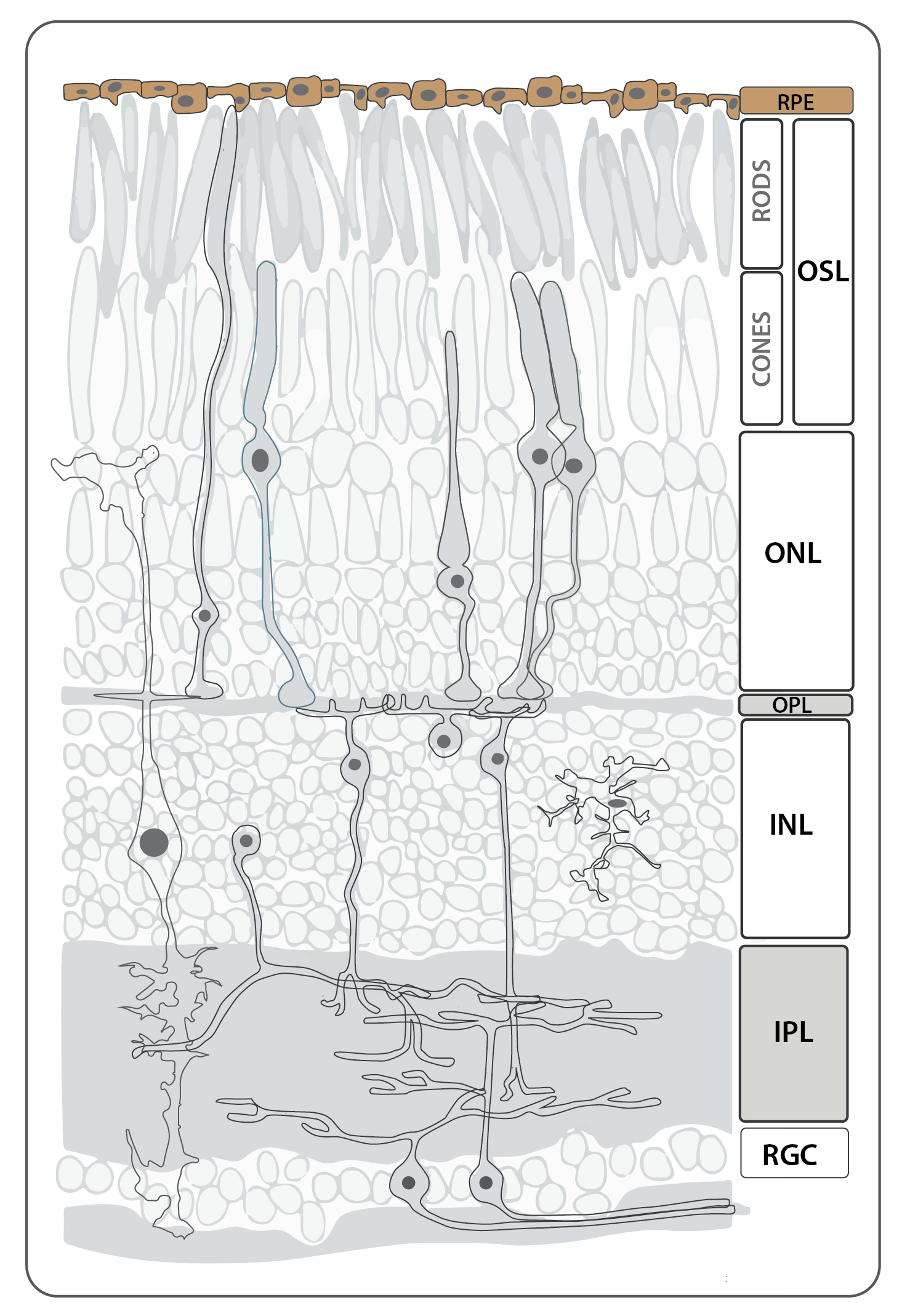
The retinal pigmented epithelium (RPE), like the neural retina, is derived from the neural ectoderm, and through complex morphogenetic movementsis during development it becomes tightly apposed to the photoreceptor cell layer of the neural retina.  This tissue is required for the function and survival of photoreceptor cells; numerous mutations affecting vacuole transport in the RPE result in non-cell autonomous photoreceptor cell degeneration. 

The RPE is a typical epithelial monolayer of tightly packed cuboidal cells forming part of the blood brain barrier between the vascular choroid and the neural retina.  The cells contain large numbers of melanosomes packed with melanin granules.  The apical microvilli of the RPE interdigitate with the photoreceptor outer segments, and the basal surface rests on Bruch’s membrane, the basement membrane between the RPE and capillaries of the choroid. 

Cones

Cones

Schematic depicting the different cell types and layers that make up the neural retina in an adult zebrafish adapted from Baden et al., 2019. Cone photoreceptors have their cell bodies in the outer nuclear layer of the retina.

Photoreceptors

Photoreceptor cells span the outer nuclear layer as well as the outer and inner segment layers. The light sensing cells of the retina, the cone and rod photoreceptors, are located in the apical-most layer of the retina, and display a stereotypical subcellular organization with their nuclei basal to the inner segment (cell body or soma) and outer segment, which abuts the retinal pigmented epithelium (RPE) and is full of membrane invaginations packed with light-sensitive cell-specific opsins.  All photoreceptors have opsin-containing outersegments, which are replaced on a daily basis.  The older membrane and protein debris is shed from the distal tip of the outersegment and phagocytosed by the adjacent RPE cells.  Because zebrafish are diurnal, their retinae contains a large number of bright-light sensitive cone subtypes in addition to dim-light sensitive rod photoreceptors, all of which are organized into regular mosaic patterns that can best be visualized in tangential sections.  Below is more detailed information about the distinct types of photoreceptors found in zebrafish.

Cones are required for bright light vision and can be subdivided into four classes based on opsin expression and morphology.  The paired long double cones express both red and green opsin, the long single cones express blue opsin, and the short single cones express uv-sensitive opsin.  Antibodies generated against each of the 4 cone opsins or rhodopsin label the outer segments of individual cones or rods, respectively; in situ hybridization of the opsin transcripts label the inner segments. Several transgenic lines, which use various opsin promoters to drive expression of fluorophores (see below), reveal the location and morphology of specific photoreceptor subtypes.

Outer photoreceptor segments stained by zpr1 (blue) and UV cones expressing GFP (magenta), Tg(opn1sw1:GFP)) in 8 dpf larvae
by Gaia Gestri

outer photoreceptor segments stained by zpr1 (magenta) and UV cones expressing GFP (green, Tg(opn1sw1:GFP)) in 8 dpf larvae by Gaia Gestri

outer photoreceptor segments stained by zpr1 (magenta) and UV cones expressing GFP (green, Tg(opn1sw1:GFP)) in 8 dpf larvae
by Gaia Gestri

Rods

Rods

Schematic depicting the different cell types and layers that make up the neural retina in an adult zebrafish adapted from Baden et al., 2019. Rod photoreceptors have their cell bodies in the outer nuclear layer. their outersegments form the most external layer of the neural retina closest to the retinal pigmented epithelium (RPE).

Photoreceptors

Photoreceptor cells span the outer nuclear layer as well as the outer and inner segment layers. The light sensing cells of the retina, the cone and rod photoreceptors, are located in the apical-most layer of the retina, and display a stereotypical subcellular organization with their nuclei basal to the inner segment (cell body or soma) and outer segment, which abuts the retinal pigmented epithelium (RPE) and is full of membrane invaginations packed with light-sensitive cell-specific opsins.  All photoreceptors have opsin-containing outersegments, which are replaced on a daily basis.  The older membrane and protein debris is shed from the distal tip of the outersegment and phagocytosed by the adjacent RPE cells.  Because zebrafish are diurnal, their retinae contains a large number of bright-light sensitive cone subtypes in addition to dim-light sensitive rod photoreceptors, all of which are organized into regular mosaic patterns that can best be visualized in tangential sections.  Below is more detailed information about the distinct types of photoreceptors found in zebrafish.

Rods are utilized mainly for dim-light vision, and their cell bodies are located vitreal to the cone nuclei, and in the light-adapted retina, the thin rod inner and outer segments project beyond the cones, interdigitating between the apical microvilli of the adjacent retinal pigmented epithelia (RPE)l cells.

Horizontal Cells

Horizontal Cells

Horizontal Cells

Horizontal cells are second order neurons that mediate the process of lateral inhibition in the outer retina, and are therefore electrically coupled by connexin-positive gap junctions.  Horizontal cell nuclei have a flattened appearance in retina sections.

Schematic depicting the different cell types and layers that make up the neural retina in an adult zebrafish adapted from Baden et al., 2019. Horizontal cells have their cell bodies in the inner nuclear layer and synapse with bipolar and photoreceptor cells (rods and cones) in the outer plexiform layer.

Bipolar Cells

Bipolar Cells

Bipolar Cells

Bipolar cells are interneurons that transmit information from photoreceptors to the inner retina.  Bipolar cells are classified by three main criteria:  the type of photoreceptor that provides input, dendritic and axonal morphologies, and the polarity of their responses (e.g., depolarizing or hyperpolarizing). 

Schematic depicting the different cell types and layers that make up the neural retina in an adult zebrafish adapted from Baden et al., 2019. Bipolar cells have their cell bodies in the inner nuclear layer. Their processes span the inner nuclear layer synapsing with horizontal cells and rods and cones in the outer plexiform layer and relaying their signals to amacrine and RGCs via connections in the inner plexiform layer.

Amacrine Cells

Amacrine Cells

Amacrine Cells

Amacrine cell are interneurons with soma that reside primarily in the inner nuclear layer and send processes into the inner plexiform layer.  Amacrine cells mediate motion selectivity and modulate input into the inner retina.  Amacrine cells are classified based upon neurotransmitter expression and morphology. 

Schematic depicting the different cell types and layers that make up the neural retina in an adult zebrafish adapted from Baden et al., 2019. Amacrine cells have their cell body in the inner nuclear layer (INL) and project dendrites into the inner plexiform layer where they synapse with RGCs and bipolar cells.

muller glia

muller glia

MÜLLER GLIA

Müller cells are radial glia that perform a wide variety of support functions for retinal neurons. Immunolabeling for carbonic anhydrase or glutamine synthetase shows that the Müller cell bodies are positioned in a row in the middle of the inner nuclear layer.  Müller glial processes, which are positive for the glial fibrillary acidic protein (GFAP), extend radially across the thickness of the retina and compose parts of the outer and inner limiting membranes.  The outer limiting membrane is formed by the tight junctions that connect Müller glia (MG) apical processes and photoreceptors cell bodies while the inner limiting membrane contains the MG end feet.  


Schematic depicting the different cell types and layers that make up the neural retina in an adult zebrafish adapted from Baden et al., 2019. Müller glia have their cell bodies in the inner nuclear layer and span the retina with their end feet forming the inner and outer limiting membranes. They elaborate processes within both the inner and outer plexiform layers.

Retinal Ganglion Cells

Retinal Ganglion Cells

RETINAL GANGLION CELLS

Ganglion cells (RGCs) are the final output neurons of the vertebrate retina.  The ganglion cell axons form the nerve fiber layer of the inner retina as they coalesce to form the optic nerve head, which is composed of ganglion cell axons and glia that migrate into the optic nerve as well as astrocytes that are derived from the optic stalk. RGC axons cross the midline at the optic chiasm and arborize in distinct arborization fields or AFs. These neuropil areas correspond to 10 different retinorecipient brain nuclei the largest of which is the midbrain optic tectum. For a complete description of all the AFs and a lot more information on the neuroanatomy and function of RGCs see Robles et al., 2014).  

Schematic depicting the different cell types and layers that make up the neural retina in an adult zebrafish adapted from Baden et al., 2019. Retinal Ganglion Cells (RGCs) have their cell bodies in the retinal ganglion cell layer and their dendrites in the inner plexiform layer where they synapse with amacrine and bipolar cells. RGC axons exit the eye as the optic nerve and innervate predominantly the optic tectum with some RGCs sending collaterals to the arborisation fields of the retino-fugal pathway.

Microglia

Microglia

Microglia are the macrophages of the brain and retina. They play important roles in tissue homeostasis, recovery from injury and in some retinal diseases In the healthy retina, microglia cells are normally located in the plexiform layers.  They exhibit elaborate ramified processes and are responsible for immune surveillance of the retina. Oxidative stress, hypoxia or inherited mutations that cause retinal injury will trigger microglia reactivity. The microglia cells will adopt an amoeboid morphology, increase proliferation and migrate to the sites of injury. This inflammatory response from the microglia can rapidly enhance tissue repair and return to homeostasis. However, sustained microglial inflammatory responses can instigate severe alterations in retinal integrity, and lead to neuronal demise . Changes in microglia inflammatory responses may play a key role in various retinopathies such as glaucoma, age-related macular degeneration and retinitis pigmentosa (Langmann et al., 2019).

Schematic depicting the different cell types and layers that make up the neural retina in an adult zebrafish adapted from Baden et al., 2019. Microglia can be found throughout the retina, they are the macrophages of the neural retina.

References

Rashid K, Akhtar-Schaefer I, Langmann T(2019)
Microglia in Retinal Degeneration
Front. Immunol. https://doi.org/10.3389/fimmu.2019.01975