


1
2
3
4
5
Eye development
The eyes enable organisms to visually perceive their surroundings. During embryogenesis, the eyes originate as outpocketings of the brain and our work aims to elucidate the key stages of eye development. These include induction of the eye field, outgrowth and morphogenesis of the optic vesicles, formation and closure of the choroid fissure, growth of the eye and differentiation of retinal neurons. In addition to deepening our understanding of normal eye development, our studies are addressing how this process can go wrong and lead to congenital eye defects. In particular, we are studying the genetic and cellular bases of MAC phenotypes (microphthalmia, anophthalmia and coloboma). These studies are helping to understand the aetiology of eye diseases and hereditary visual system conditions in human patients.
Complementing our studies of eye formation and differentiation we are also studying cell behaviour in a retinal stem cell niche. In the post-embryonic zebrafish eye, all the stages of progression from stem cell to differentiated neuron are found near the margin of the eye, in a region termed the ciliary marginal zone (CMZ). We are interested in exploring the molecular mechanisms that govern neurogenesis in the CMZ and how specific intrinsic and extrinsic signals are integrated to carry out this task and allow the growth of the eye.
Neuroanatomy
The developing zebrafish is rapidly becoming a leading vertebrate model for studies of circuit function and behaviour. An essential prerequisite for such studies is an understanding of the neuroanatomy of the brain. In collaboration with Jon Clarke's, lab in KCL, we have been developing an online, high-resolution atlas of the developing zebrafish brain. Zebrafishbrain.org aims to be a user-friendly and highly flexible resource for presenting information about the neuroanatomy of the developing zebrafish brain.
The embryonic zebrafish has outstanding optical properties that permit imaging of the entire intact brain at high resolution by confocal microscopy and neuron-targeted transgenes are excellent tools for resolving neuroanatomy.
To date we have imaged many beautiful transgenic lines. We are currently working with colleagues to morph this data onto standard neuroanatomical frameworks to make it more accessible to the community. Please visit Zebrafishbrain.org for more details.
The asymmetric brain
Structural and functional asymmetries in the nervous system are found throughout the animal kingdom. In humans, for instance, aspects of language processing occur predominantly in the left hemisphere. Brain lateralization is thought to increase cognitive performance, whereby specialization of one hemisphere leaves the other free to perform different tasks. Compromised brain asymmetries have been linked to several neuropathologies including schizophrenia, autism, and neuronal degenerative diseases. Yet despite the prevalence and importance of nervous system asymmetries, our knowledge of the mechanisms that underlie the development and functional consequences of asymmetry is far from complete.
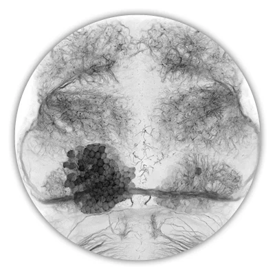
The best-described neuroanatomical asymmetries in vertebrates are in the epithalamus, a dorsally positioned structure in the diencephalic forebrain. Asymmetries consist of differences in size, neuronal organisation, neurochemistry, connectivity and functional properties of neurons. In recent years, we and others have established the zebrafish as a model to study the genetic and developmental mechanisms underlying the establishment of asymmetry. Our studies are now addressing how asymmetry is encoded within circuitry and are beginning to address the behavioural consequences of asymmetric circuitry.
Modelling human disease
Zebrafish are increasingly becoming a popular model for translational research aimed at understanding and treating human disease. With the development of gene editing systems such as CRISPR/Cas9, targeted knock-ins, knock-outs and other specific genetic modifications are becoming more straightforward. Our lab has established collaborations with clinicians (and indeed we have clinicians studying for PhDs in our lab) and together we are studying fish orthologues of candidate disease genes from human patients. We aim to use studies in fish to determine if genetic mutations are really causative of disease phenotypes, help place genes in genetic pathways through epistasis analyses and understand the cellular and molecular bases of disease phenotypes.
The lab currently works on developing zebrafish models for quite a diverse range of human conditions including ciliopathies, neurodegenerative disorders, coloboma, microphthalmia and other eye abnormalities and, in collaboration with the Rihel, Dreosti and Hoffman labs, autism spectrum disorders.