

Description
Derived from prosomere
Ontology
is part of: brain
has parts: optic tectum, tegmentum, torus longitudinalis, torus semicircularis.
ZFIN
OLS Tree Diagram
Key Publications
Back to Explore the Brain


Derived from prosomere
is part of: brain
has parts: optic tectum, tegmentum, torus longitudinalis, torus semicircularis.
ZFIN
OLS Tree Diagram

Back to Explore the Brain
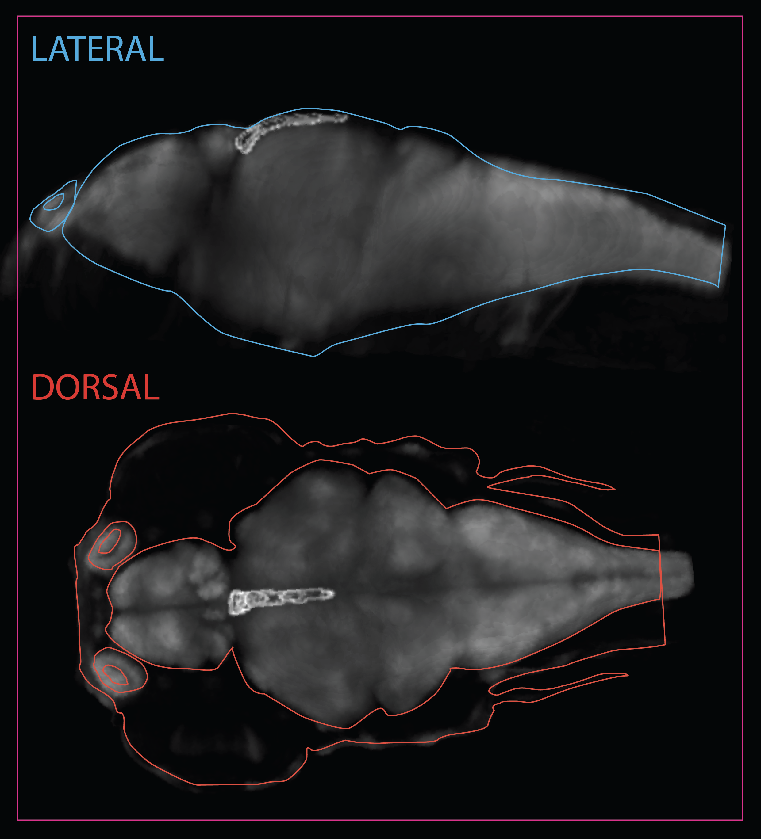
Midbrain > Tegmentum

Schematic showing the position of the tegmentum in sagittal, horizontal and coronal sections through the zebrafish brain.
Based on the anatomical segmentation of 3 day old zebrafish larval brain by Thomas Müller, Olaf Ronneberger, Wolfgang Driever and colleagues. For details see Ronneberger et al., Nat. Meth. 2012 and http://vibez.informatik.uni-freiburg.de
Abbreviations: Ce, cerebellar plate; D, dorsal telencephalon/pallium; E, epiphysis; EmT, eminentia thalami; Hb, habenula; Hyp, hypothalamus; MO, medulla oblongata; OB, olfactory bulb; OT, optic tectum; PG, preglomerular complex; PO, preoptic area;PrT, pretectum; PTd, posterior tuberculum dorsal part; PTh, prethalamus; PTv posterior tuberculum ventral part; Teg, tegmentum; Th, thalamus; TS, torus semicircularis; V, ventral telencephalon/subpallium; Va, valvula cerebelli.
The tegmentum is a multi-tissue structure that forms the ventral part of the midbrain.
is part of: midbrain
has parts: cranial nerve III, cranial nerve VI, dorsal tegmental nucleus, interpeduncular nucleus, oculomotor nucleus, rostral tegmental nucleus, superior reticular formation, tegmental nucleus, trochlear motor nucleus, nucleus ruber, lateral lemniscus nucleus
OLS Tree Diagram

Back to Midbrain
Midbrain > Torus Semicircularis

Schematic showing the position of thetorus semicircularis in sagittal, horizontal and coronal sections through the zebrafish brain.
Based on the anatomical segmentation of 3 day old zebrafish larval brain by Thomas Müller, Olaf Ronneberger, Wolfgang Driever and colleagues. For details see Ronneberger et al., Nat. Meth. 2012 and http://vibez.informatik.uni-freiburg.de
Abbreviations: Ce, cerebellar plate; D, dorsal telencephalon/pallium; E, epiphysis; EmT, eminentia thalami; Hb, habenula; Hyp, hypothalamus; MO, medulla oblongata; OB, olfactory bulb; OT, optic tectum; PG, preglomerular complex; PO, preoptic area;PrT, pretectum; PTd, posterior tuberculum dorsal part; PTh, prethalamus; PTv posterior tuberculum ventral part; Teg, tegmentum; Th, thalamus; TS, torus semicircularis; V, ventral telencephalon/subpallium; Va, valvula cerebelli.

Schematic showing the position of the torus semicircularis at 6dpf based on the 3D anatomical segmentation used by the Zebrafish Brain Browser by Gupta et al., 2018.
The torus semicircularis is part of the auditory pathway in zebrafish. The auditory system in zebrafish is similar to that of mammals. fish sense sound waves via the otoliths of the inner ear and convey this auditory information via the VIIIth cranial nerve to the octavolateralis nuclei in the hindbrain then through the torus semicircularis (the equivalent of the inferior colliculus in mammals) to the thalamus (Vanwalleghem et al 2017).
is part of: midbrain
has parts:
OLS Tree Diagram

Vanwalleghem, G., Heap, L.A., Scott, E.K. (2017)
A profile of auditory-responsive neurons in the larval zebrafish brain.
The Journal of comparative neurology. 525(14):3031-3043. DOI: 10.1002/cne.24258
Back to Midbrain
Midbrain > Optic Tectum

Schematic showing the position of the optic tectum in sagittal, horizontal and coronal sections through the zebrafish brain.
Based on the anatomical segmentation of 3 day old zebrafish larval brain by Thomas Müller, Olaf Ronneberger, Wolfgang Driever and colleagues. For details see Ronneberger et al., Nat. Meth. 2012 and http://vibez.informatik.uni-freiburg.de
Abbreviations: Ce, cerebellar plate; D, dorsal telencephalon/pallium; E, epiphysis; EmT, eminentia thalami; Hb, habenula; Hyp, hypothalamus; MO, medulla oblongata; OB, olfactory bulb; OT, optic tectum; PG, preglomerular complex; PO, preoptic area;PrT, pretectum; PTd, posterior tuberculum dorsal part; PTh, prethalamus; PTv posterior tuberculum ventral part; Teg, tegmentum; Th, thalamus; TS, torus semicircularis; V, ventral telencephalon/subpallium; Va, valvula cerebelli.
The optic tectum (OT) and its mammalian equivalent, the superior colliculus (SC), is a key processing centre for sensory information. The OT receives the majority of its inputs from the retina and constructs an image of the physical surroundings. Through connections with several other regions of the brain, it integrates visually acquired information with motor inputs and outputs to initiate appropriate behavioural responses.

Hodology
The main input of the OT comes from the retina in the form of the topographically mapped retinotectal projections of the retinal ganglion cells (RGCs), but it also has bilateral connections with the pretectum, dorsal thalamus (which relays the tectal signals to the telencephalon), dorsal tegmentum, nucleus isthmi, reticular formation and controlateral tectum. The OT also receives input from the torus longitudinalis (TL), a structure that serves as a relay of telencephalic inputs. Most of the afferents from these different sources terminate in different, characteristic layers of the OT, a feature important for its coordinating and integrating roles.
Development
During embryogenesis, the tectum develops from the simple neuroepithelium of the mesencephalic alar plate into a complex, multilayered structure, one of the most conserved sections of the vertebrate brain.
Cytoarchitecture
In most vertebrates three tectal layers can be distinguished: the superficial and central zones, where the tectal afferents end, and the periventricular zone (SPV), where the majority of the tectal cell bodies reside. The superficial and central zones can be subdivided into further layers, but the number of these varies among phyolgenetic groups. In fish the superficial zone consists of the stratum marginale (SM) and stratum opticum (SO); the central zone consists of the stratum fibrosum et griseum superficiale (SFGS), stratum griseum centrale (SGC) and stratum album centrale (SAC).
In a typical teleost, the OT contains between 11-15 morphologically distinct cell types are distinguishable, with the most abundant cells (over 90% of the total) being piriform neurons. These observations based on classical staining procedures, however, most likely understate the true diversity of the tectal neurons, as morphologically similar neurons can express very different transcription factor combinations.
Function
The OT/SC coordinates the saccadic/microsaccadic eye movements of vertebrates and through the integration of topographic optic- and somatosensory information it regulates the motor functions of prey-catching and avoidance behaviours in frogs, and possibly humans. In teleosts tectal size and complexity varies depending on the behaviour and ecological niche of the fish: species that process more visual information have larger tecta.
Tectal Growth
Fish continuously grow throughout their life, thus unlike in mammals, where the neurogenic capacity of the SC diminishes postnatally, the neural stem cell niche of the OT in teleosts is preserved and the proliferation of tectal progenitor cells continues well into adulthood and most likely throughout the life of the animals.
The progenitor cells of the OT in zebrafish reside at the mediocaudal edge of the tectal hemispheres, adjacent to the tectal ventricles. Similarly to the stem cells located in the zebrafish cerebellum, and unlike the neural progenitors of the mammalian brain, these cells are neuroepithelial in their character and do not express glial markers.
The retinotectal pathway: formation and maintenance of topographic visual maps
One of the hallmark features of the vertebrate visual system is the topographic organization of the retinotectal projections. Retinal ganglion cells (RGCs) from the retina project through the optic chiasm to the controlateral tectal hemisphere, and through a seemingly simple matching rule Cartesian coordinates of the eye are projected on the OT. RGCs located in the nasal part of the retina project project to the caudal part of the controlateral OT, whereas temporal RGCs project rostrally. Similarly, the medial/dorsal parts of the controlateral tectal hemisphere receive projections from ventral RGCs, while the lateral/ventral tectal neurons are innervated by the dorsal retina. Intermediate RGCs terminate in the apporpriate intermediate position of the OT.
A further layer of complexity of retintotectal projections is added by the presence of multiple RGC subtypes in the retina. Although the axons of these ganglion cells traverse the tectum together through the SO, at their appropriate termination sites they will project to subtype-characteristic layers of the OT/SC.
Authors: Kara Cerveny and Máté Varga.
is part of: midbrain
has parts:
Butler, A. B. and Hodos, W. (2005).
Comparative vertebrate neuroanatomy : evolution and adaptation.
Hoboken, N.J.: Wiley-Interscience.
Ito, Y., Tanaka, H., Okamoto, H. and Ohshima, T. (2010).
Characterization of neural stem cells and their progeny in the adult zebrafish optic tectum.
Dev Biol 342, 26-38.
Nevin, L. M., Taylor, M. R. and Baier, H. (2008).
Hardwiring of fine synaptic layers in the zebrafish visual pathway.
Neural Dev 3, 36.
Robles, E., Smith, S.J., and Baier, H. (2011)
Characterization of genetically targeted neuron types in the zebrafish optic tectum.
Frontiers in neural circuits. 5:1.
Back to Midbrain
Midbrain > Torus Longitudinalis
Schematic showing the position of torus longitudinalis at 6dpf based on the 3D anatomical segmentation used by the Zebrafish Brain Browser by Gupta et al., 2018.
The torus longitudinalis (TL) is a structure exclusive to ray-finned fish. It is a paired elongated neural structure lying along the medial margins of the optic tectum, suspended from the intertectal commissure and protruding into the tectal ventricle (Ito, 1971; Wullimann, 1994; Northmore, 2011). (Folgueira et al., 2020)
The adult TL can be roughly subdivided into three regions based on morphology and location along the rostro-caudal axis: rostral, intermediate and caudal. The rostral TL sits on top of the posterior commissure with its two halves fused forming a slightly flattened structure . The intermediate TL is longer than the other two, with both halves still fused at the midline. Rostrally, intermediate TL hangs into the tectal ventricle, while a bit more caudally it is flanked by the cerebellar valvula laterally and/or ventrally." (Folgueira et al., 2020)
Connectivity of TL
TL is reciprocally connected to the optic tectum. Toral cell axons exit the TL, then reach the stratum album centrale (SAC) of the optic tectum and finally ascend in separate bundles to the marginal layer, where they run parallel to the brain surface and form terminals. Other efferents project to the thalamic nucleus rostrolateralis.
Afferents to the TL come from visual and cerebellum-related nuclei in the pretectum, namely the central, intercalated and the paracommissural pretectal nuclei, as well as from the subvalvular nucleus in the isthmus. (Folgueira et al., 2020). A single type of tectal neuron projects to the TL in zebrafish that morphologically corresponds to the type X cell described in goldfish (Meek and Schellart, 1978) and in zebrafish (De-Marco et al., 2019).
is part of: midbrain
has parts:
OLS Tree Diagram

Folgueira M, Riva-Mendoza S, Ferreño-Galmán N, Castro A, Bianco IH, Anadón R and Yáñez J. Anatomy and Connectivity of the Torus Longitudinalis of the Adult Zebrafish.
Front. Neural Circuits (2020) 14:8. doi:10.3389/fncir.2020.00008
Back to Midbrain
Midbrain > Tegmentum > Dorsal Tegmental Nucleus
is part of: midbrain
has parts:
ZFIN
OLS Tree Diagram
Back to Midbrain
is part of: midbrain
has parts:
ZFIN
OLS Tree Diagram
is part of: midbrain
has parts:
ZFIN
OLS Tree Diagram
Mueller, T., Vernier, P., and Wullimann, M.F. (2004)
The adult central nervous cholinergic system of a neurogenetic model animal, the zebrafish Danio rerio.
Brain research. 1011(2):156-169.
Oculomotor nerve innervates four extraocular muscles, consisting of the inferior oblique (IO), inferior rectus (IR), medial rectus (MR), and superior rectus (SR) (Clarke et al., 2013).
Oculomotor nerve neurons and the occulomotor nerve root are labelled with choline acetyltransferase the acetylcholine synthesising enzyme indicating that this nucleus forms part of the cholinergic system in zebrafish (Mueller et al., 2004).


is part of: midbrain
has parts:
Shin-ichi Higashijima, Yoshiki Hotta, and Hitoshi Okamoto
Visualization of Cranial Motor Neurons in Live Transgenic Zebrafish Expressing Green Fluorescent Protein Under the Control of the Islet-1 Promoter/Enhancer.
The Journal of Neuroscience, January 1, 2000, 20(1):206–218
Clark, C., Austen, O., Poparic, I., and Guthrie, S. (2013)
alpha2-Chimaerin Regulates a Key Axon Guidance Transition during Development of the Oculomotor Projection.
The Journal of neuroscience. 33(42):16540-16551.
David Schoppik, Isaac H. Bianco, David A. Prober, Adam D. Douglass, Drew N. Robson, Jennifer M.B. Li, Joel S.F. Greenwood. Edward Soucy, Florian Engert, and Alexander F. Schier,
Gaze-Stabilizing Central Vestibular Neurons Project Asymmetrically to Extraocular Motoneuron Pools.
The Journal of Neuroscience, November 22, 2017 • 37(47):11353–11365 • 11353
Thomas Mueller, Philippe Vernier, Mario F. Wullimann
The adult central nervous cholinergic system of a neurogenetic model animal, the zebrafish Danio rerio
Brain Research 1011 (2004) 156–169
is part of: midbrain
has parts:
ZFIN
OLS Tree Diagram
is part of: midbrain
has parts:
ZFIN
OLS Tree Diagram
Mueller, T., Vernier, P., and Wullimann, M.F. (2004)
The adult central nervous cholinergic system of a neurogenetic model animal, the zebrafish Danio rerio.
Brain research. 1011(2):156-169.
M.F. Wullimann, B. Rupp, H. Reichert,
Neuroanatomy of the Zebrafish Brain. A Topological Atlas
Birkhauser Verlag, Basel, 1996, 144 pp.
is part of: midbrain
has parts:
ZFIN
OLS Tree Diagram
is part of: midbrain
has parts:
ZFIN
OLS Tree Diagram
is part of: midbrain
has parts:
Kimmel, C.B., Powell, S.L., and Metcalfe, W.K. (1982)
Brain neurons which project to the spinal cord in young larvae of the zebrafish.
The Journal of comparative neurology. 205:112-127.
Joanna YN Lau, Isaac H Bianco and Kristen E Severi
Cellular-level understanding of supraspinal control: what can be learned from zebrafish?
Current Opinion in Physiology 2019, 8:141–145
Orger MB, Kampff AR, Severi KE, Bollmann JH, Engert F:
Control of visually guided behavior by distinct populations of spinal projection neurons.
Nat Neurosci 2008, 11:327-333.
Thiele, T.R., Donovan, J.C., Baier, H. (2014)
Descending Control of Swim Posture by a Midbrain Nucleus in Zebrafish.
Neuron. 83(3):679-691.
Severi, K.E., Portugues, R., Marques, J.C., O'Malley, D.M., Orger, M.B., Engert, F. (2014)
Neural Control and Modulation of Swimming Speed in the Larval Zebrafish.
Neuron. 83(3):692-707.
Gahtan. E., Tanger. P., and Baier, H. (2005)
Visual prey capture in larval zebrafish is controlled by identified reticulospinal neurons downstream of the tectum.
The Journal of neuroscience : the official journal of the Society for Neuroscience. 25(40):9294-9303.


