Viewing entries tagged
subpallium

Tg(-8.0cldnb:lynGFP)zf106

Tg(-8.0cldnb:lynGFP)zf106

About

construct:
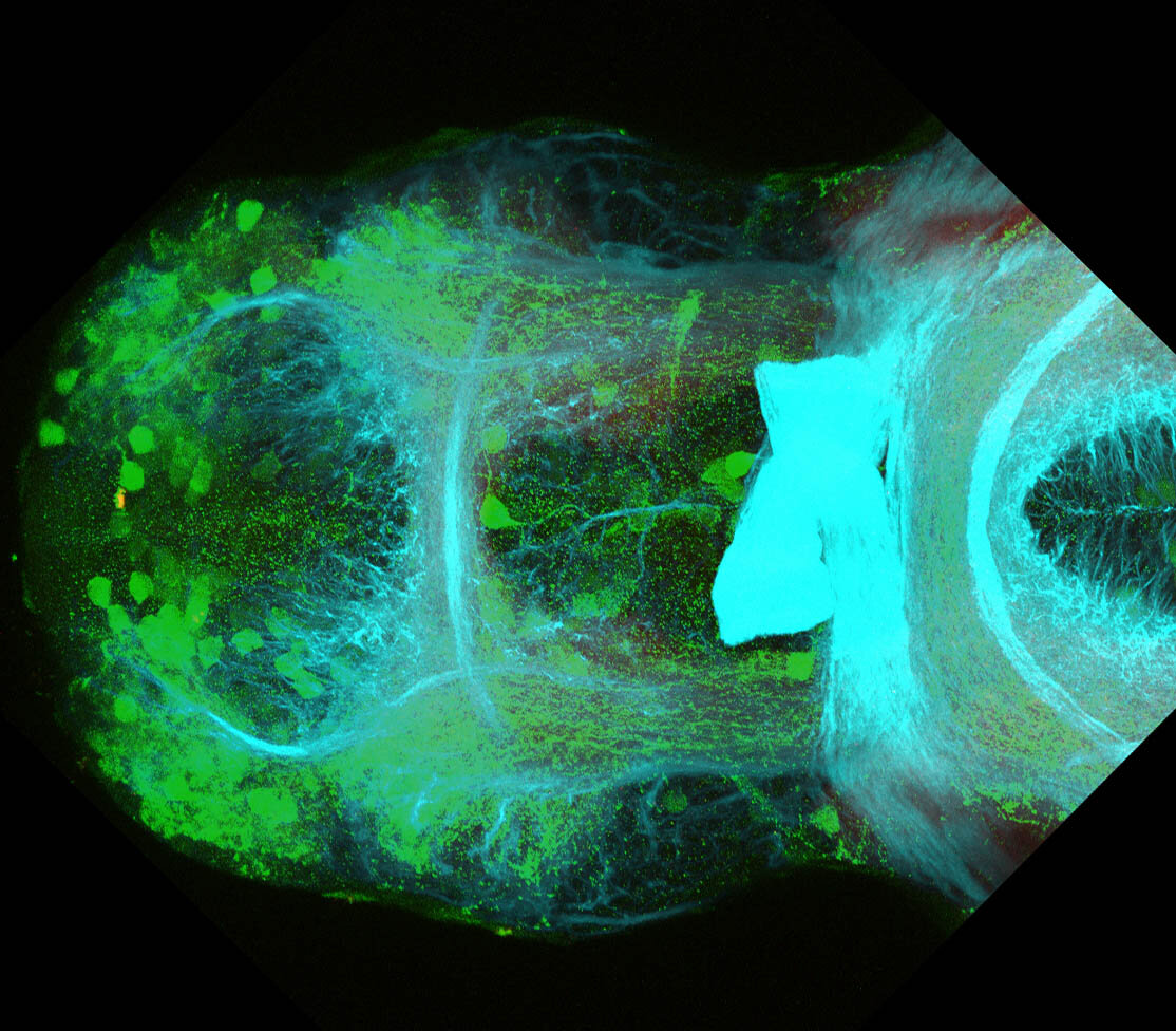
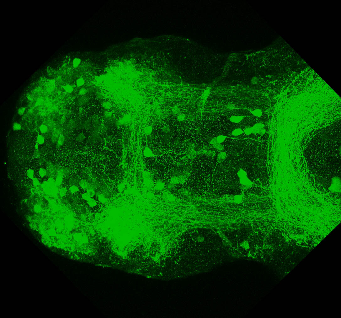
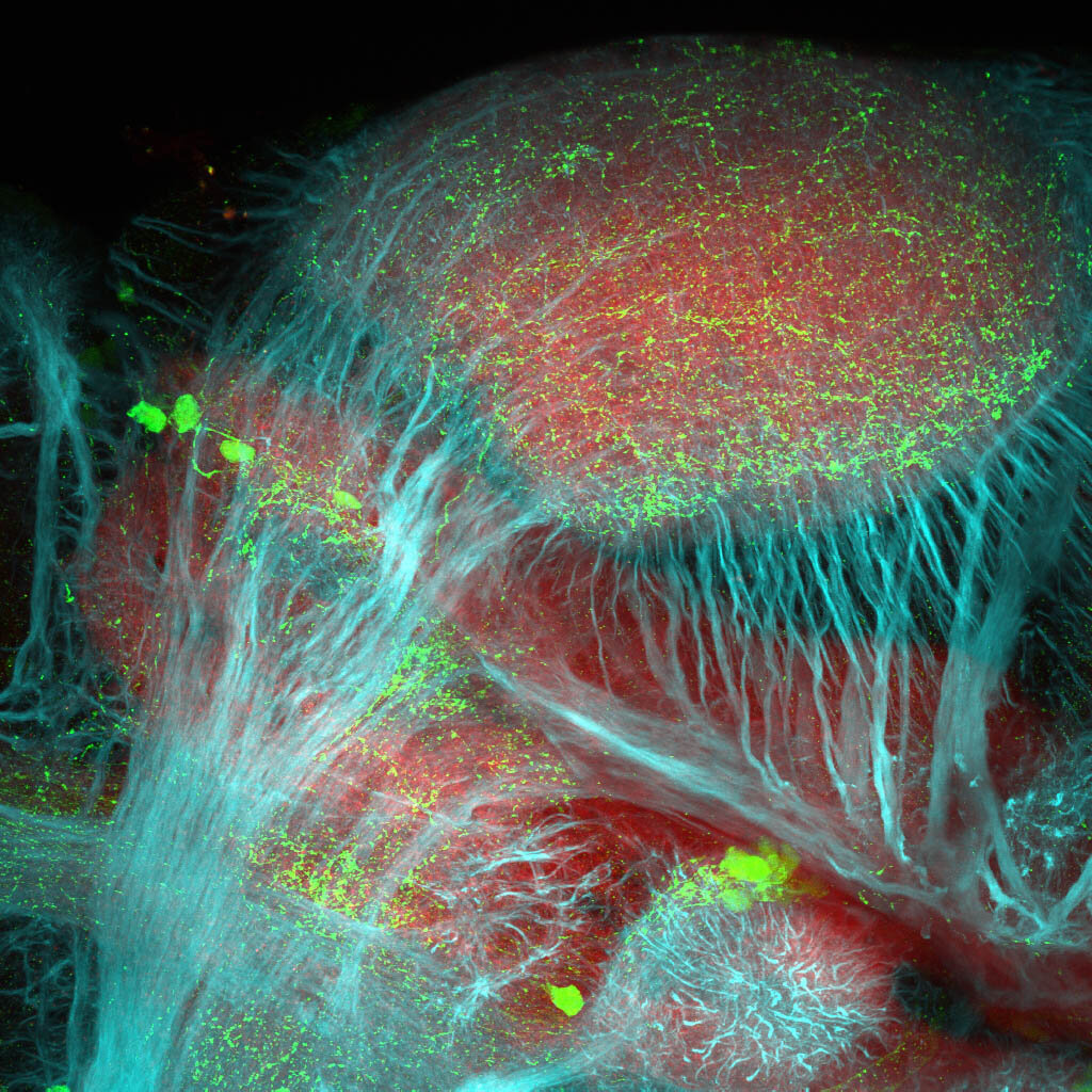
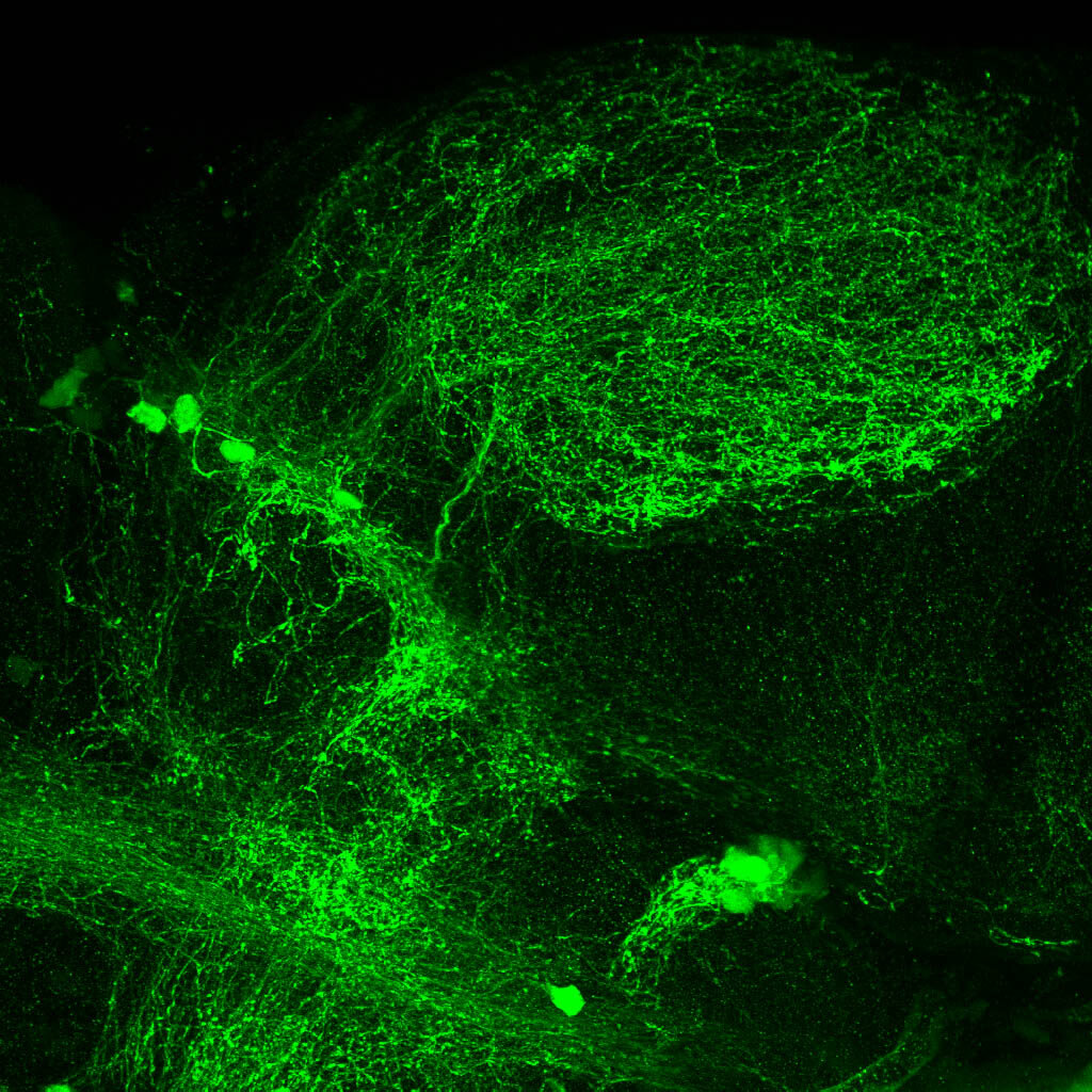
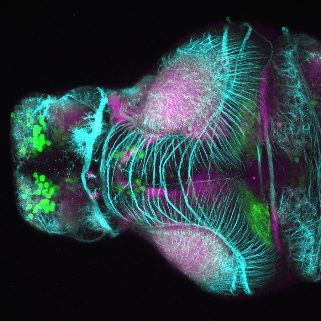
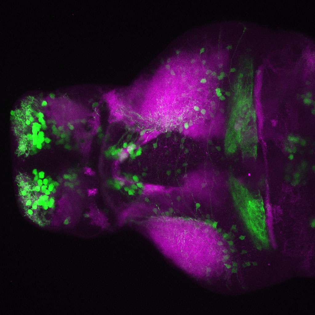
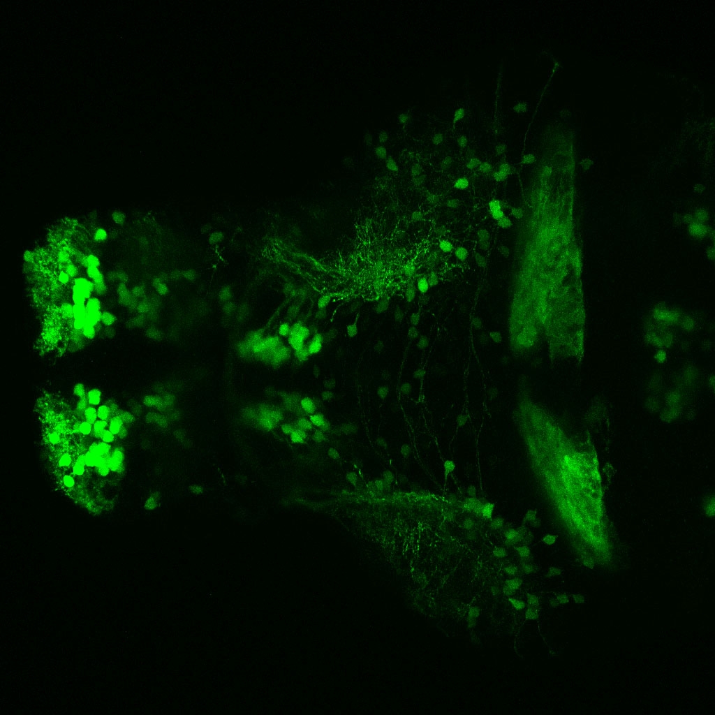
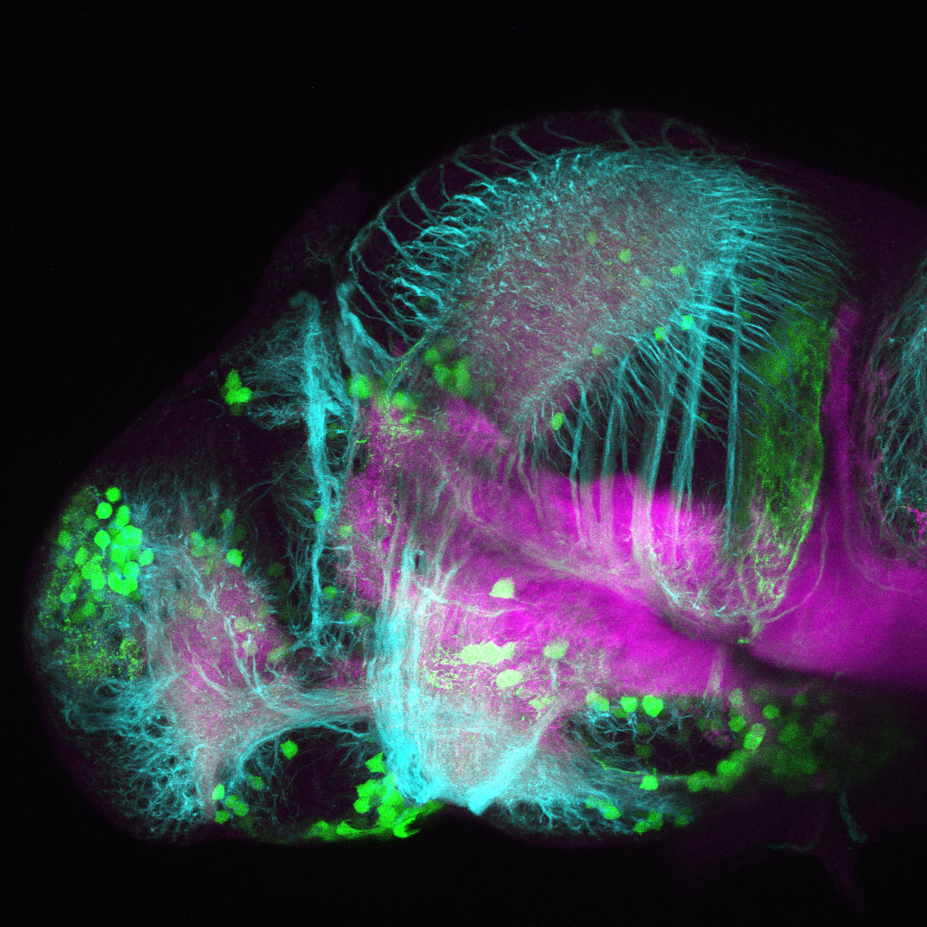
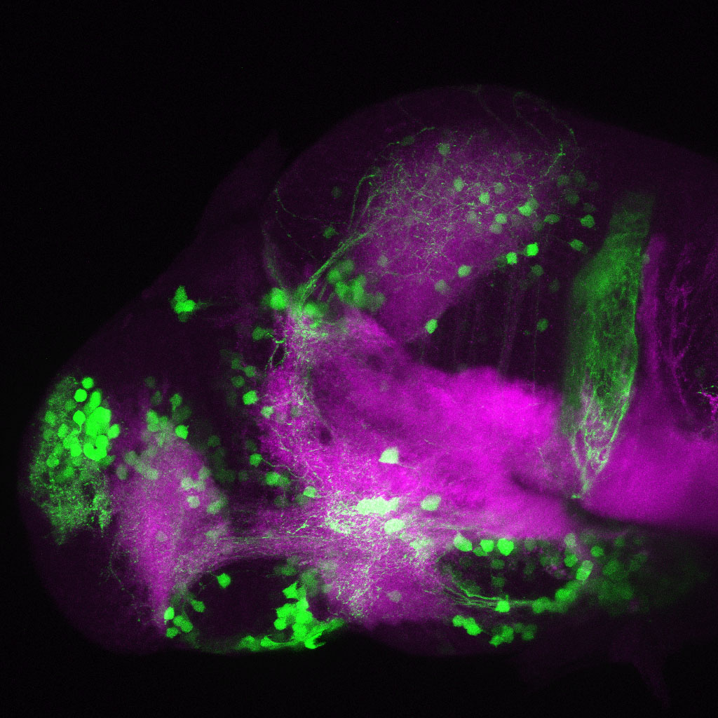
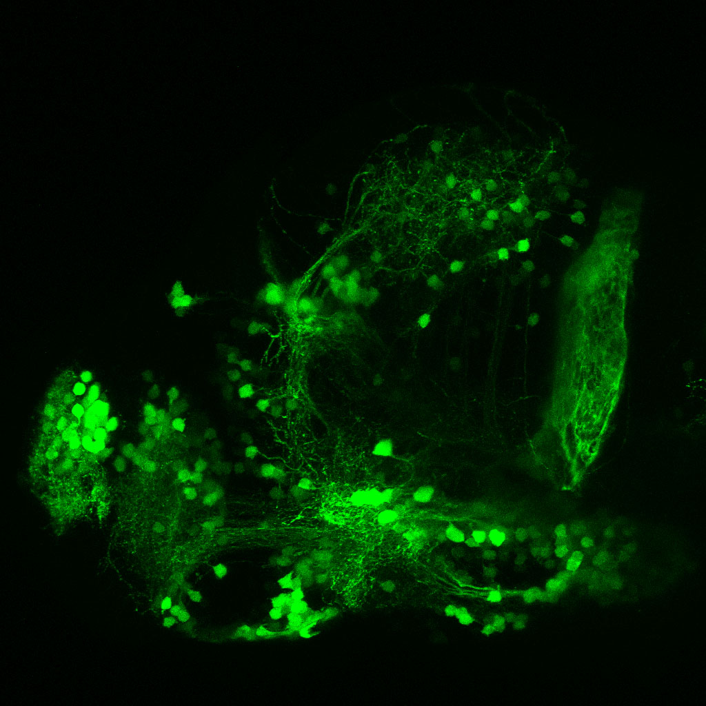
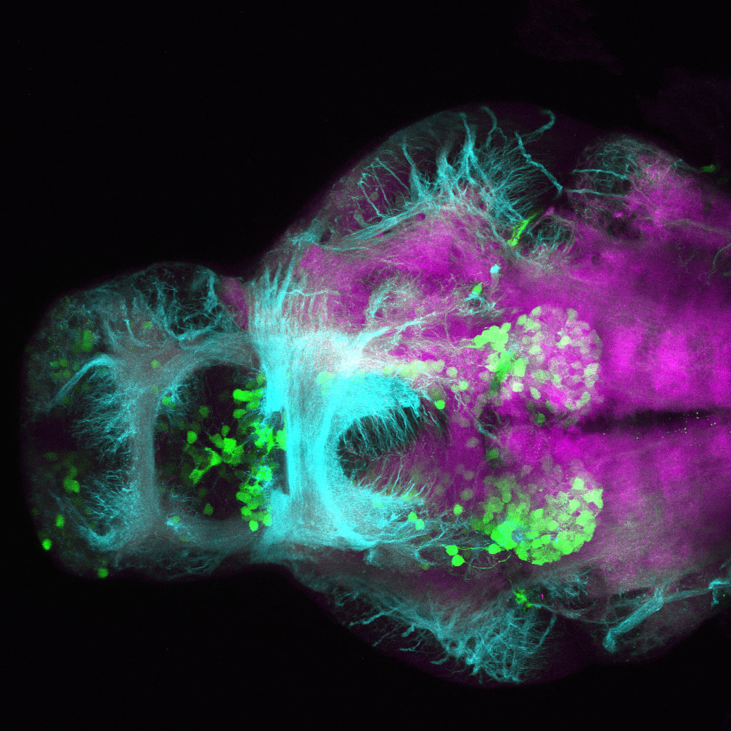
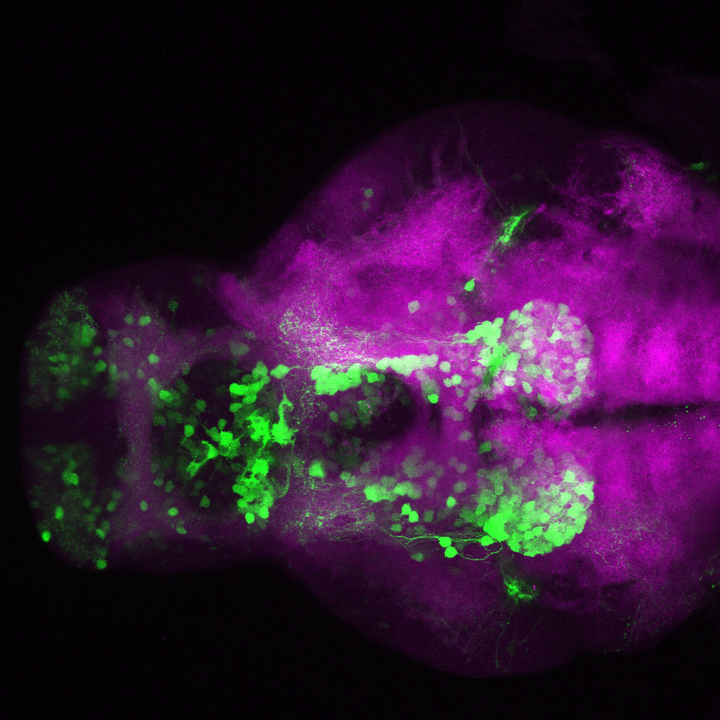
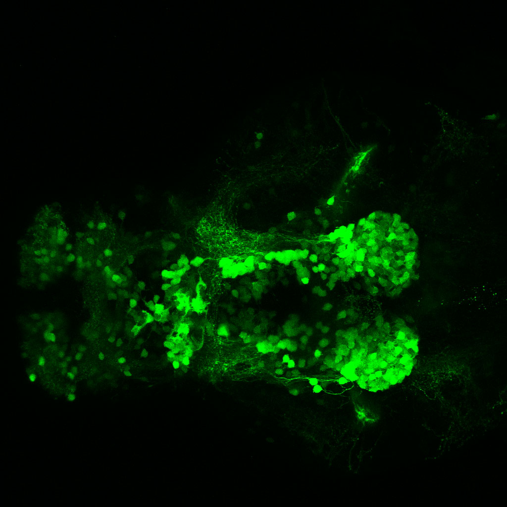
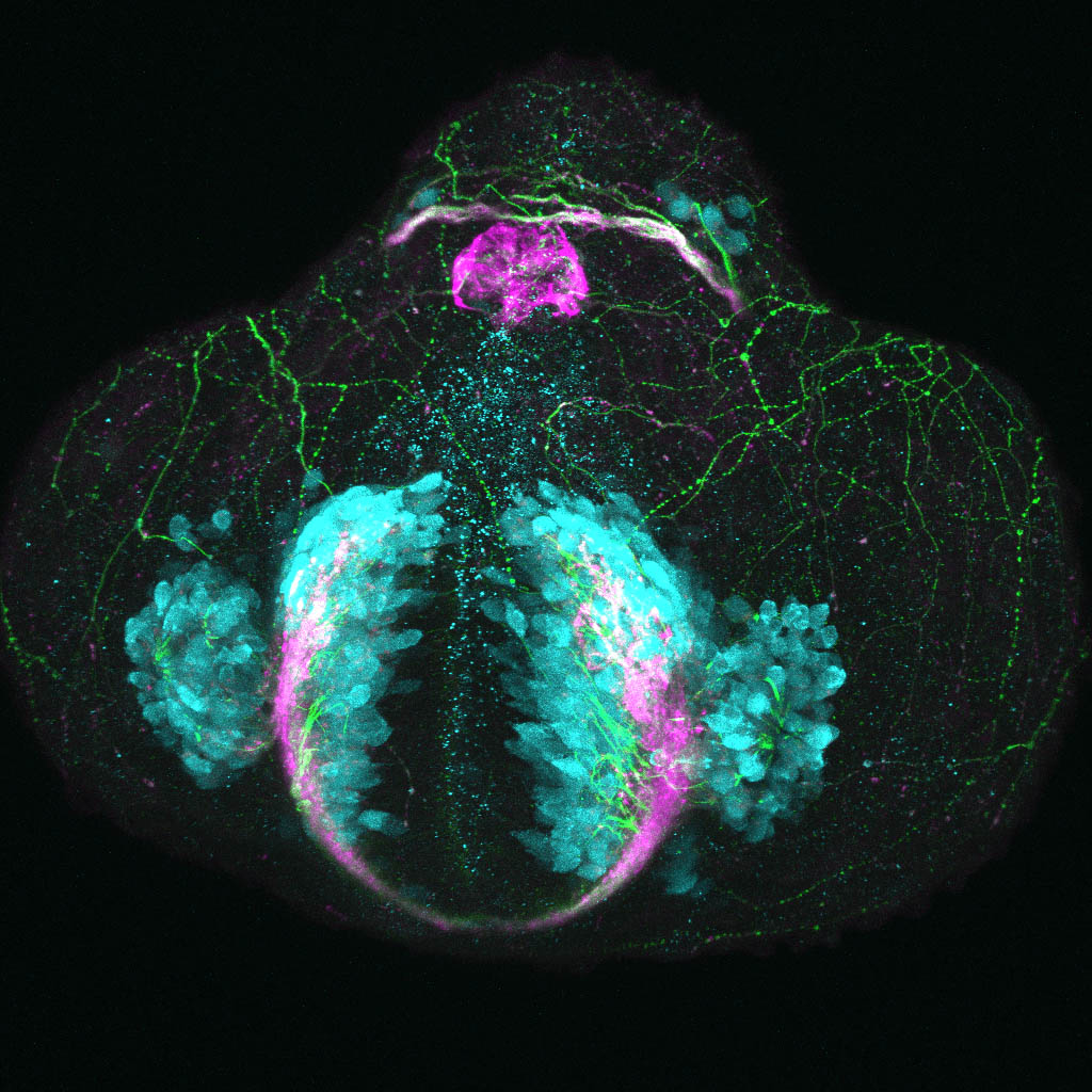
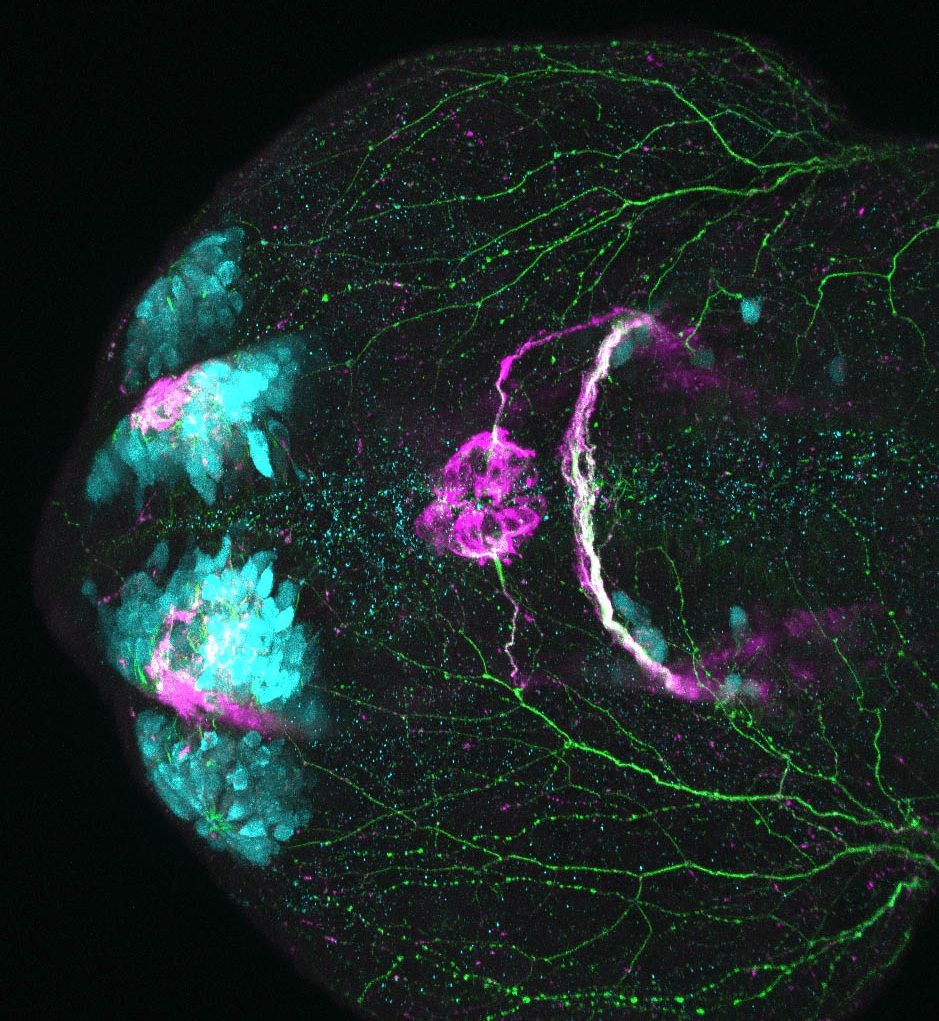
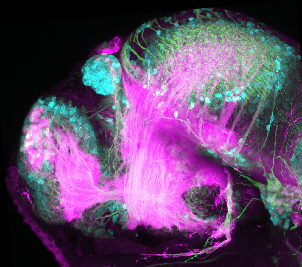
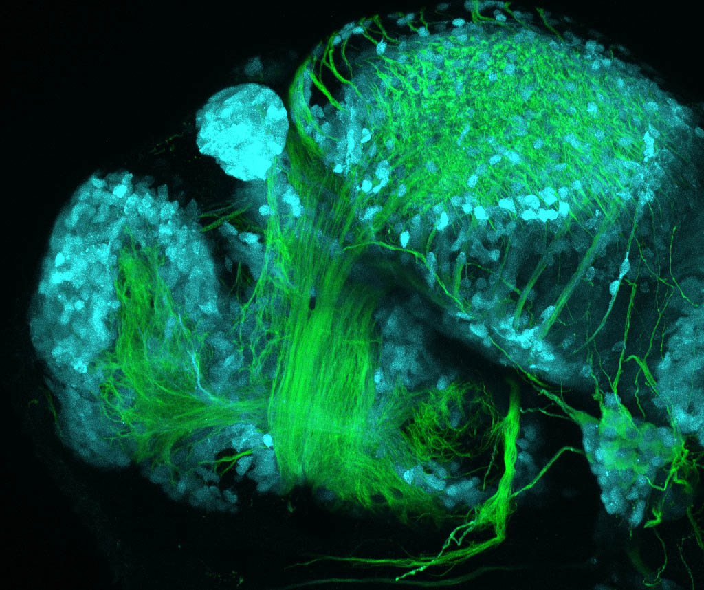
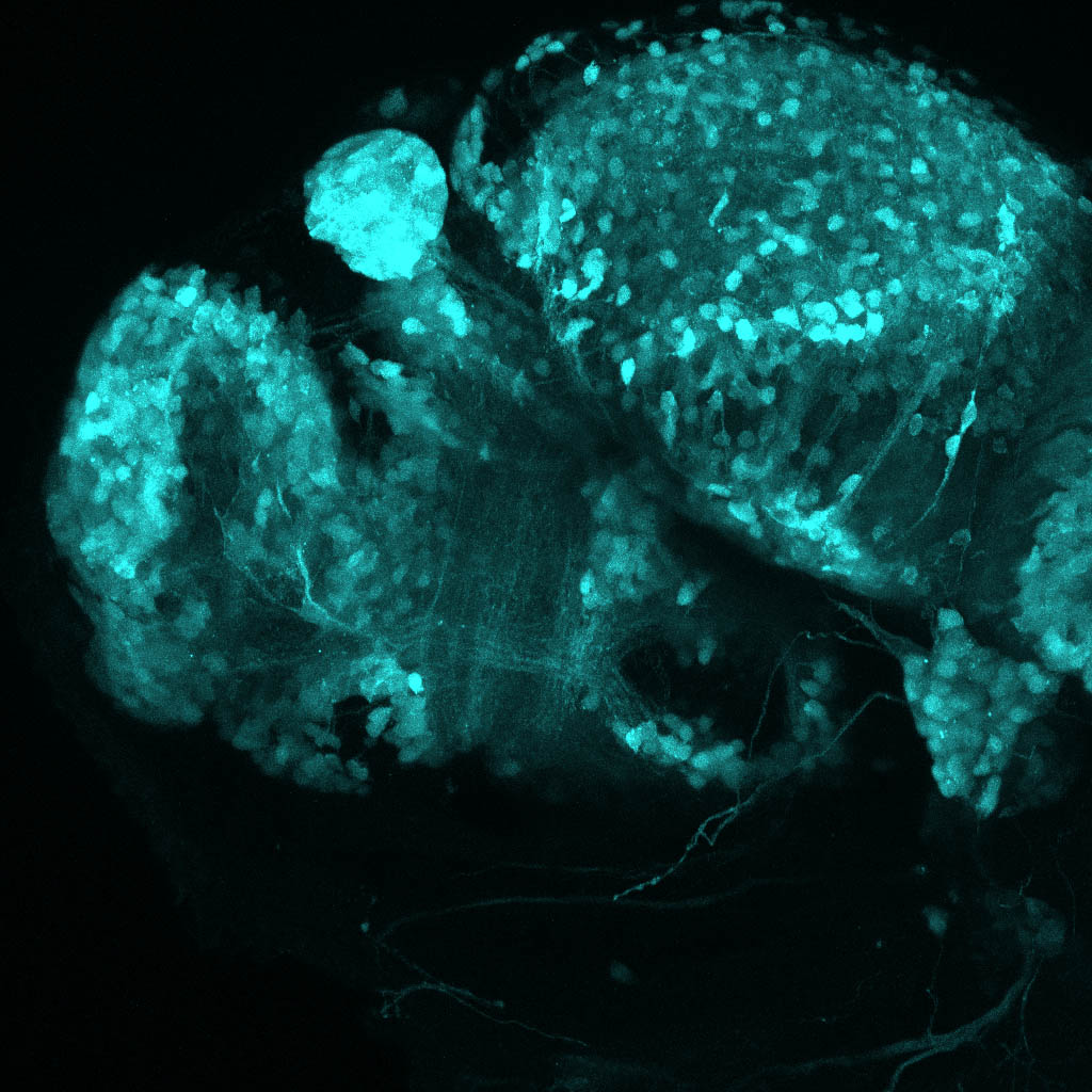
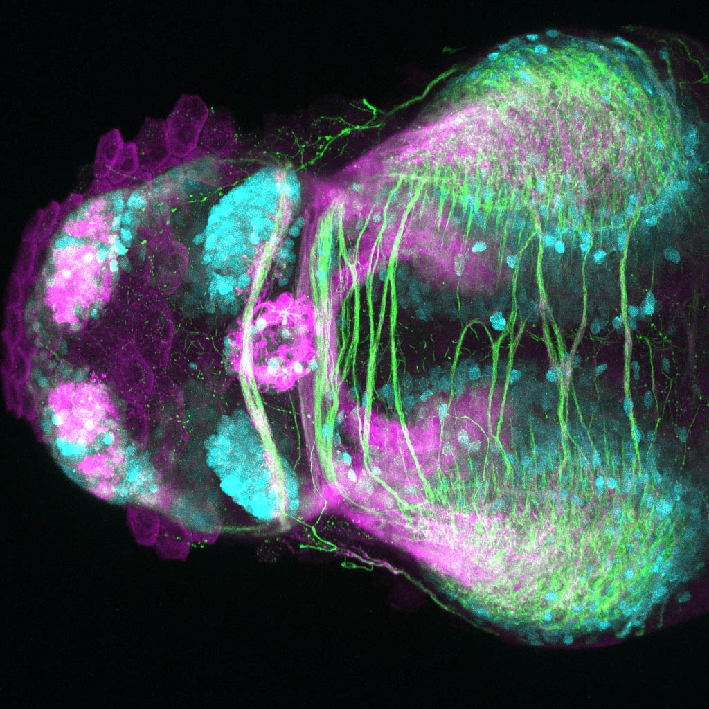
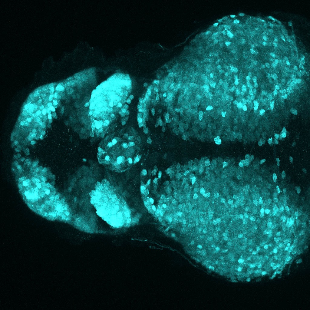
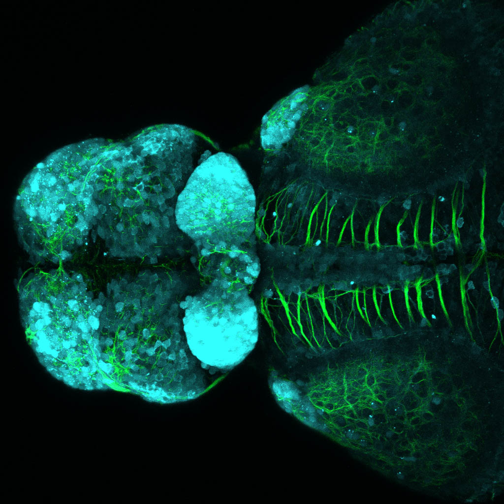
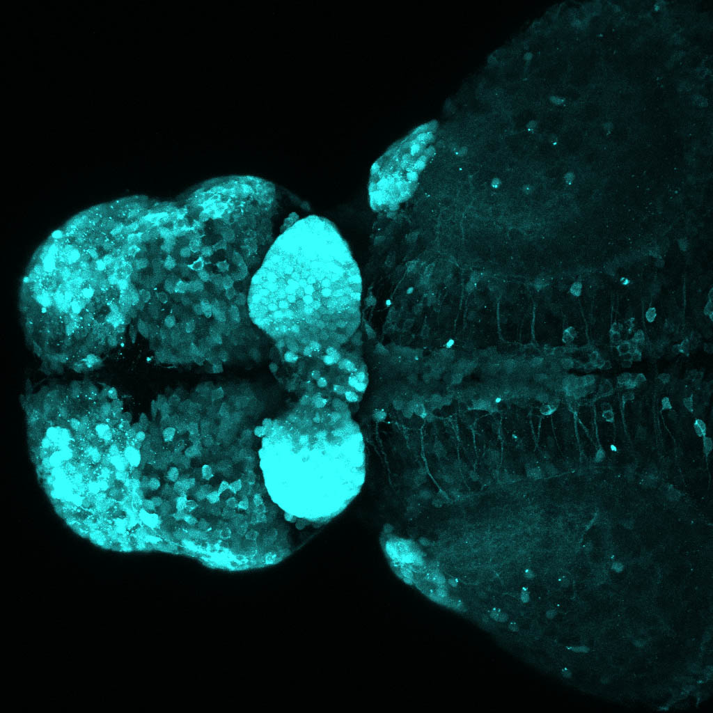
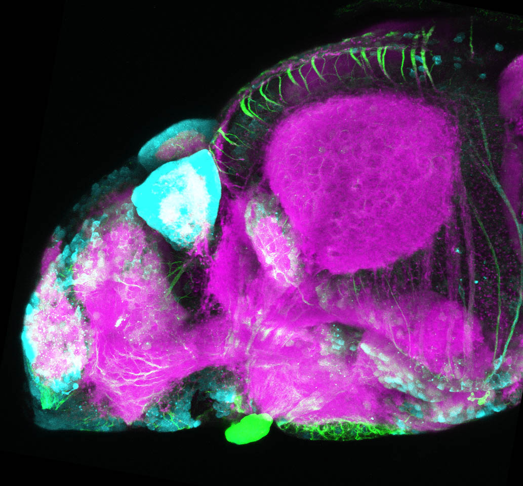
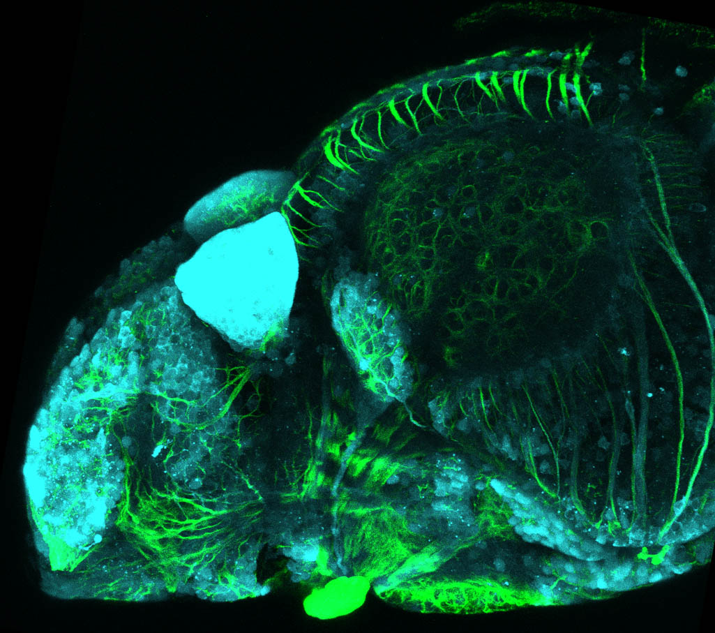
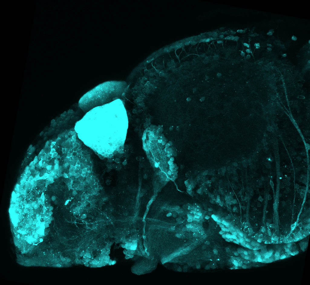
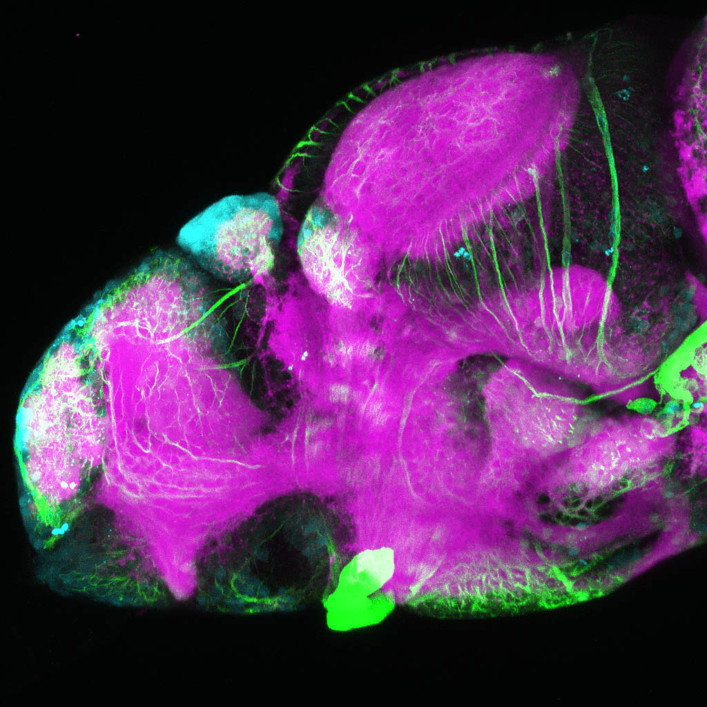
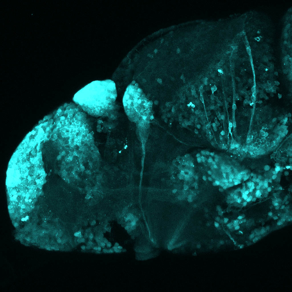
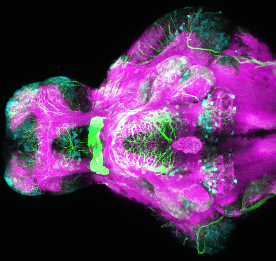
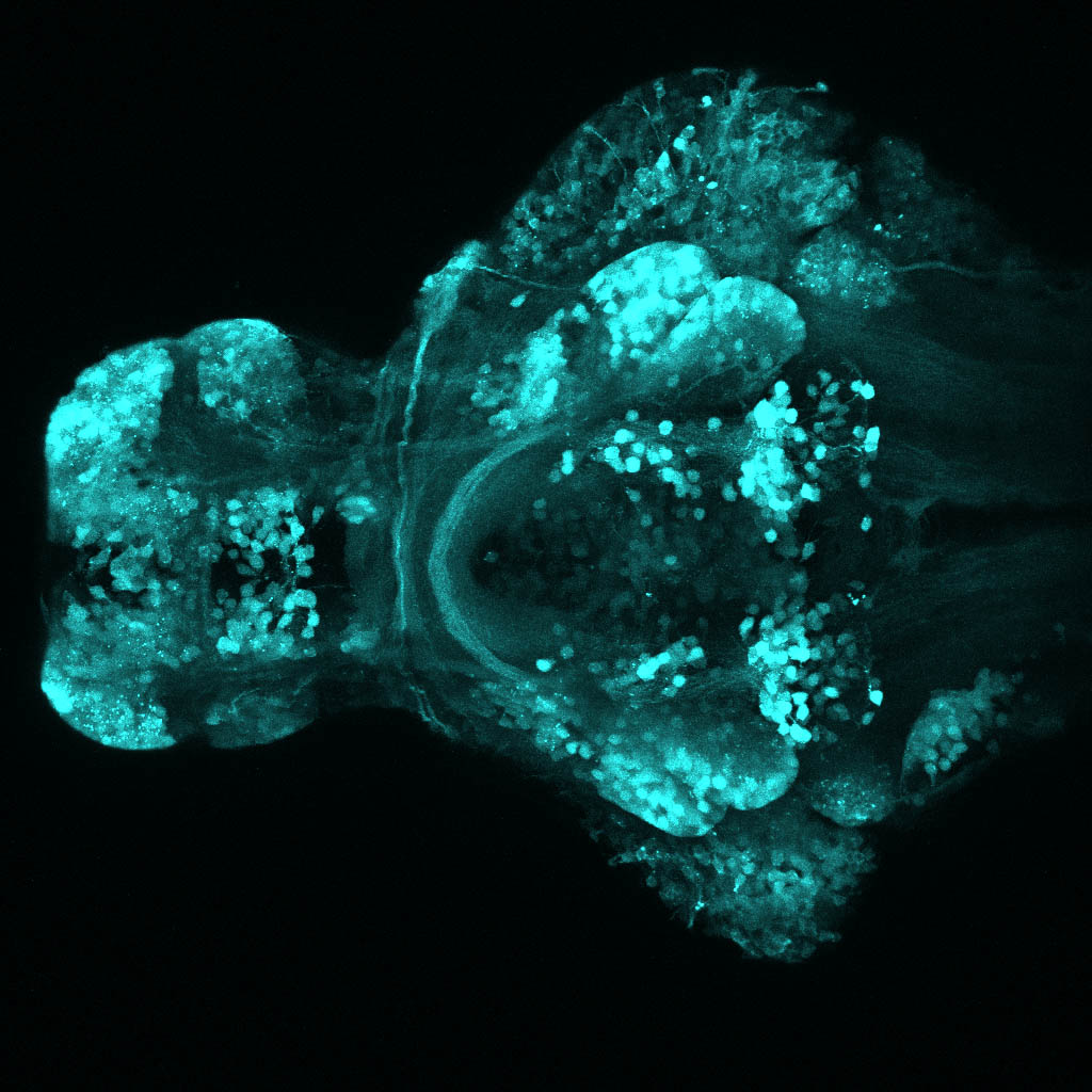
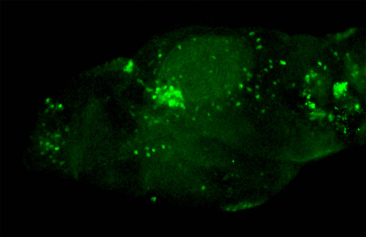
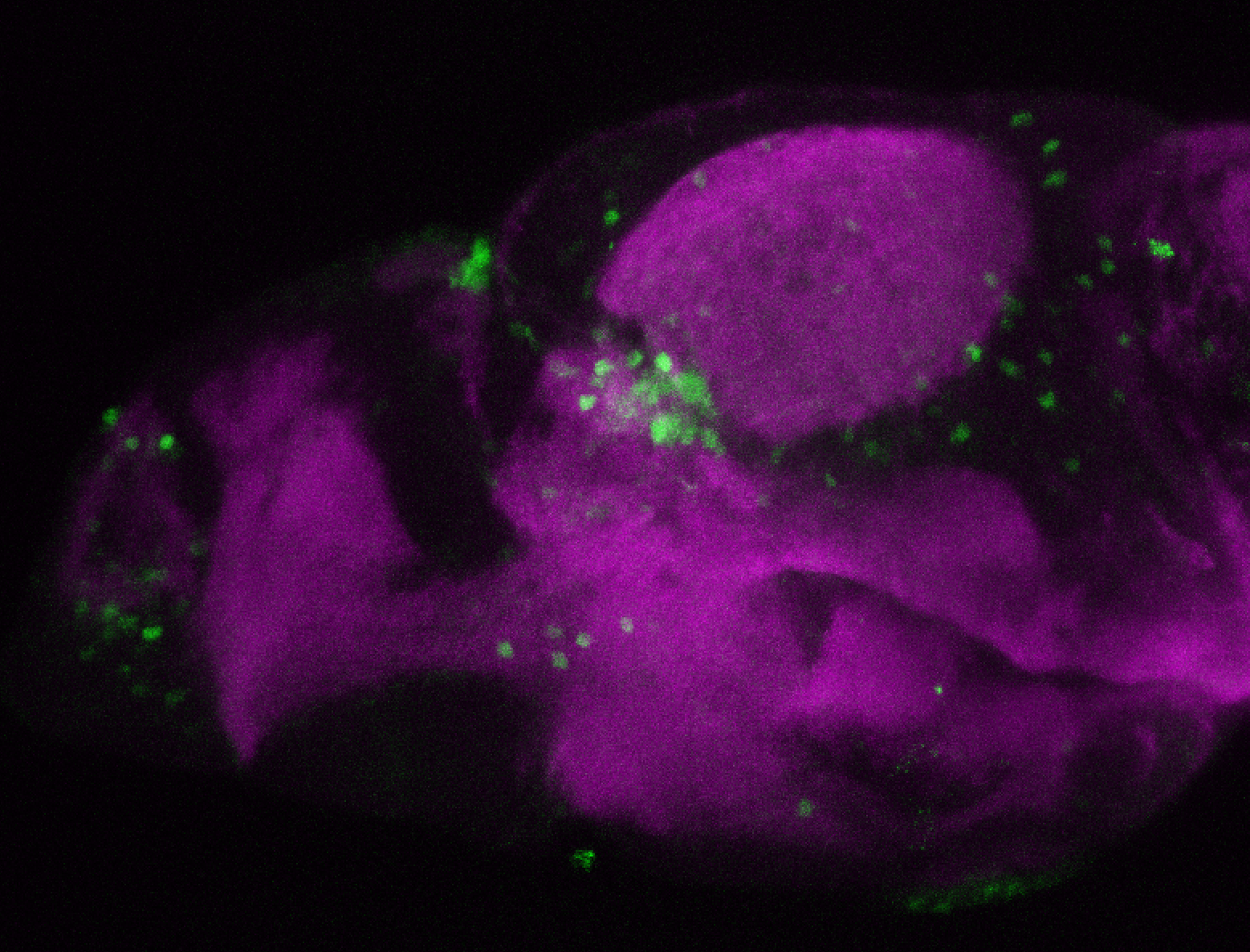
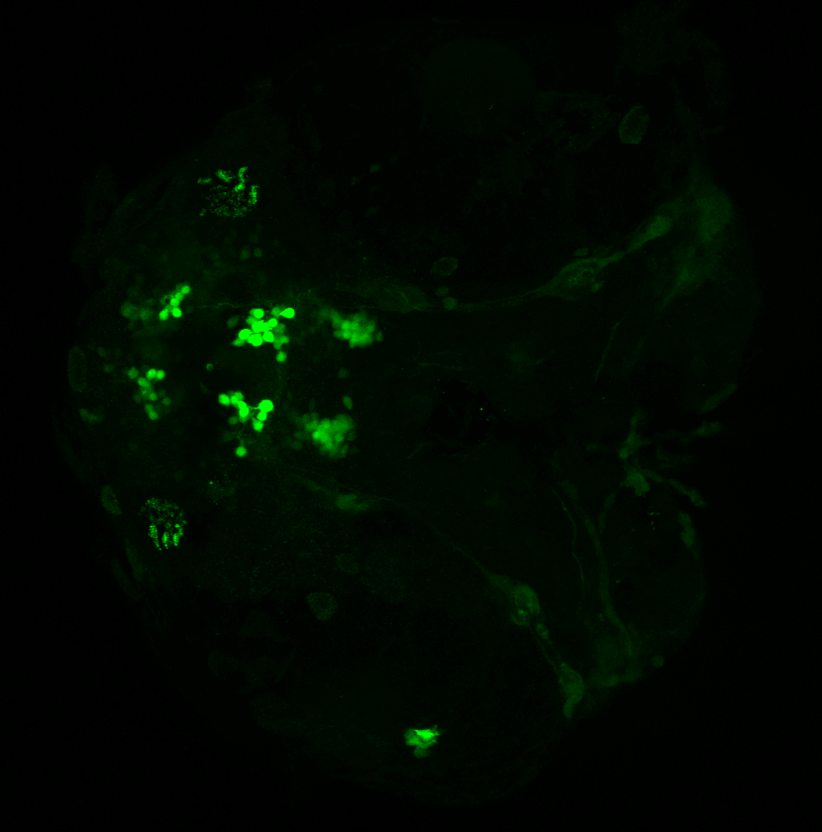
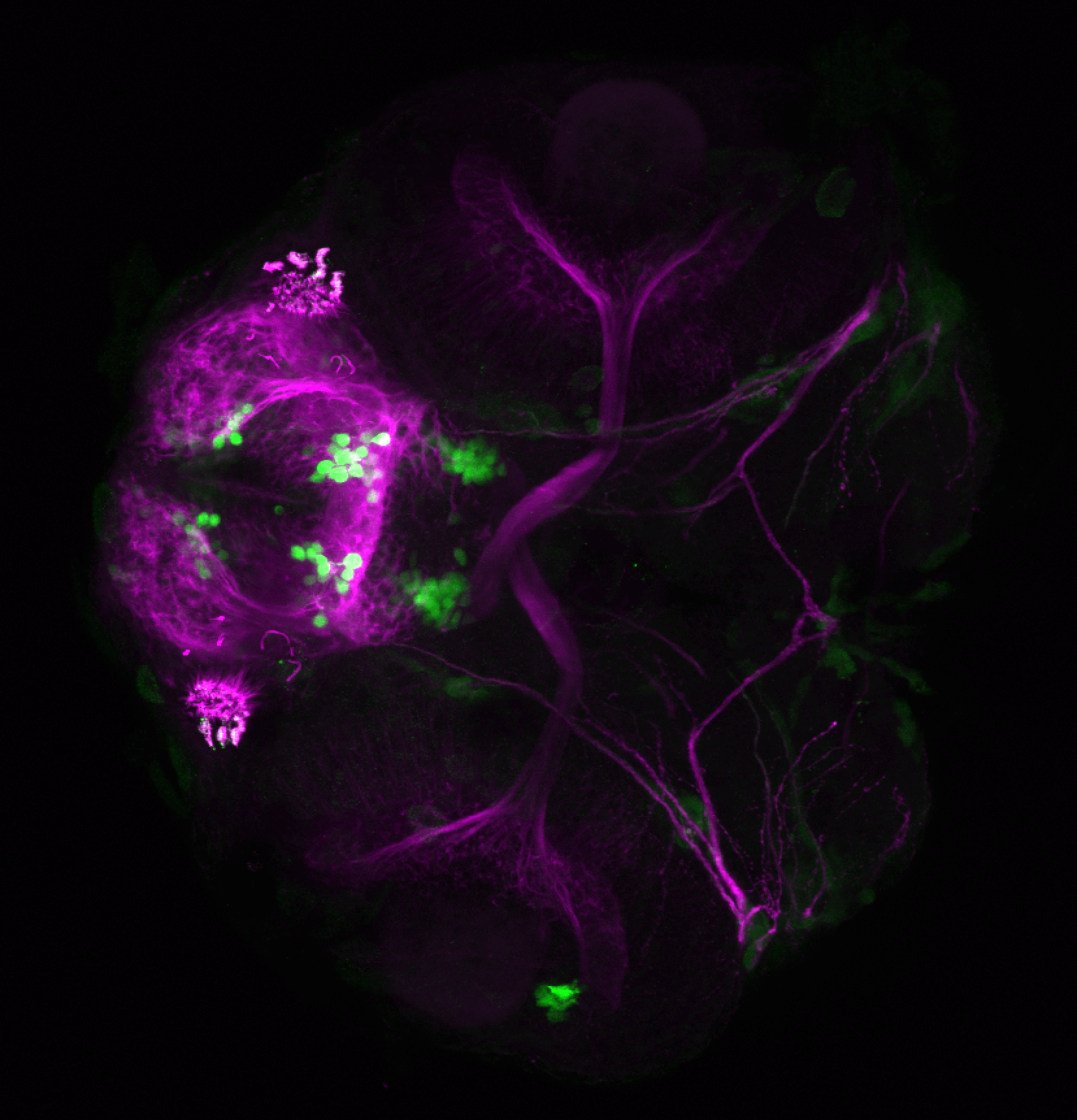
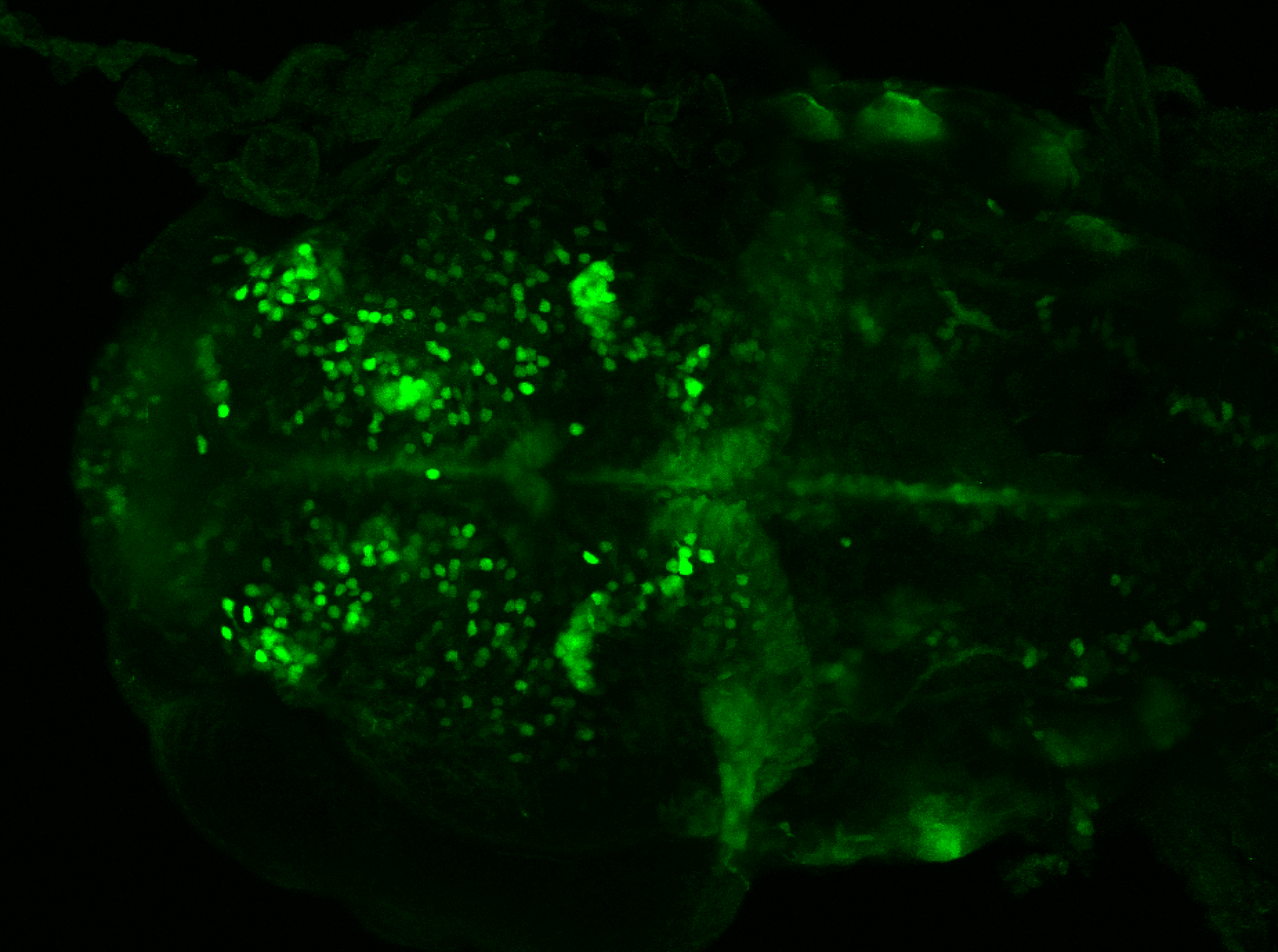
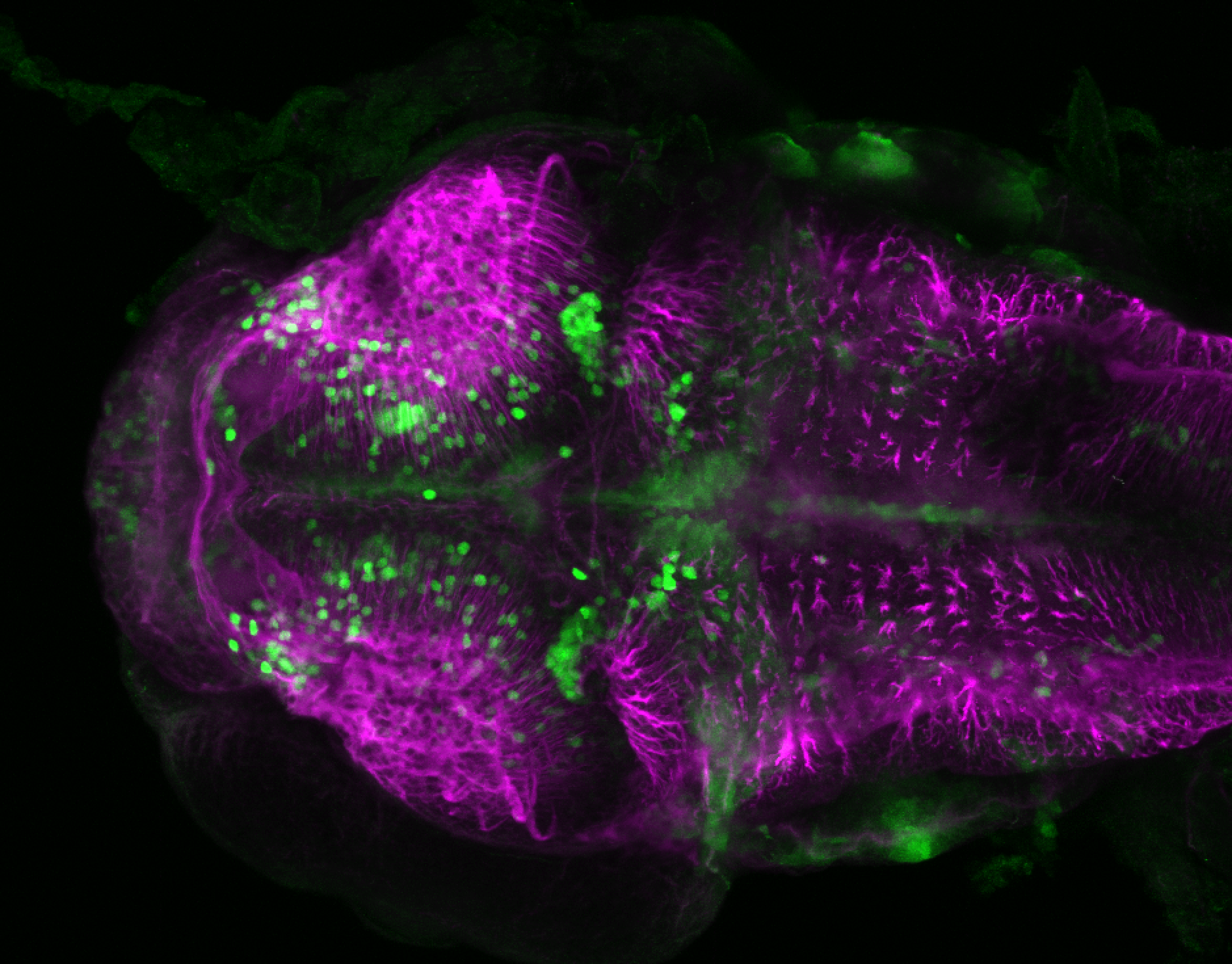
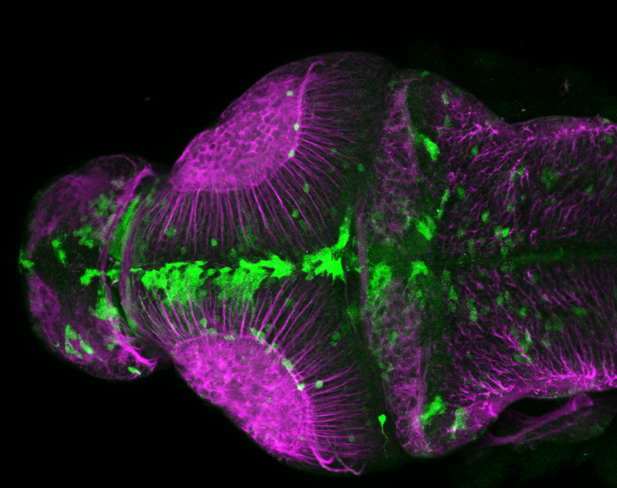
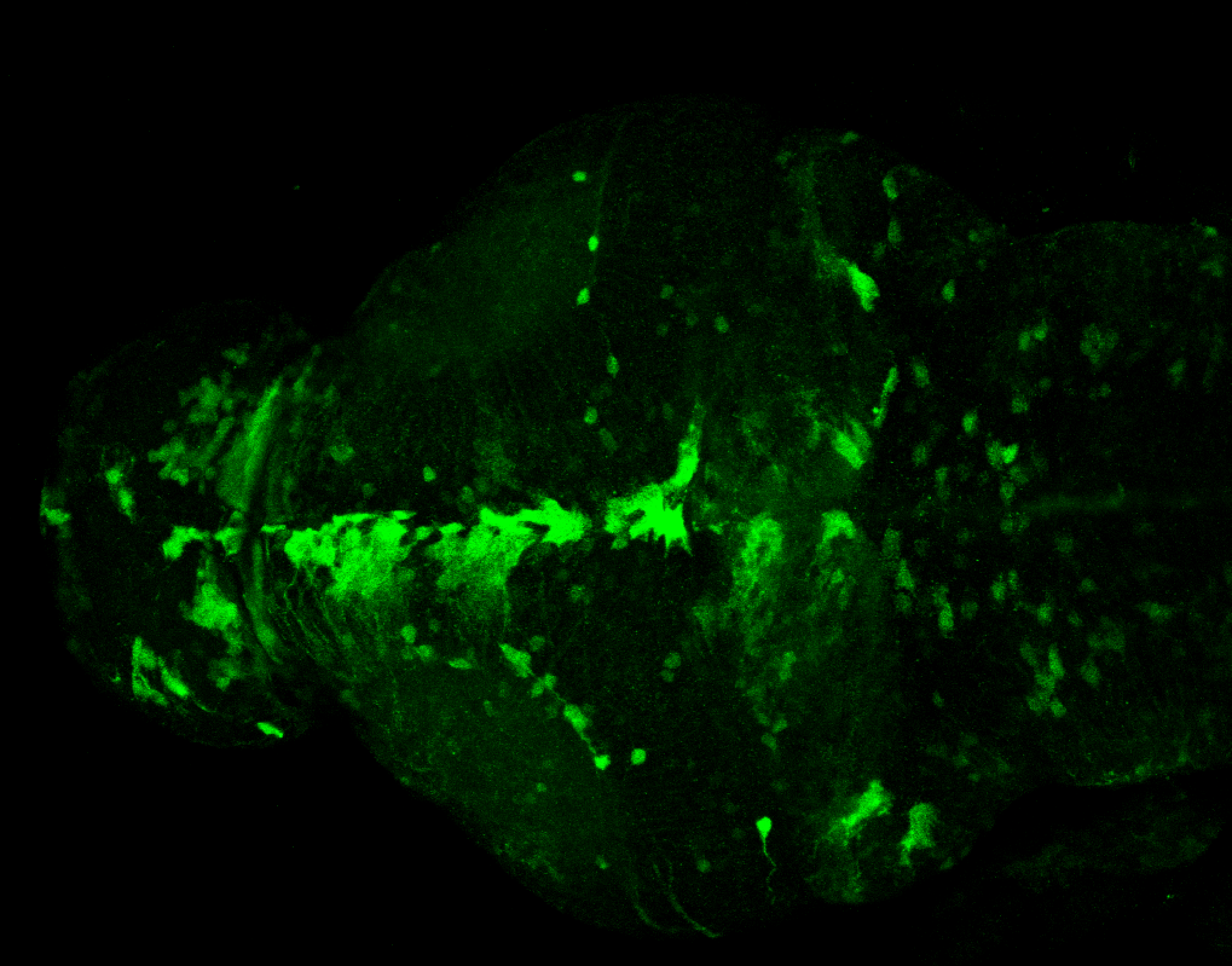
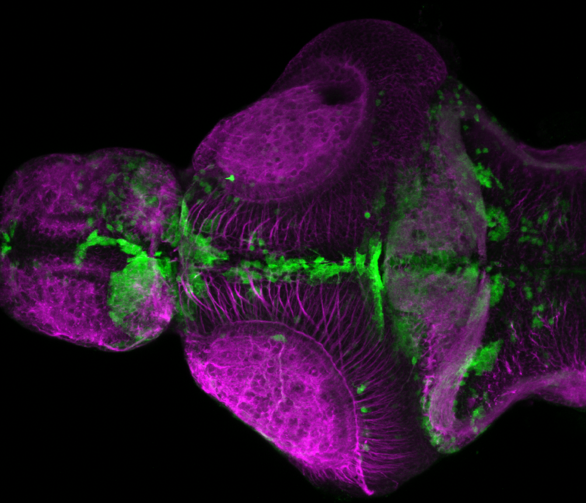
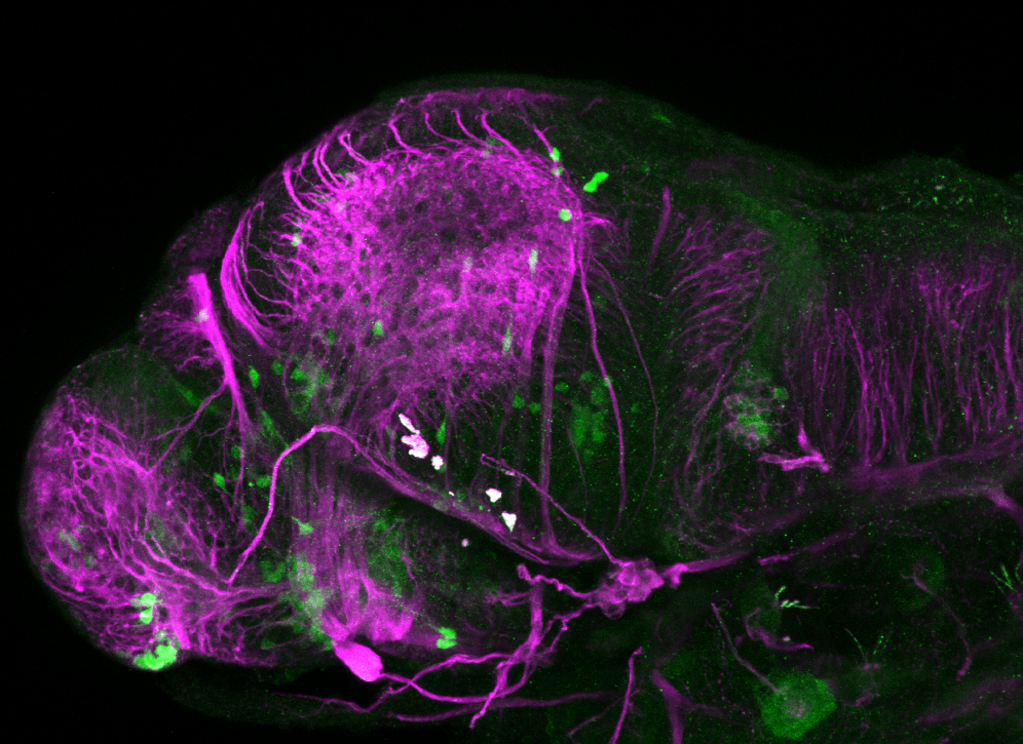
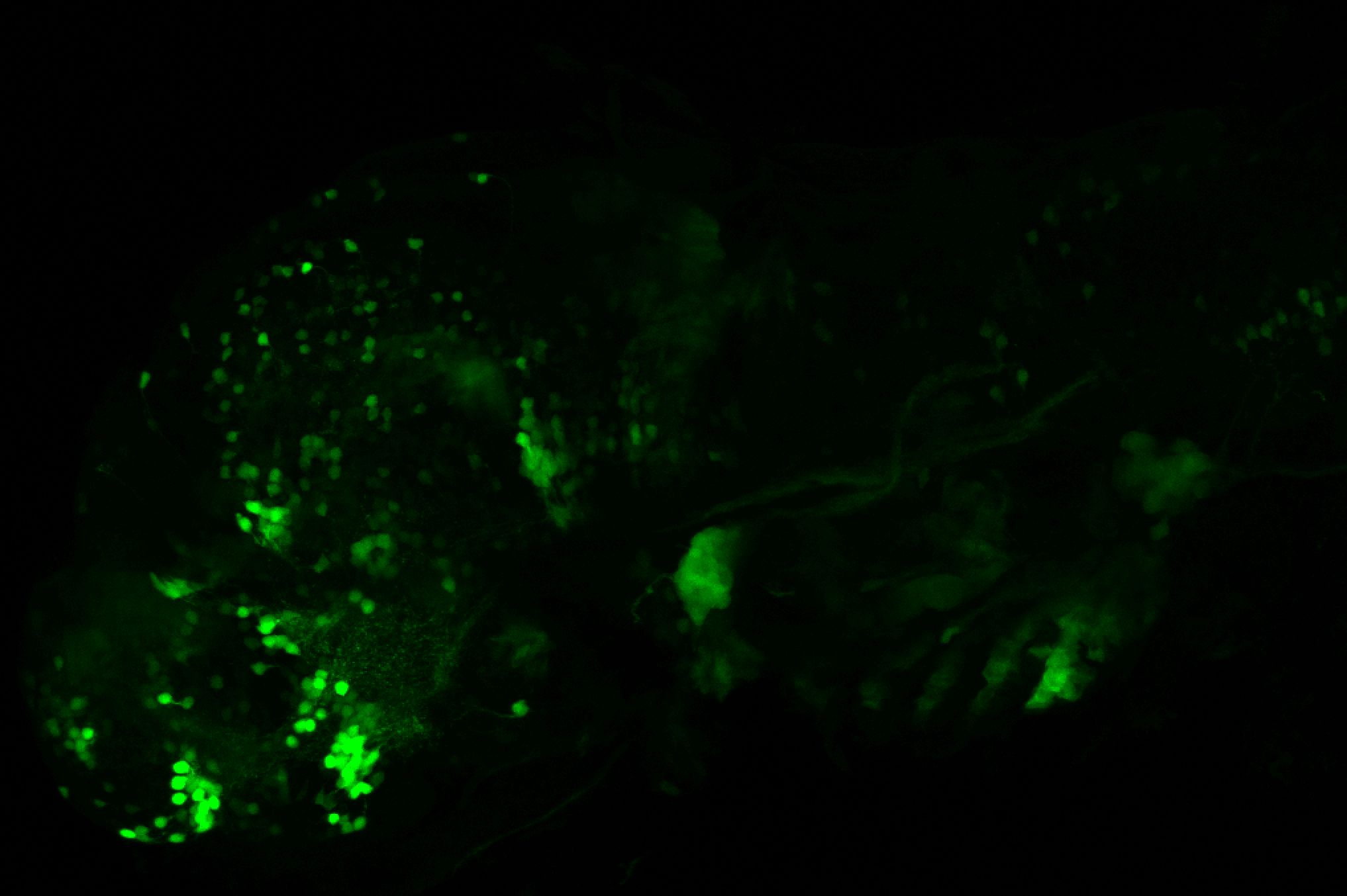
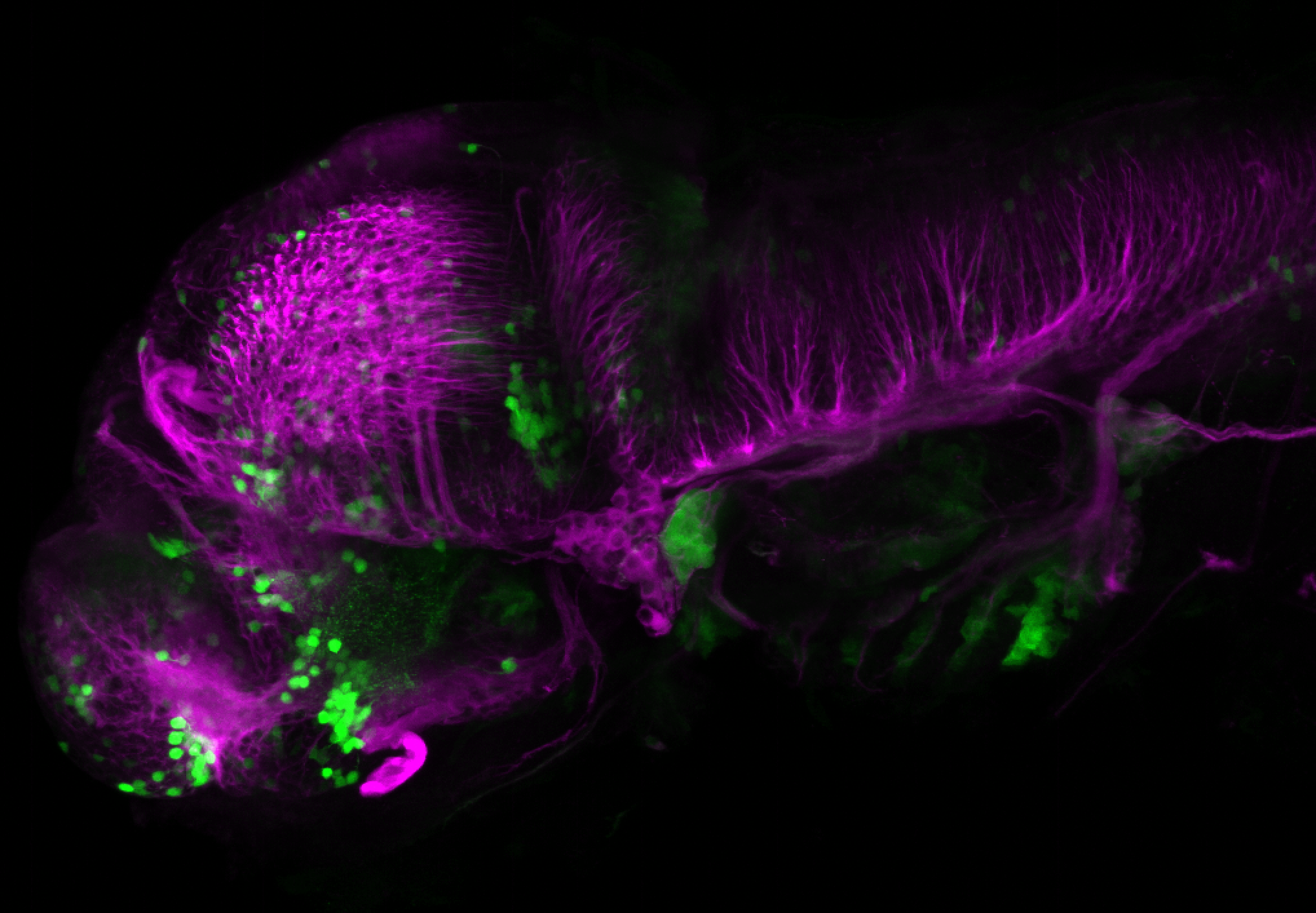
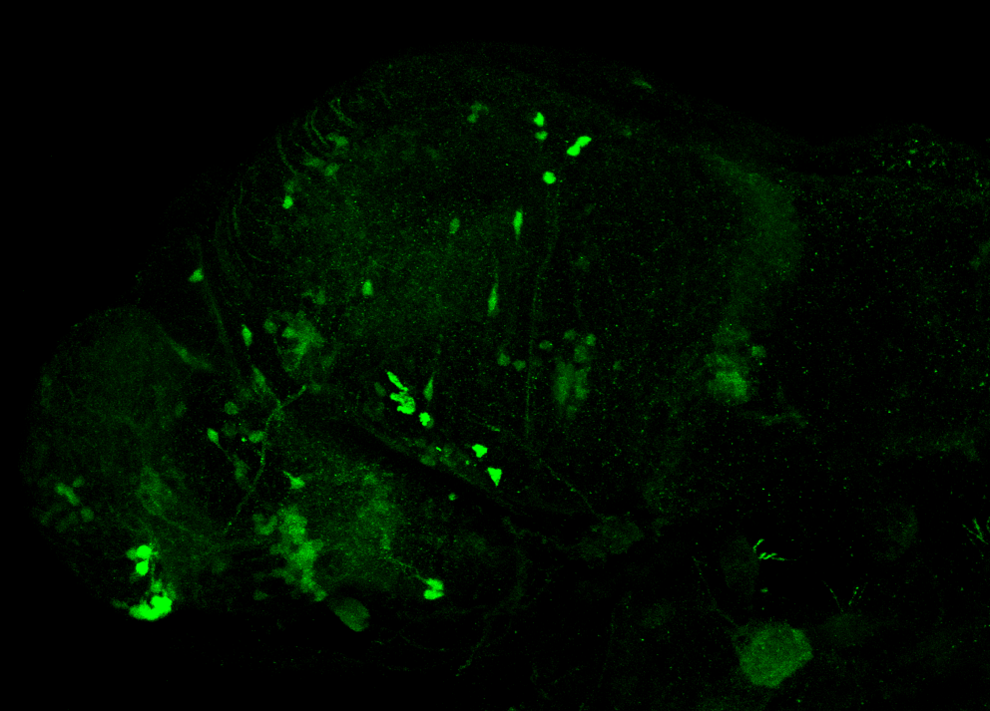
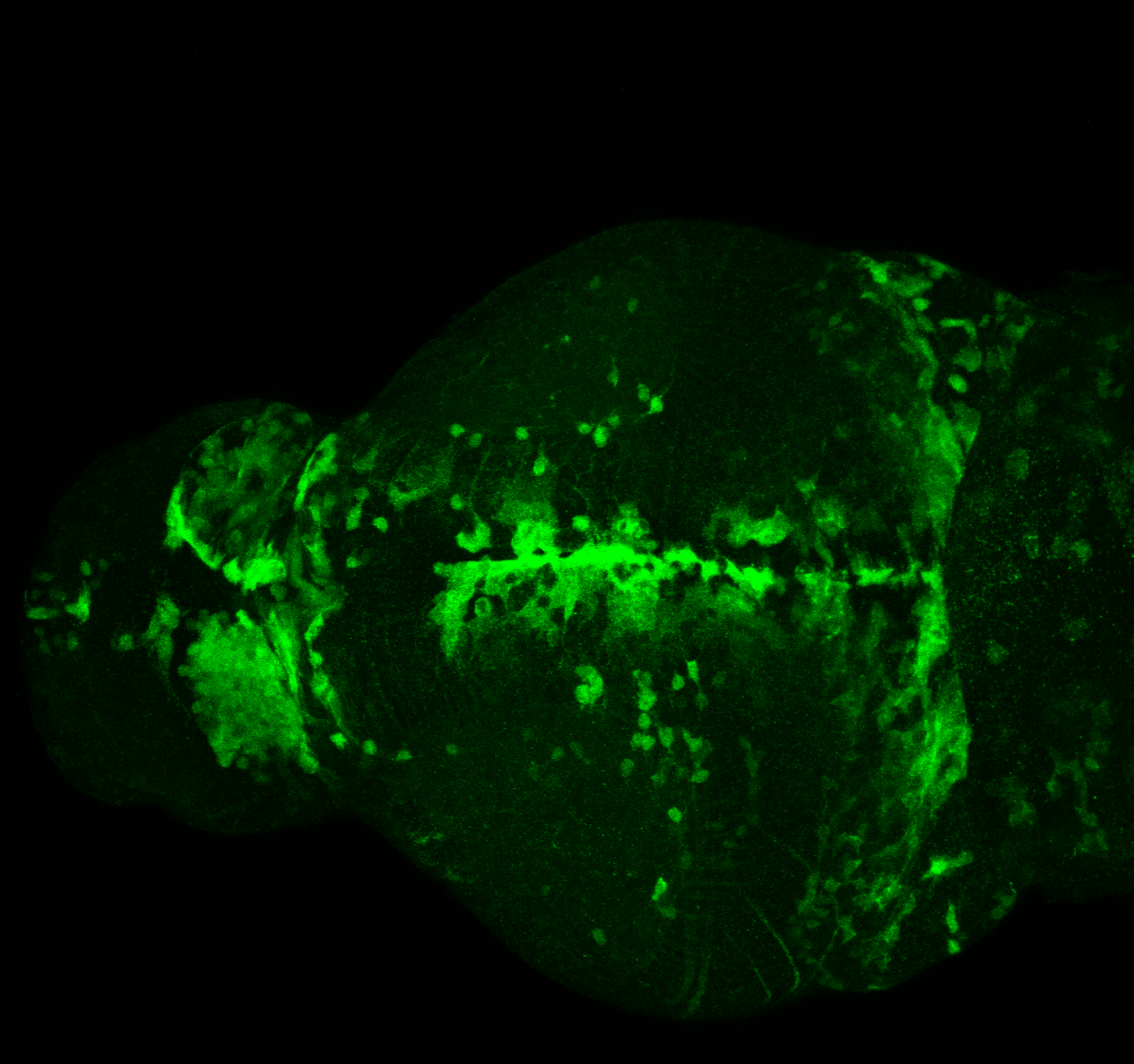
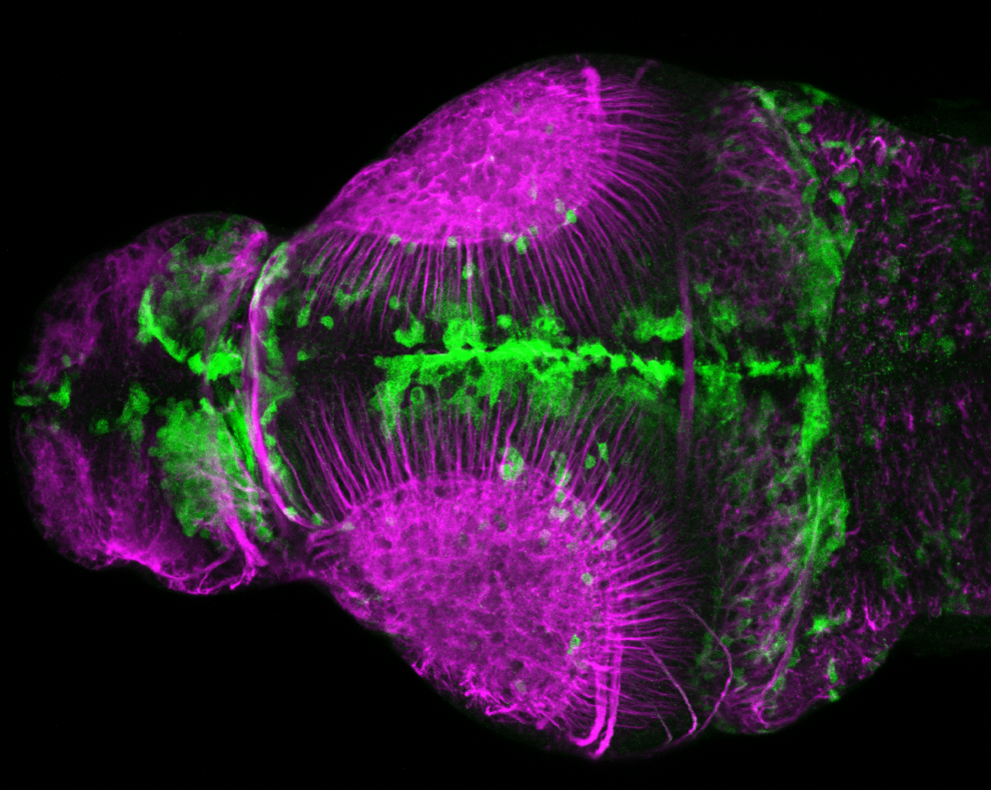
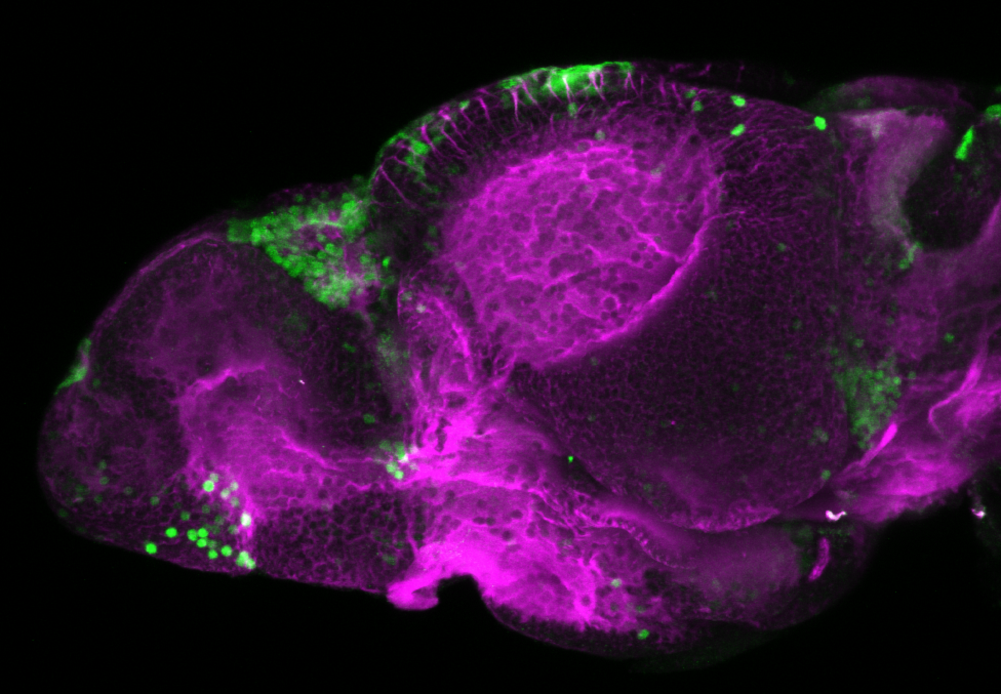
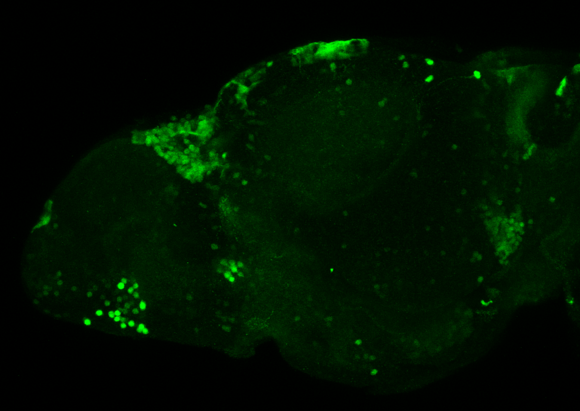
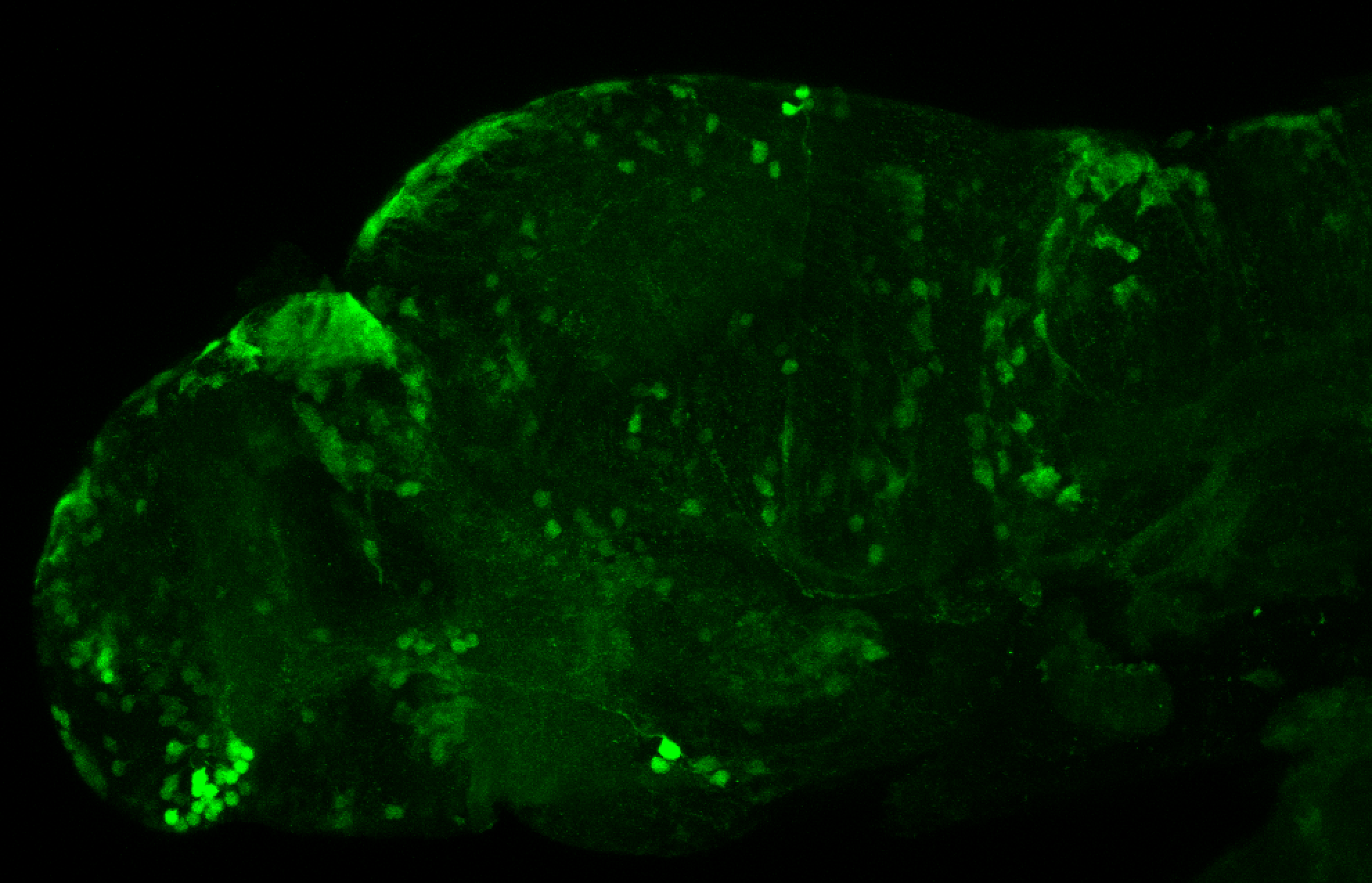
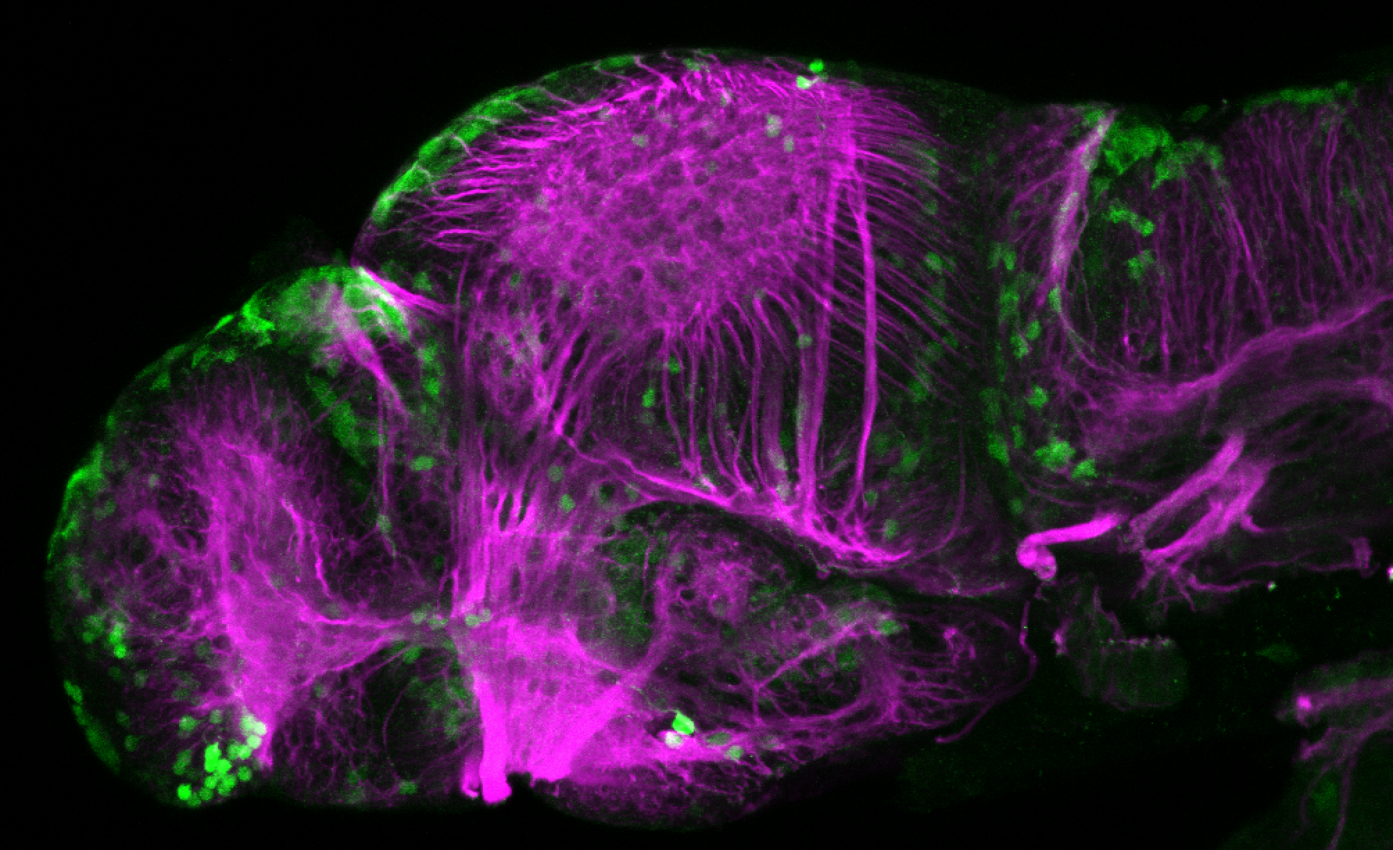
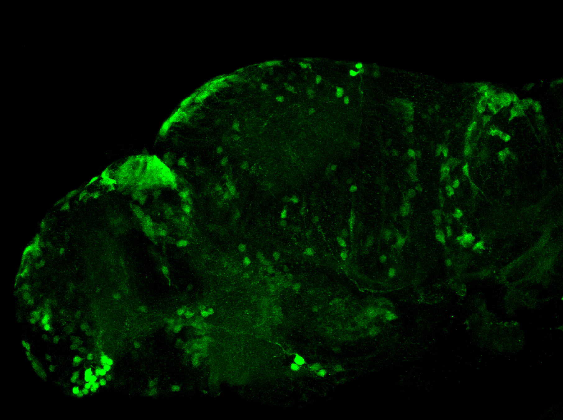
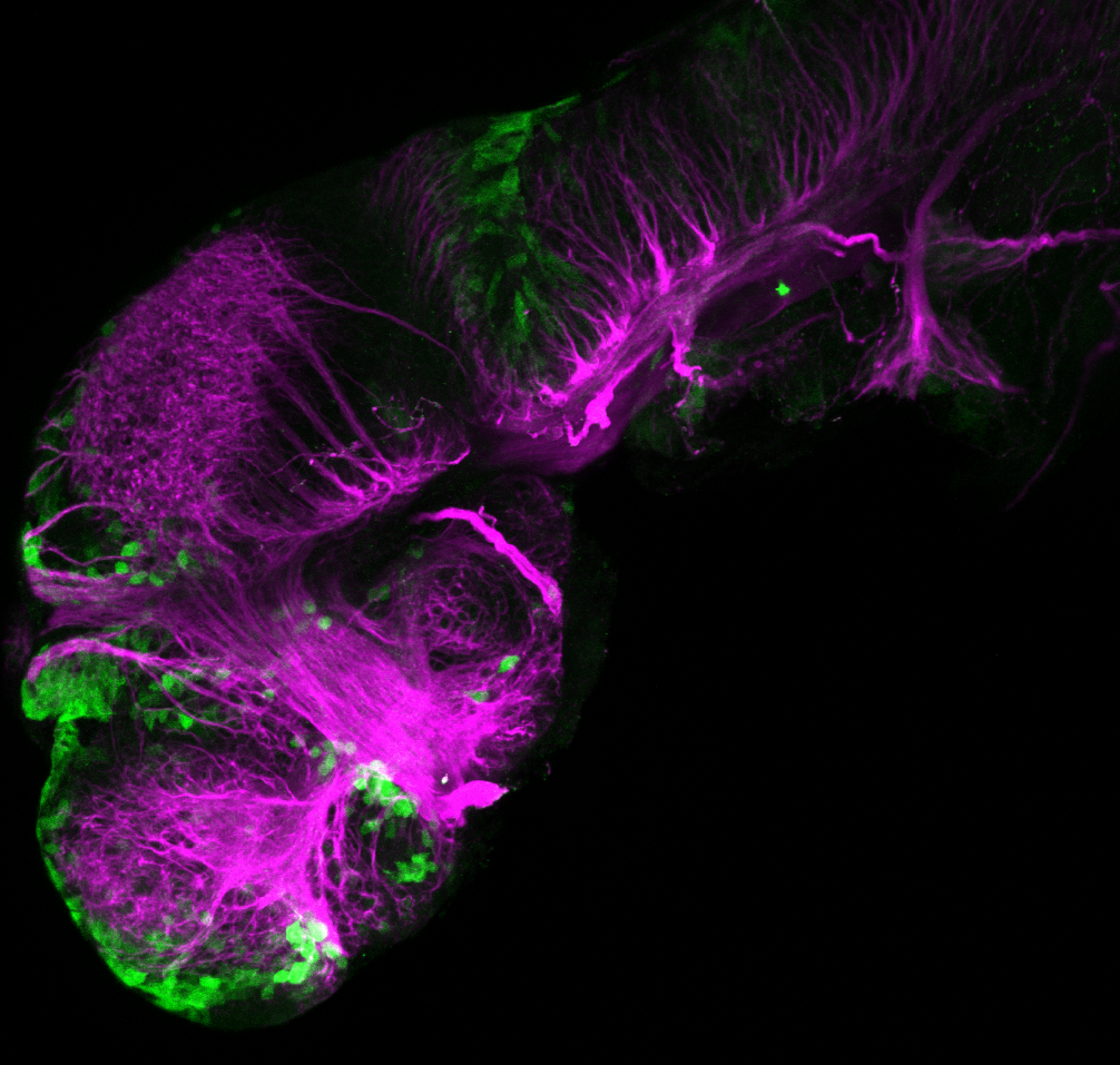
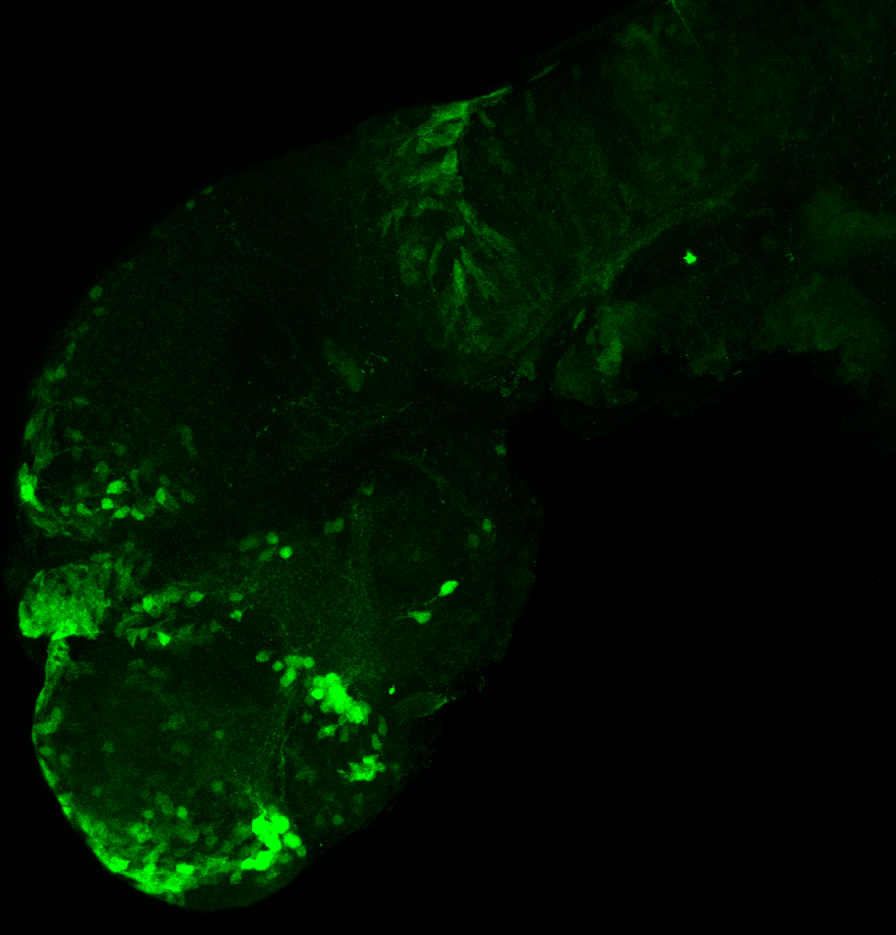
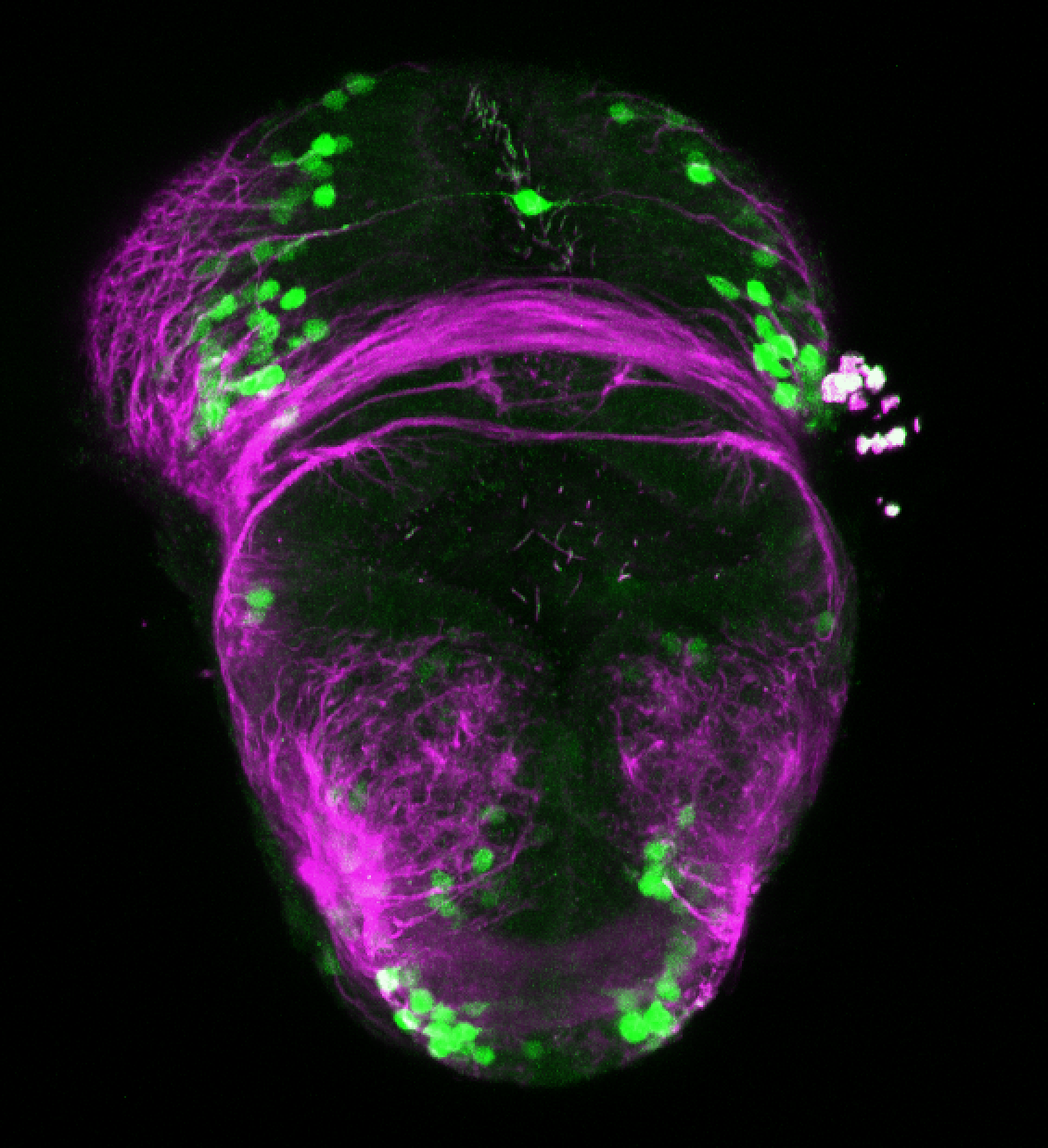
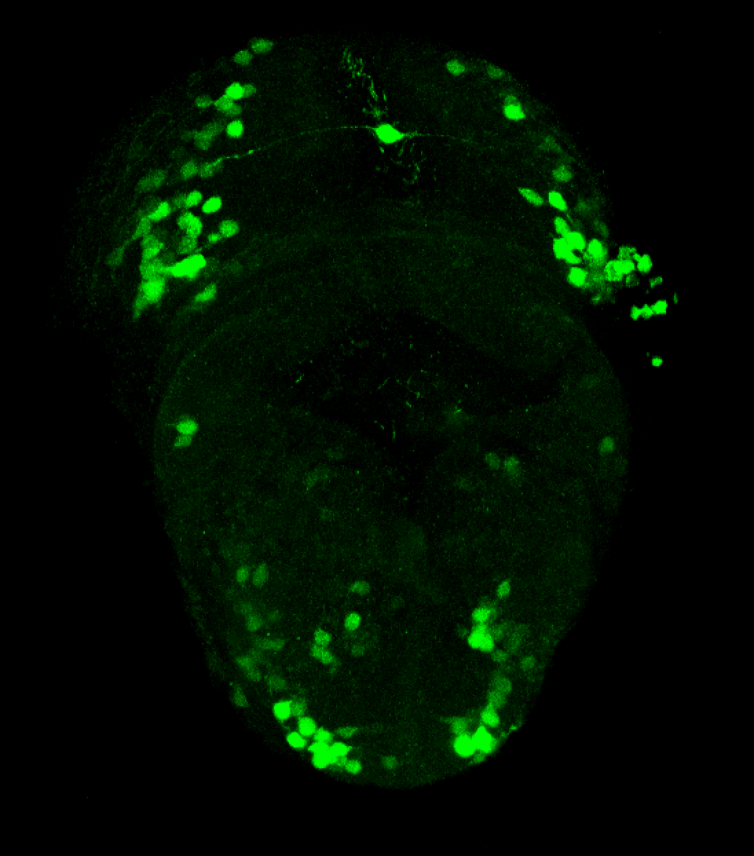
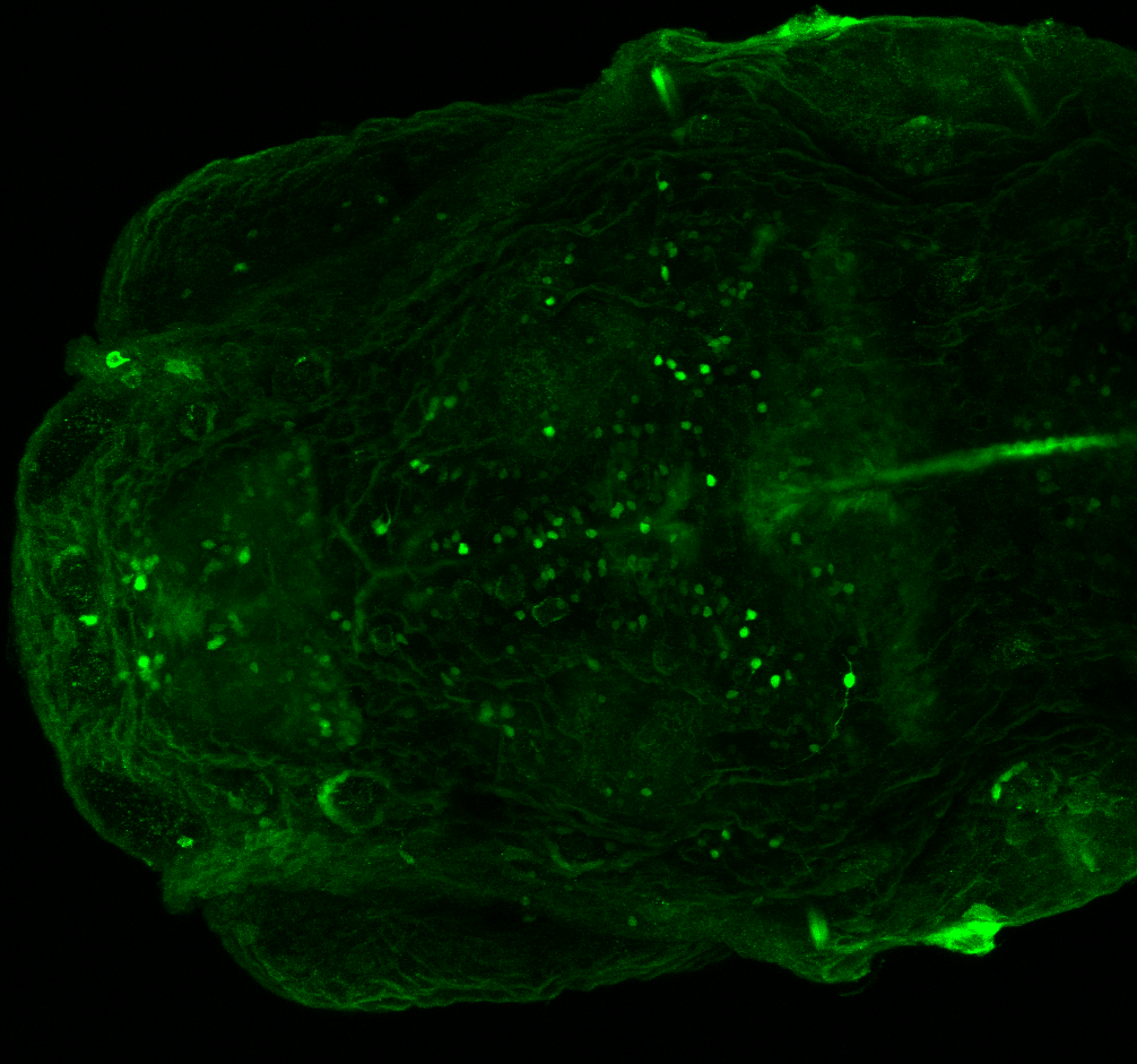
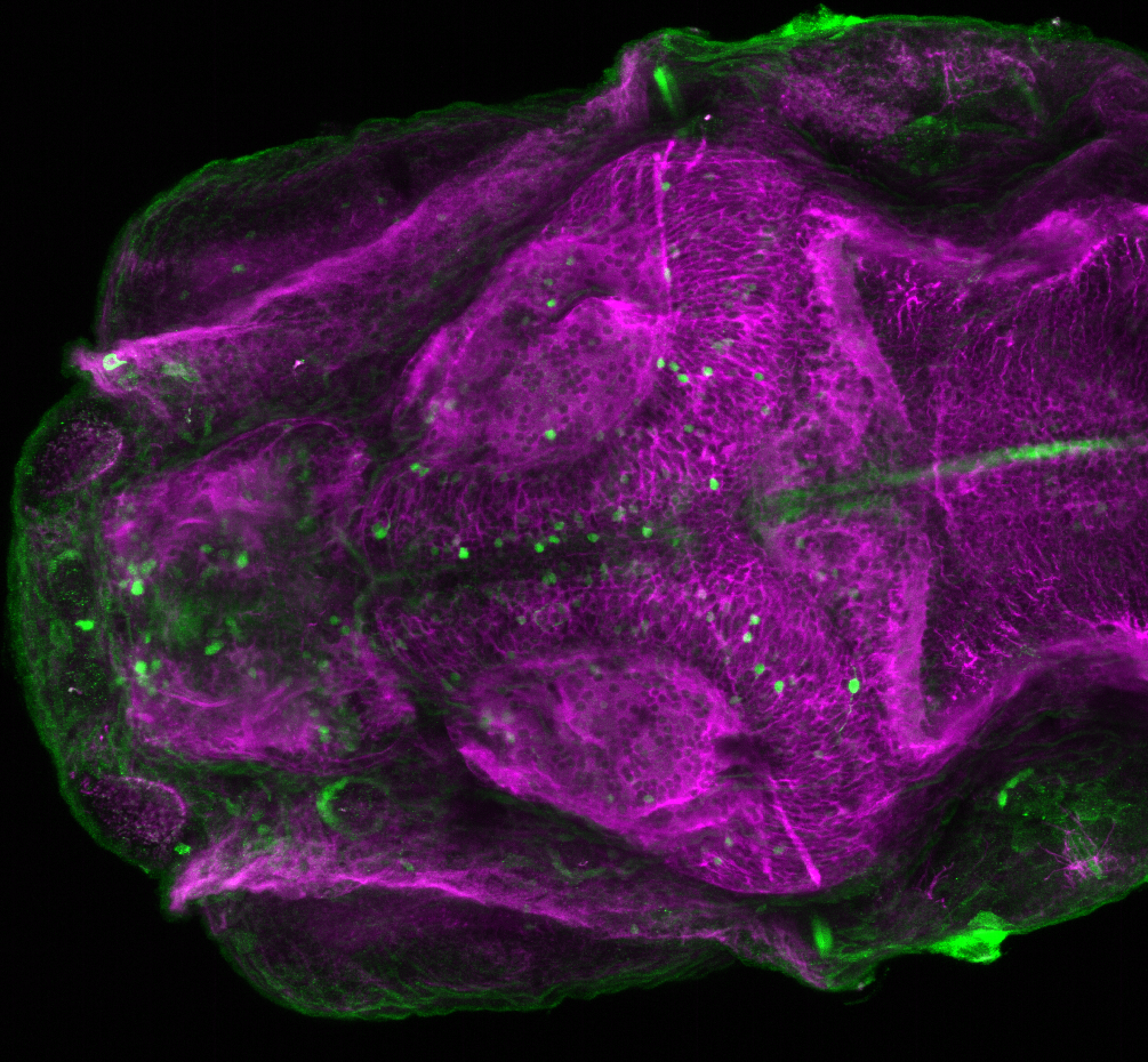
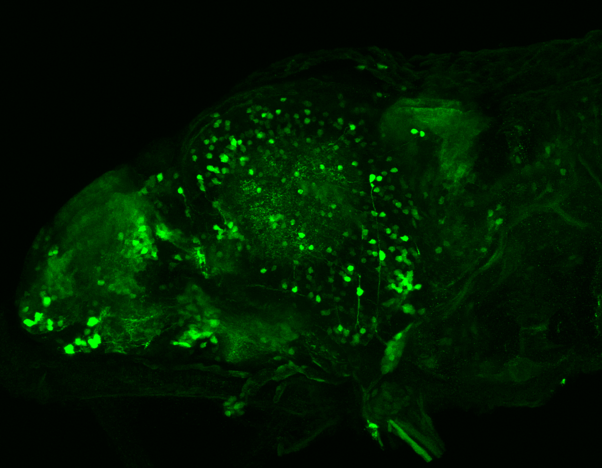
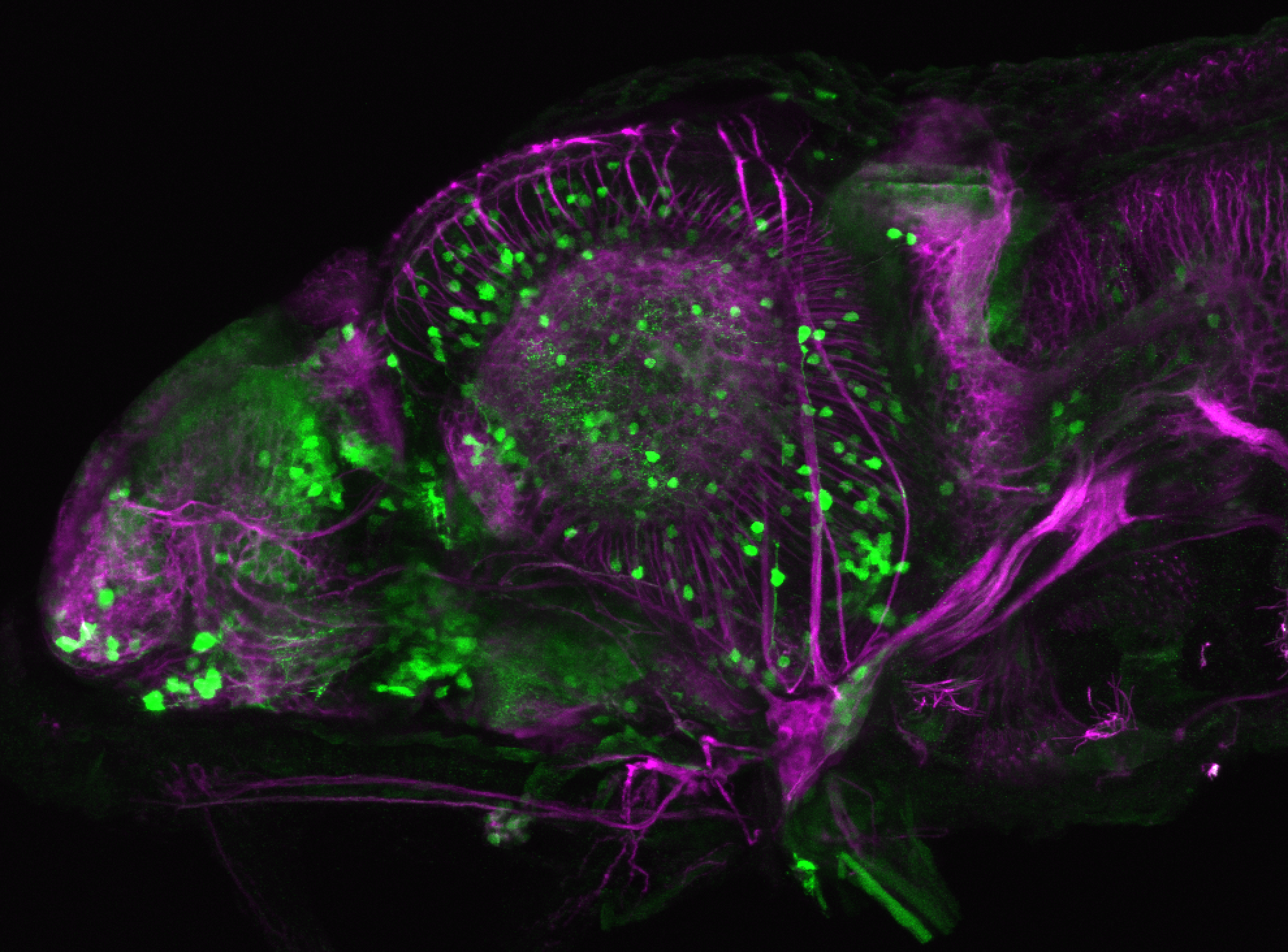
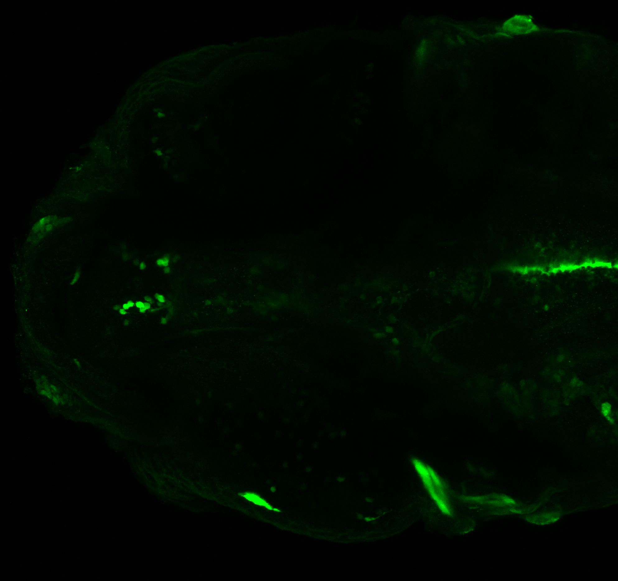
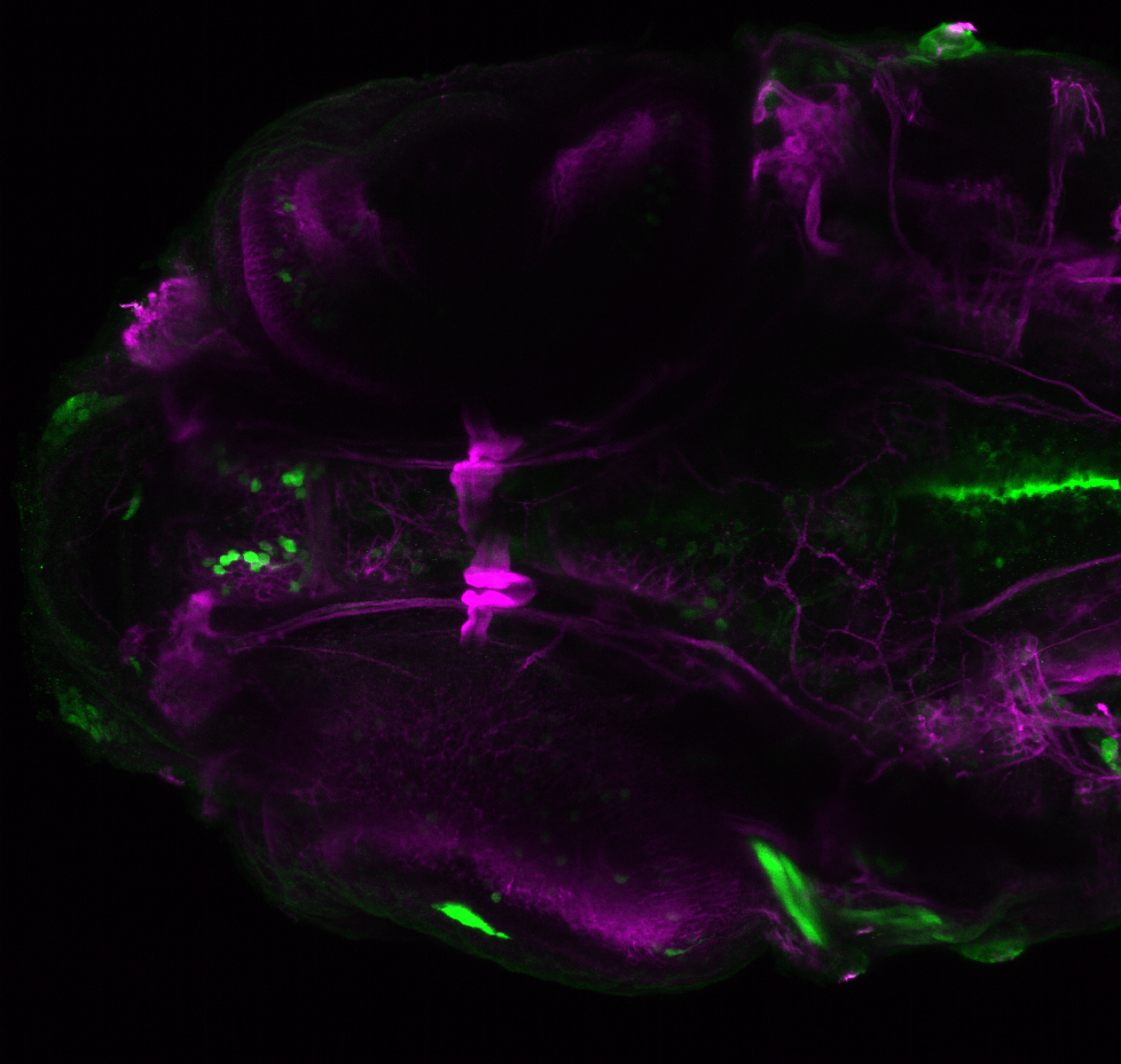
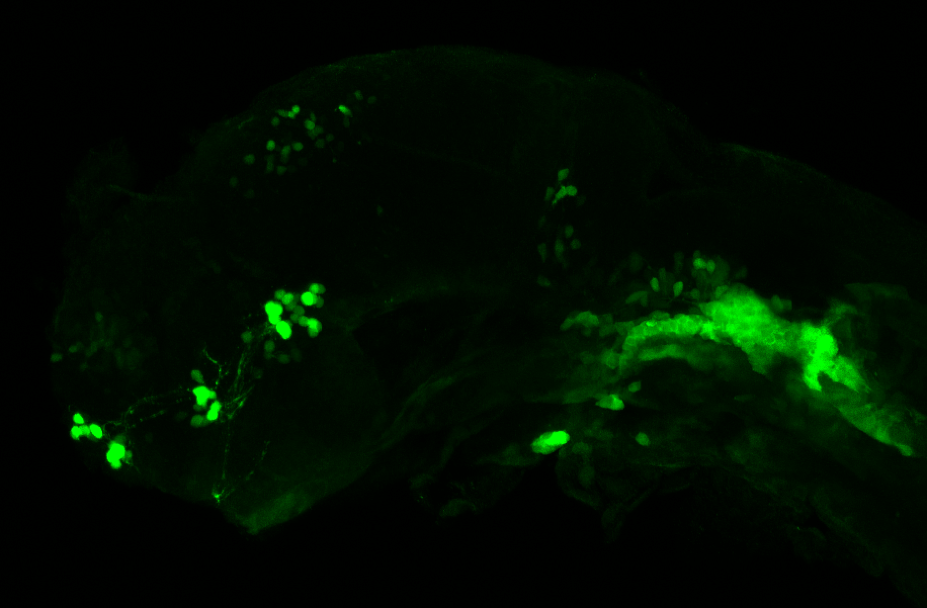
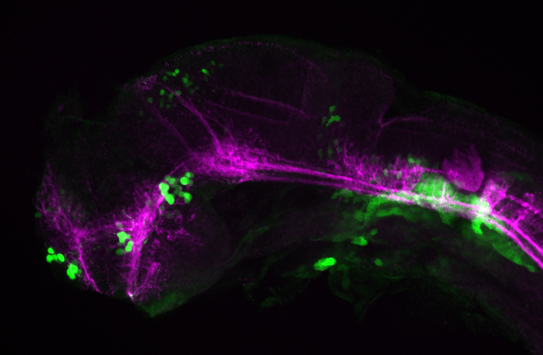
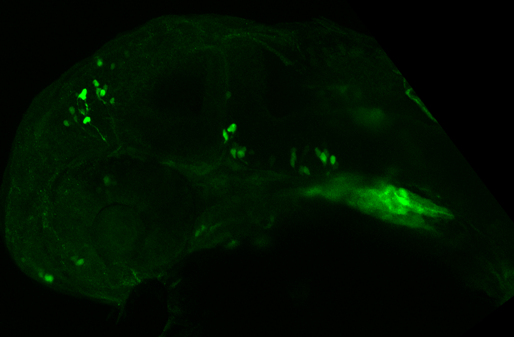
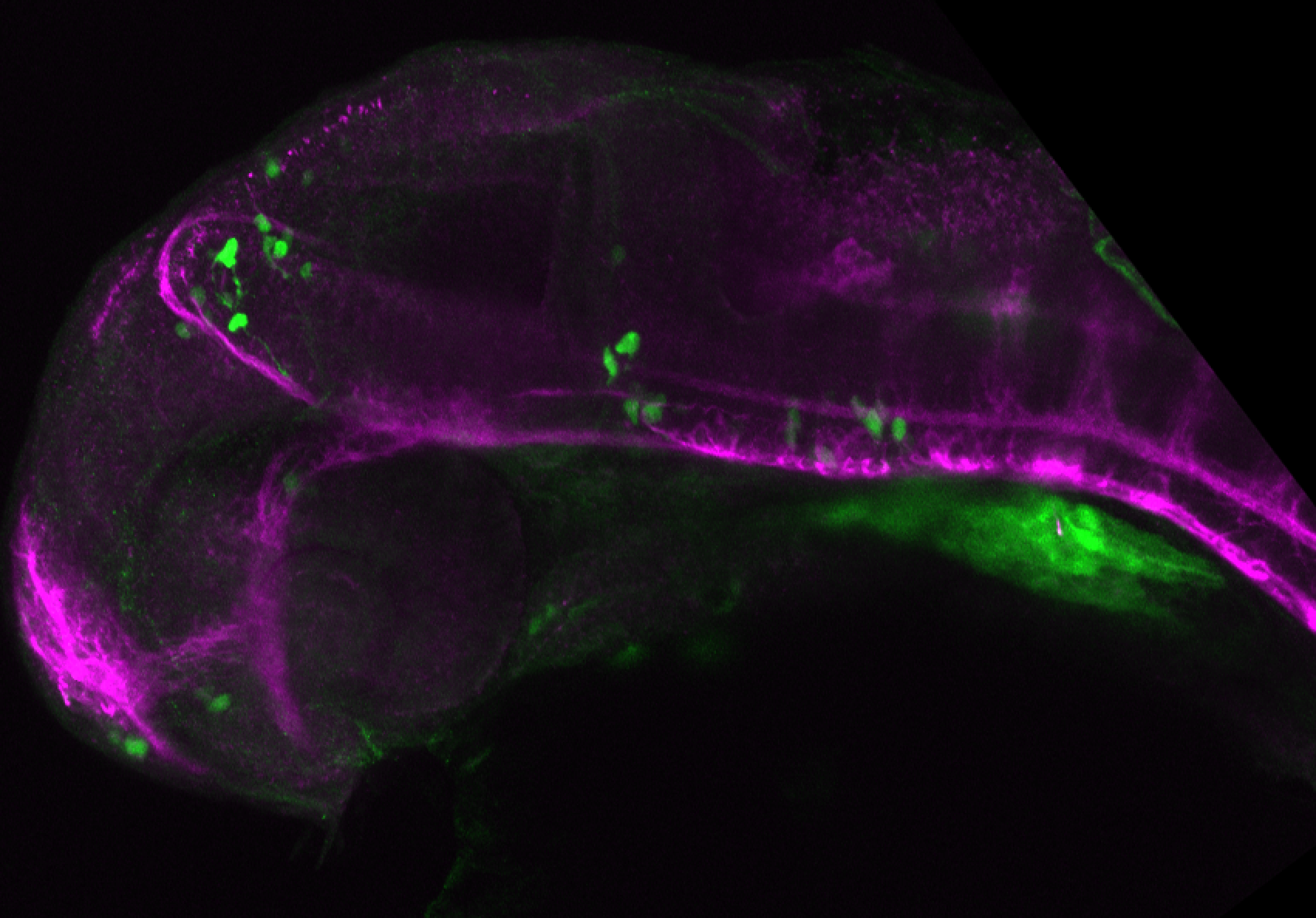
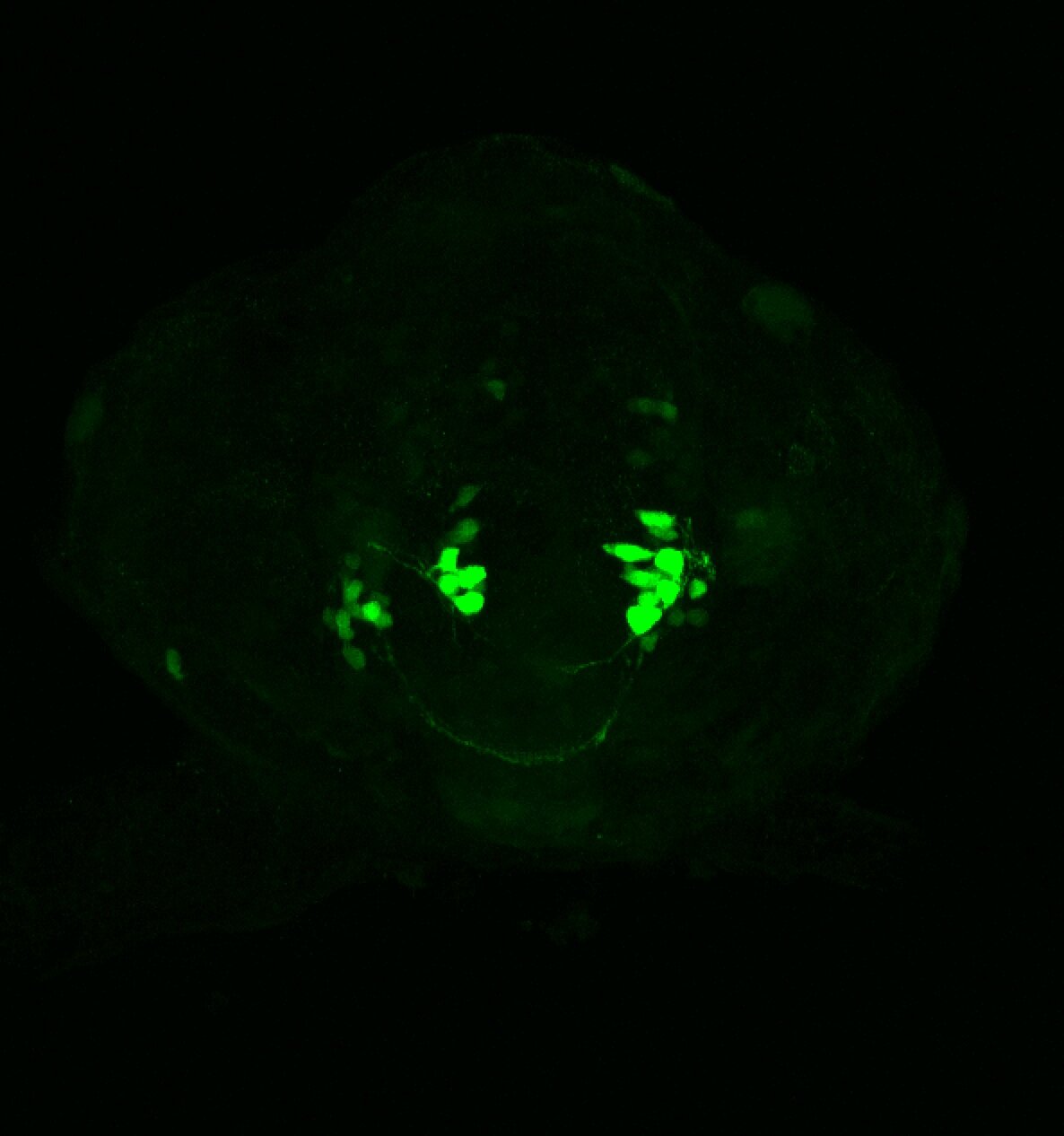
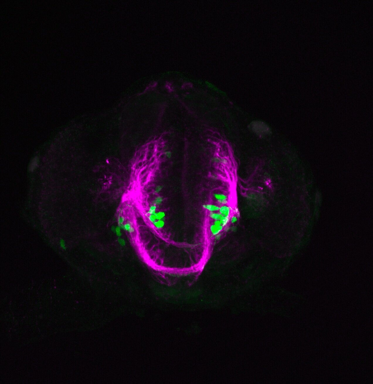
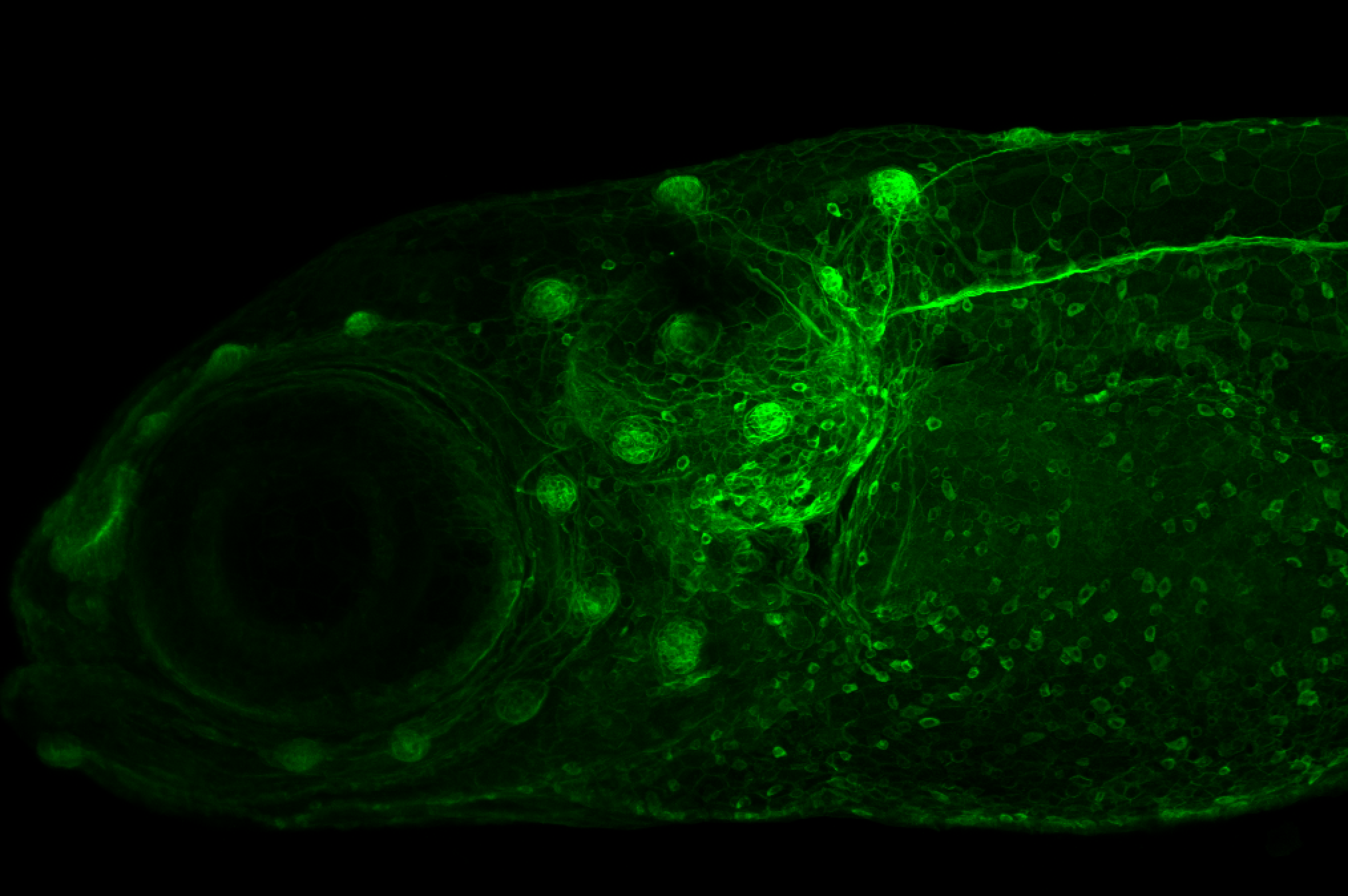
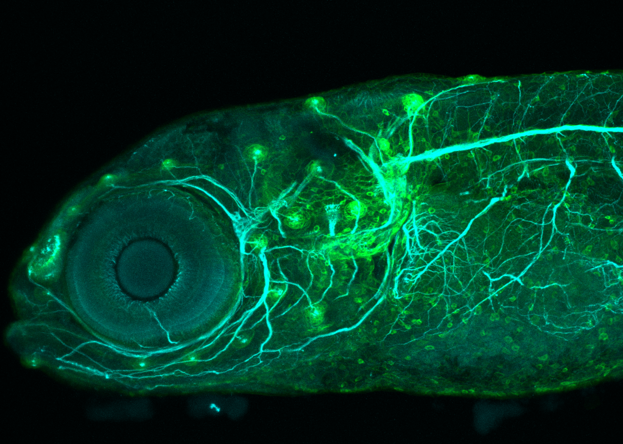
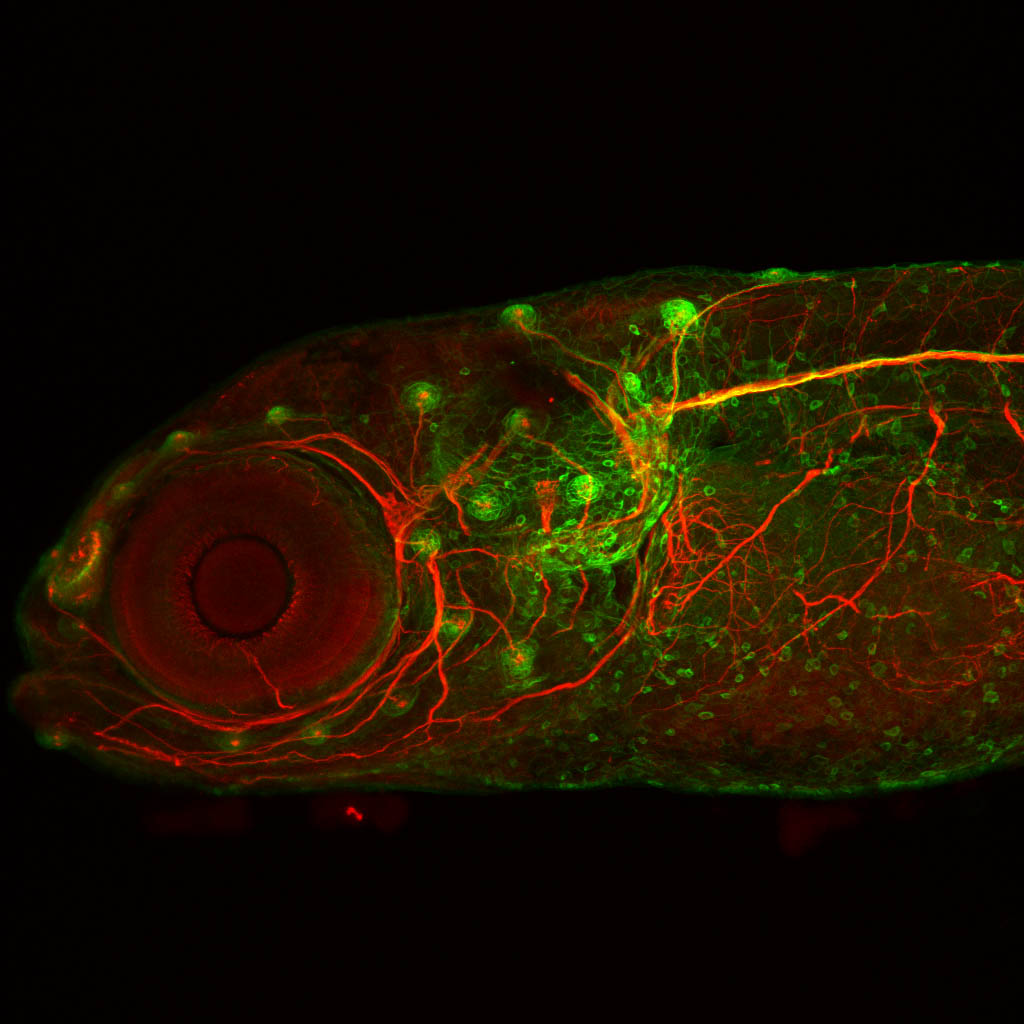
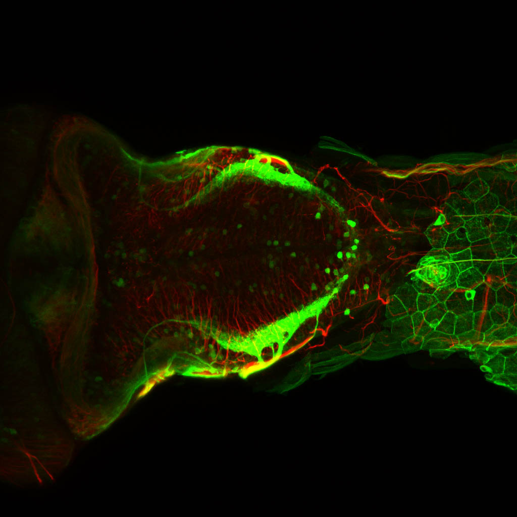
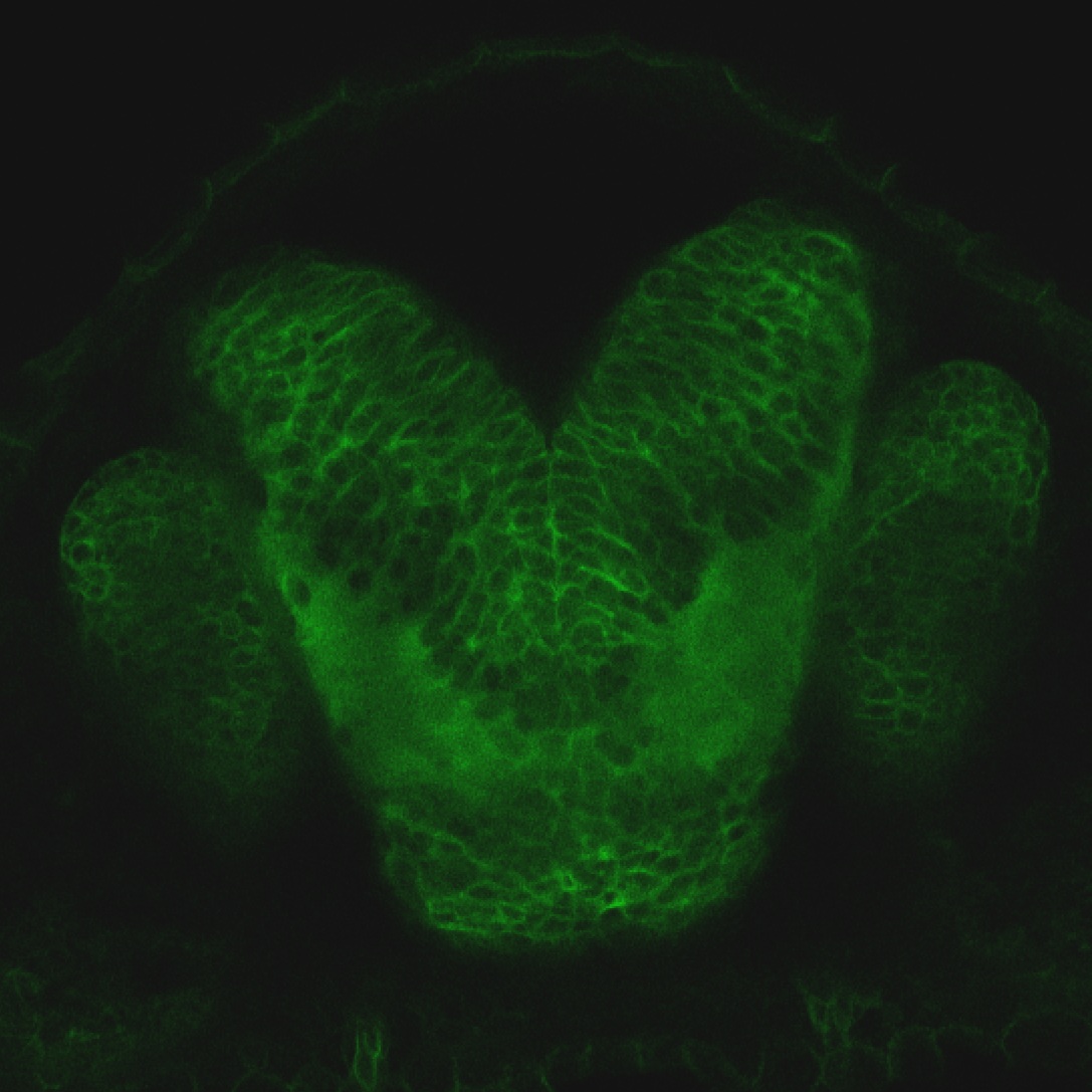
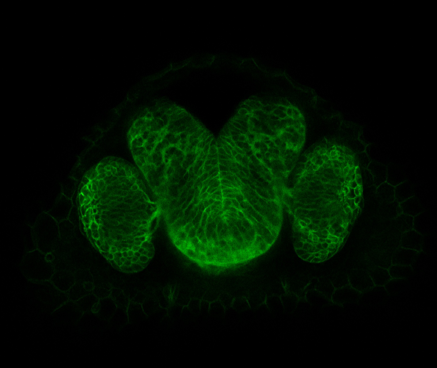
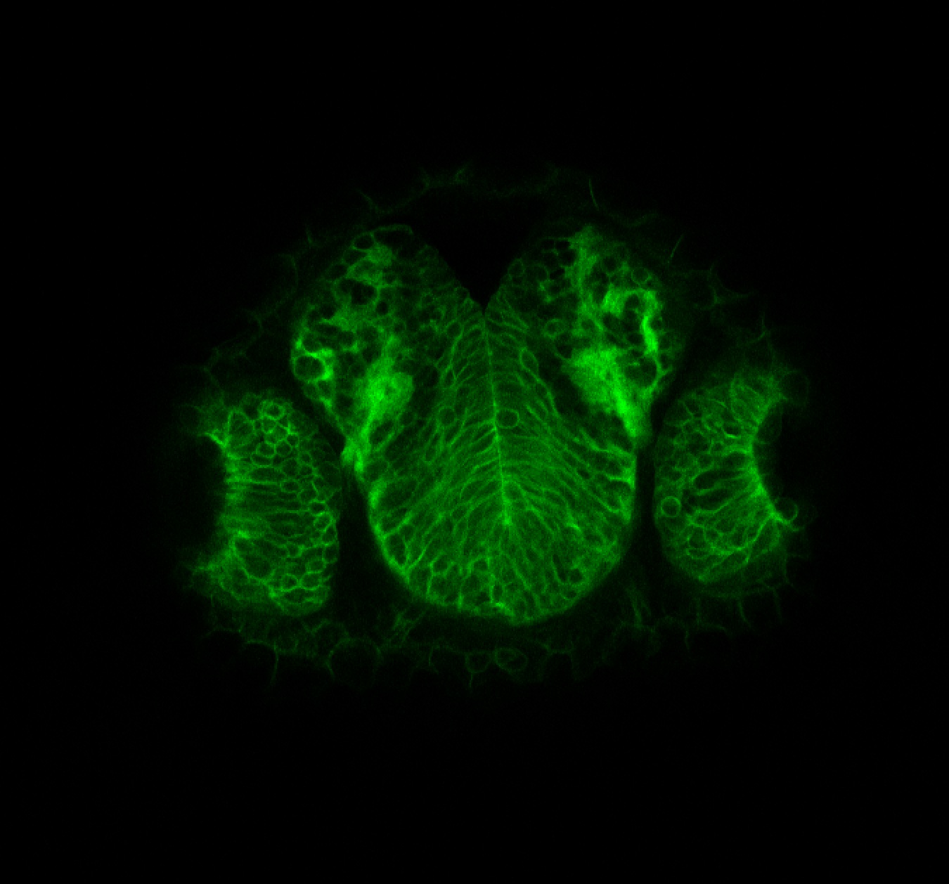
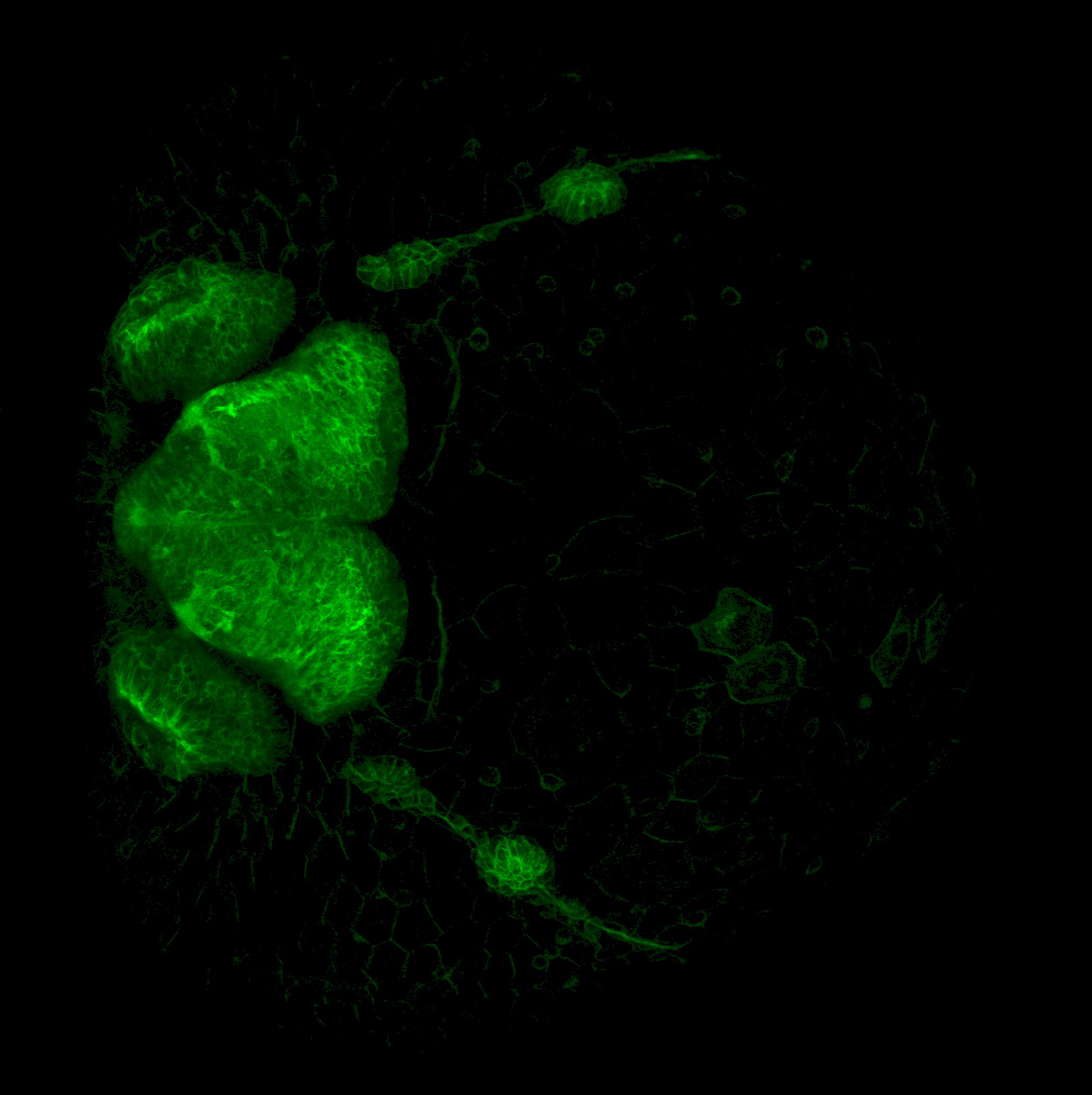
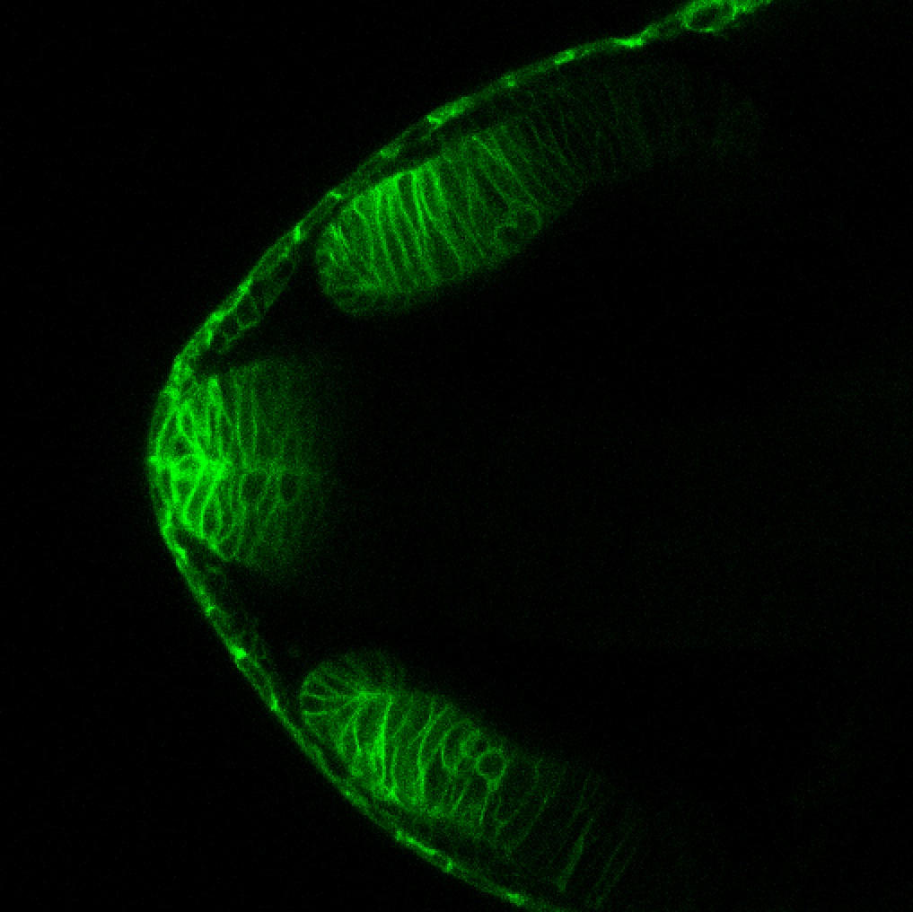
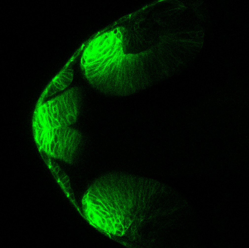
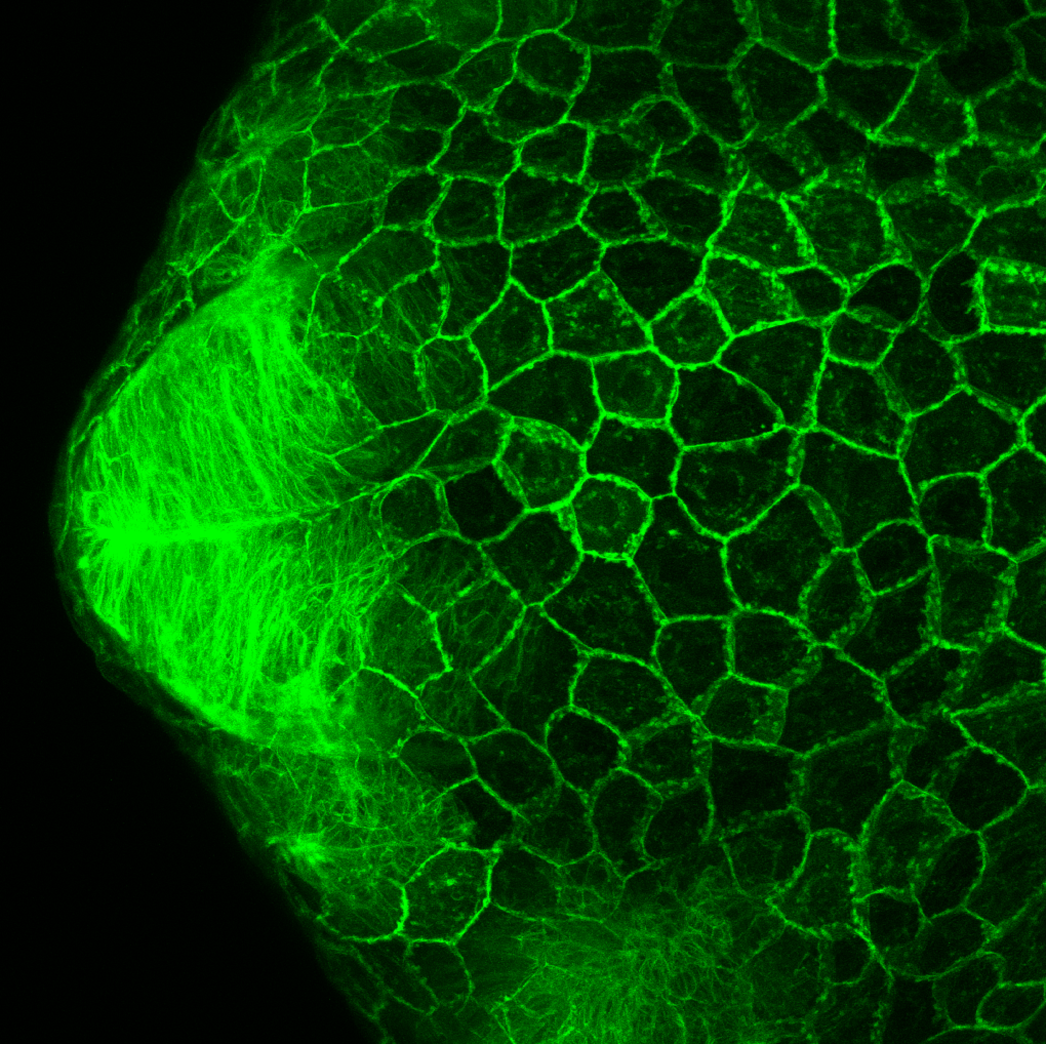
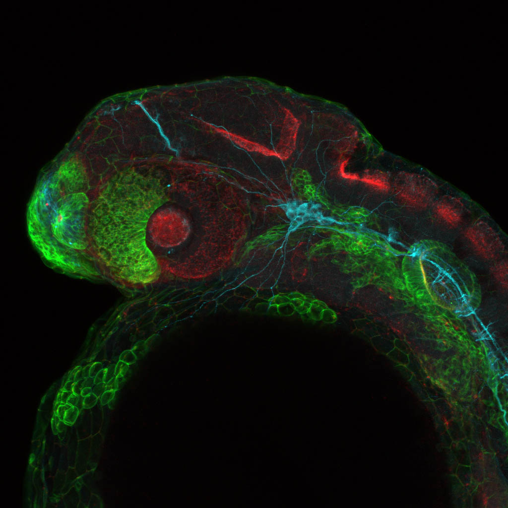
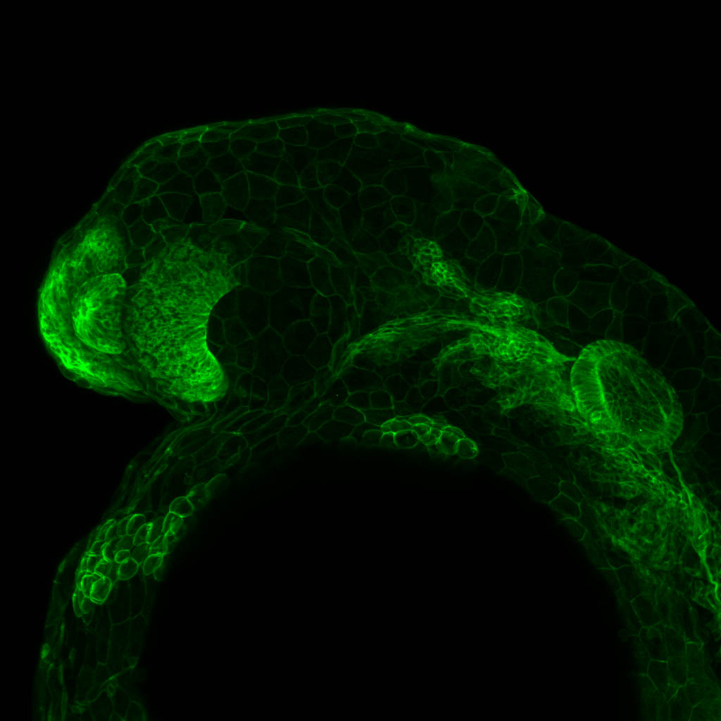
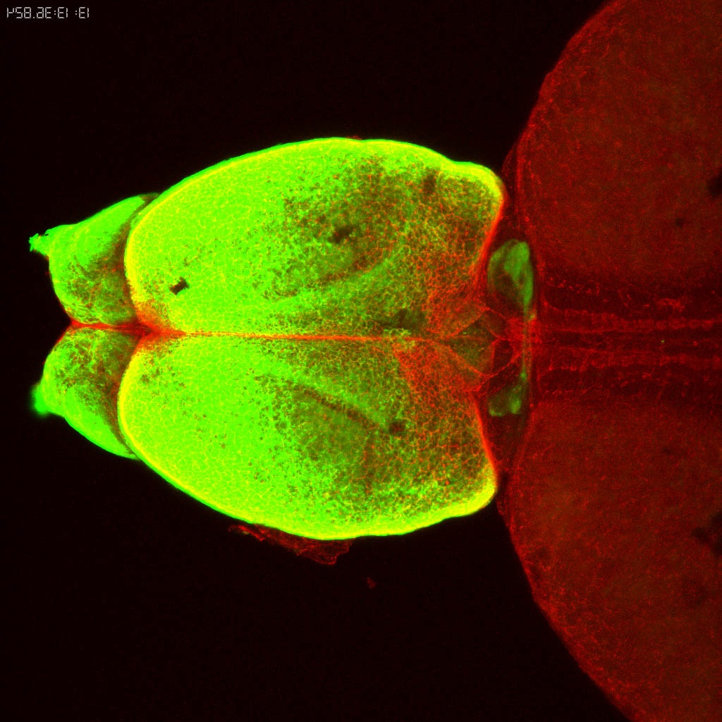
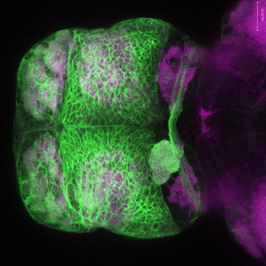
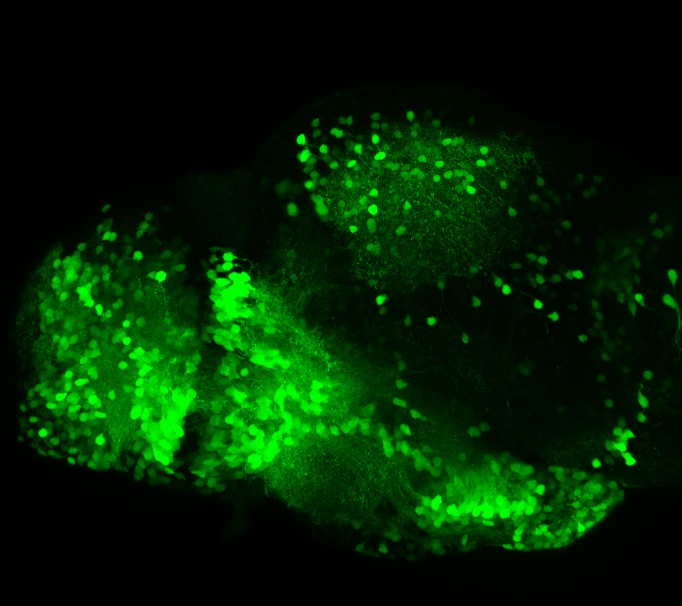
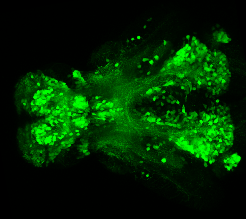
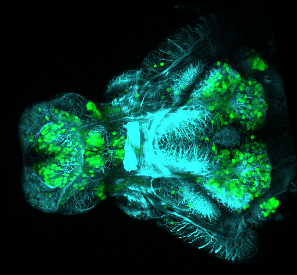
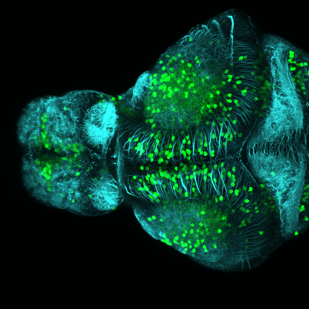
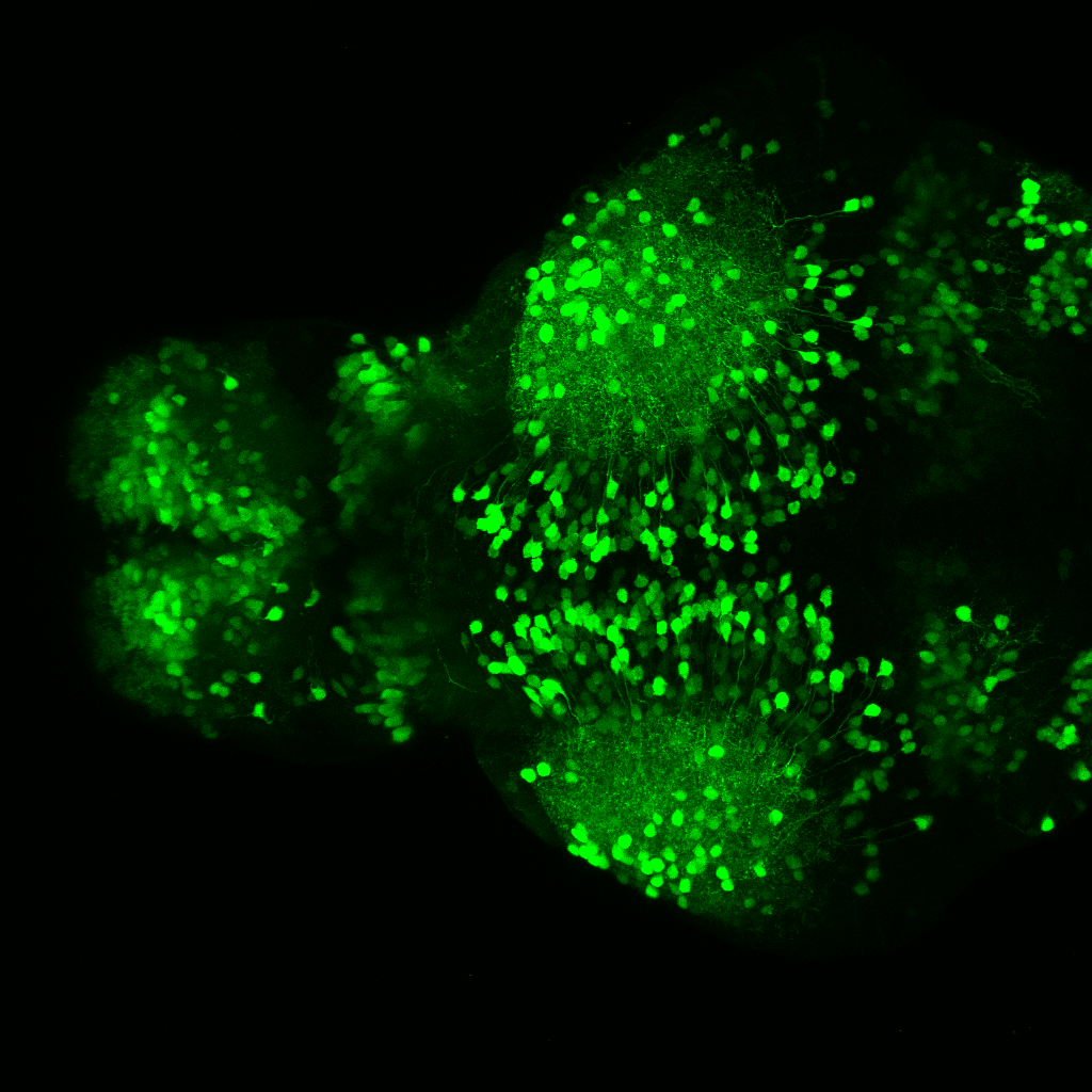
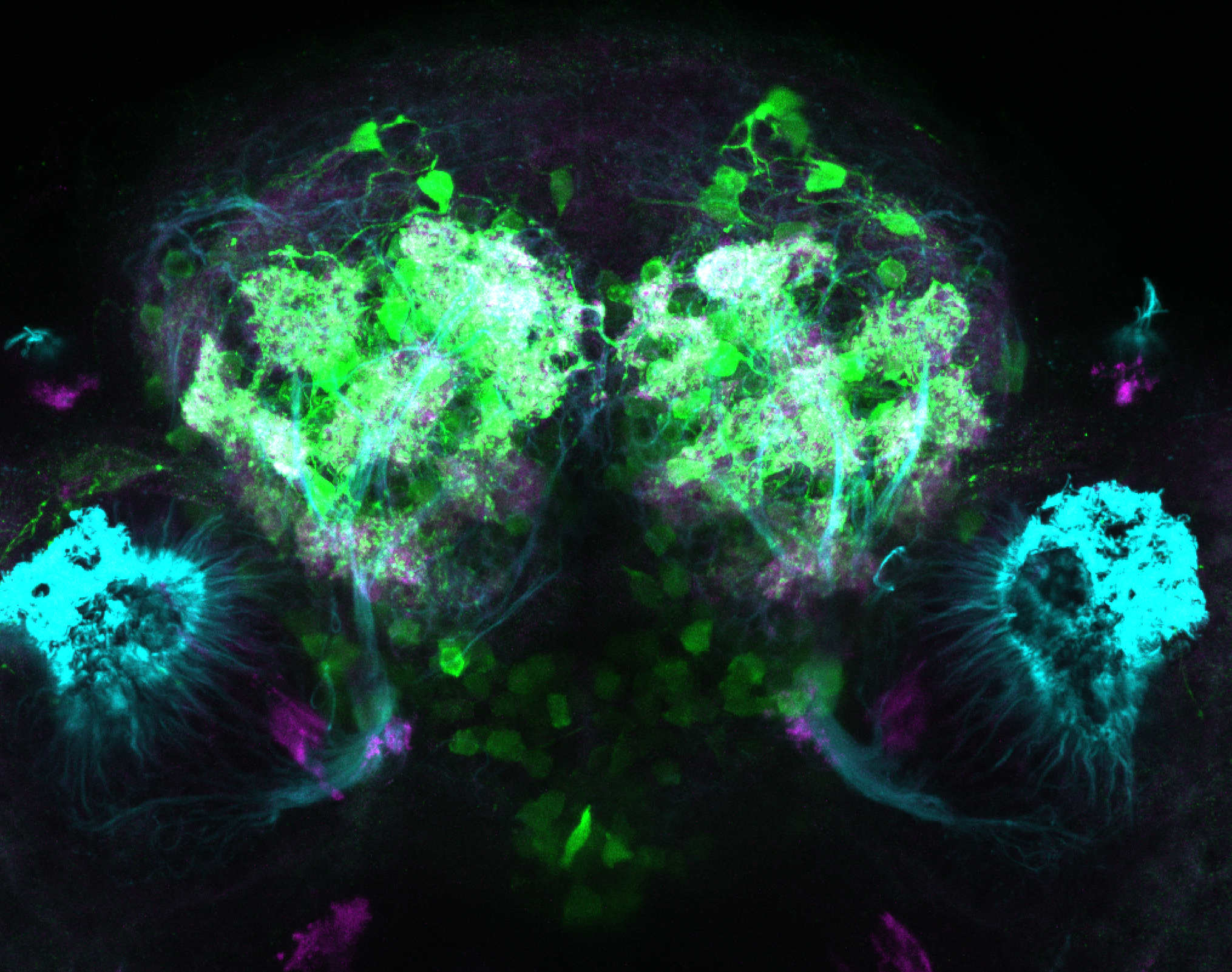
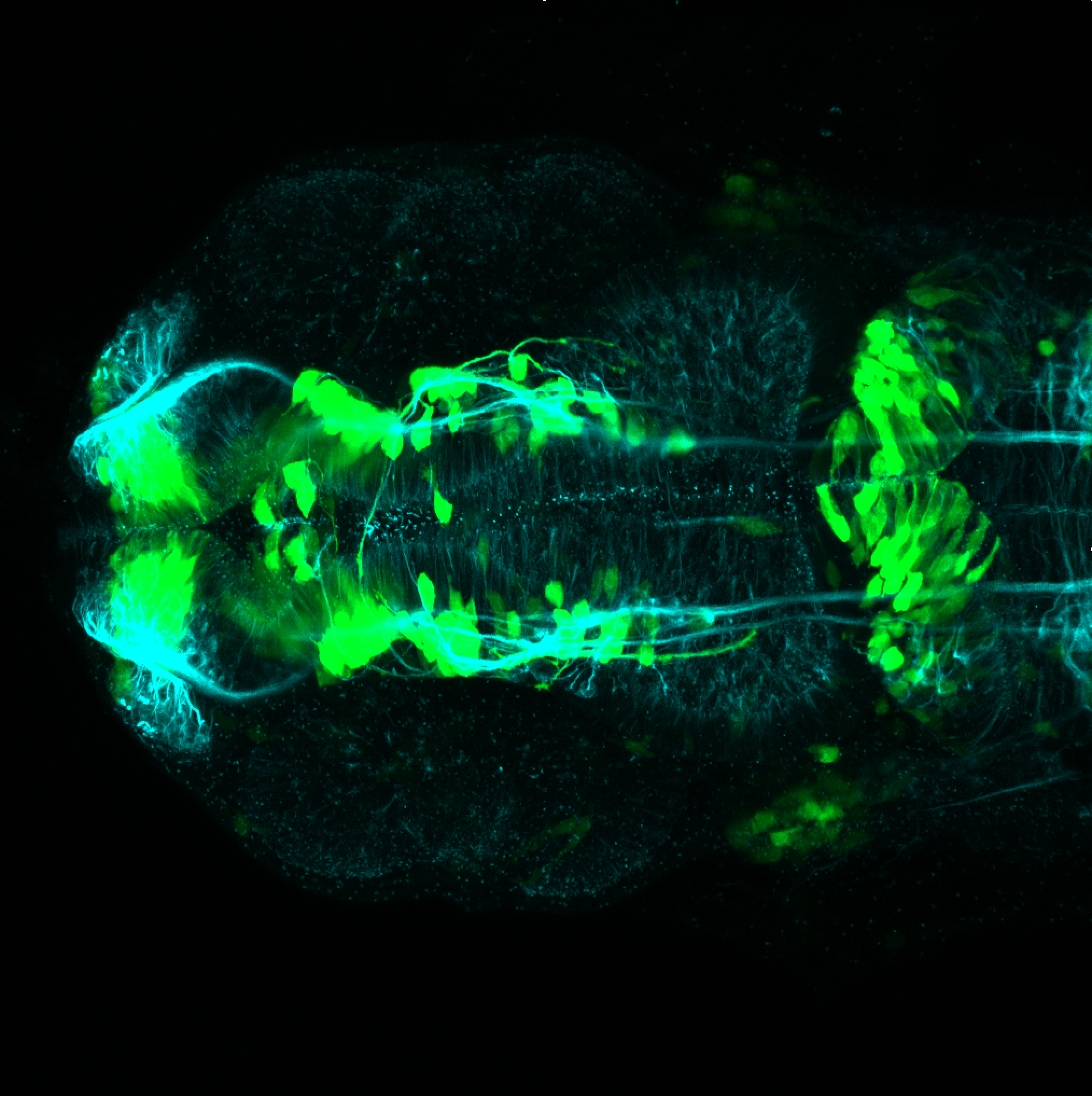
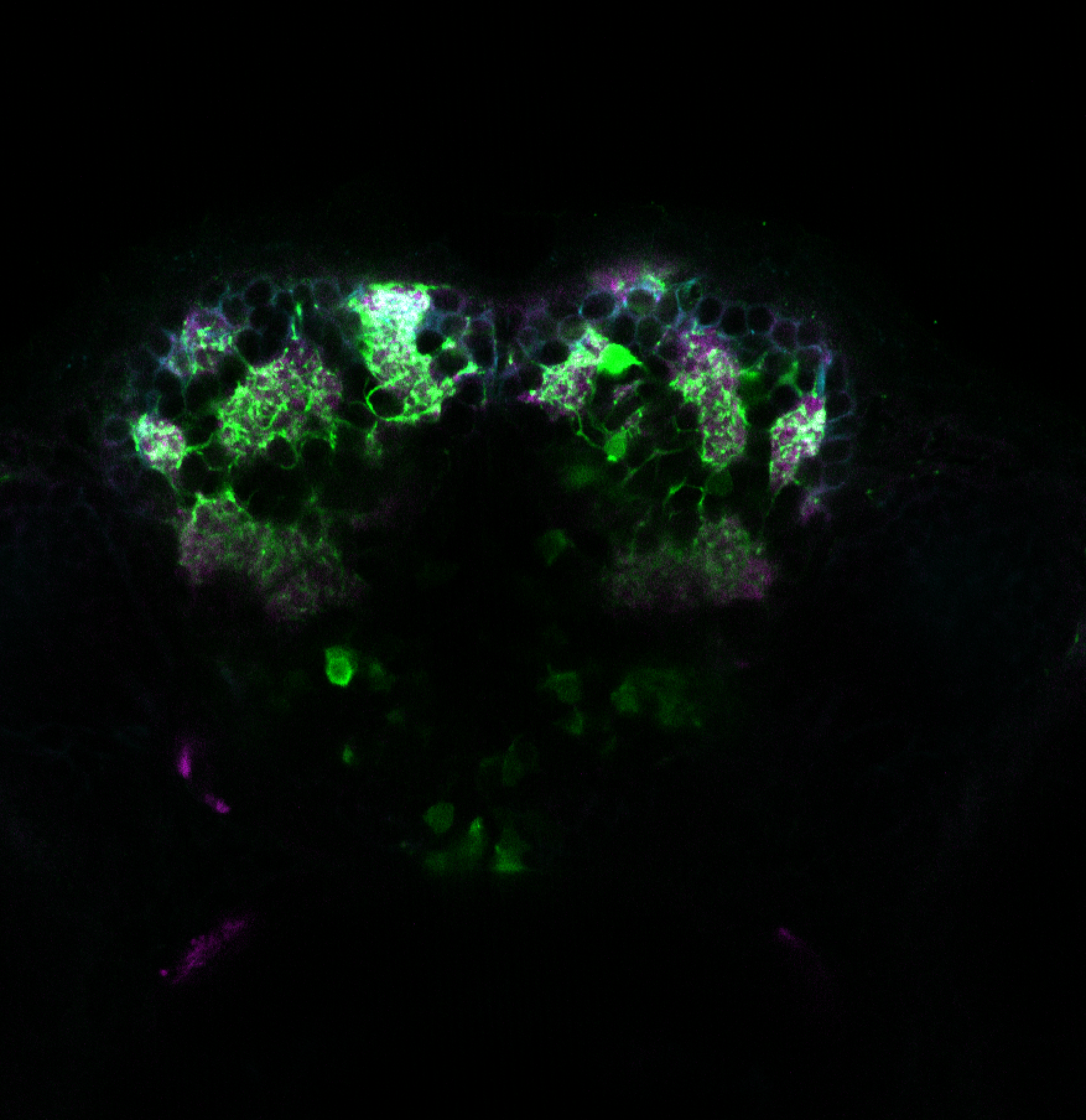
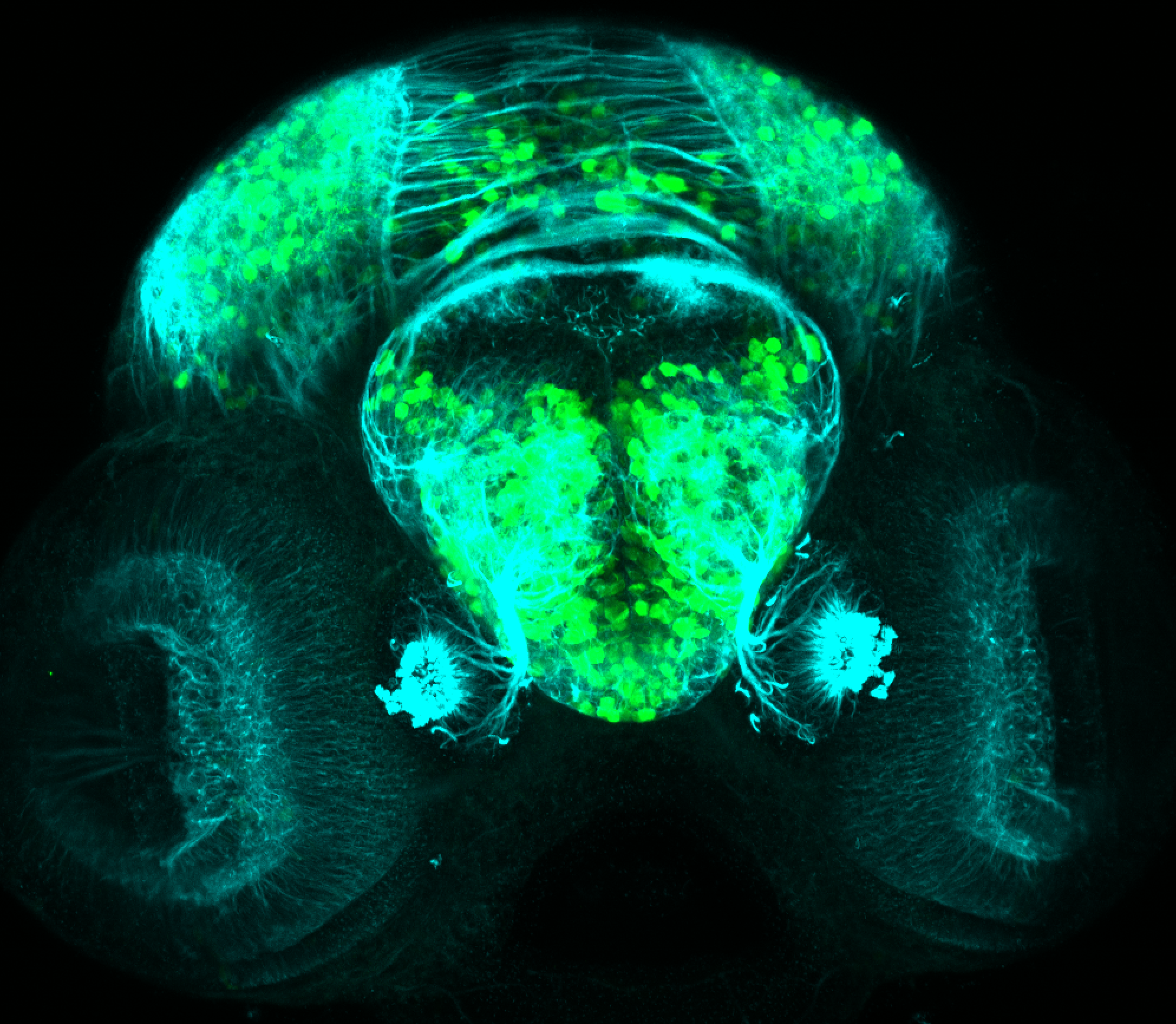
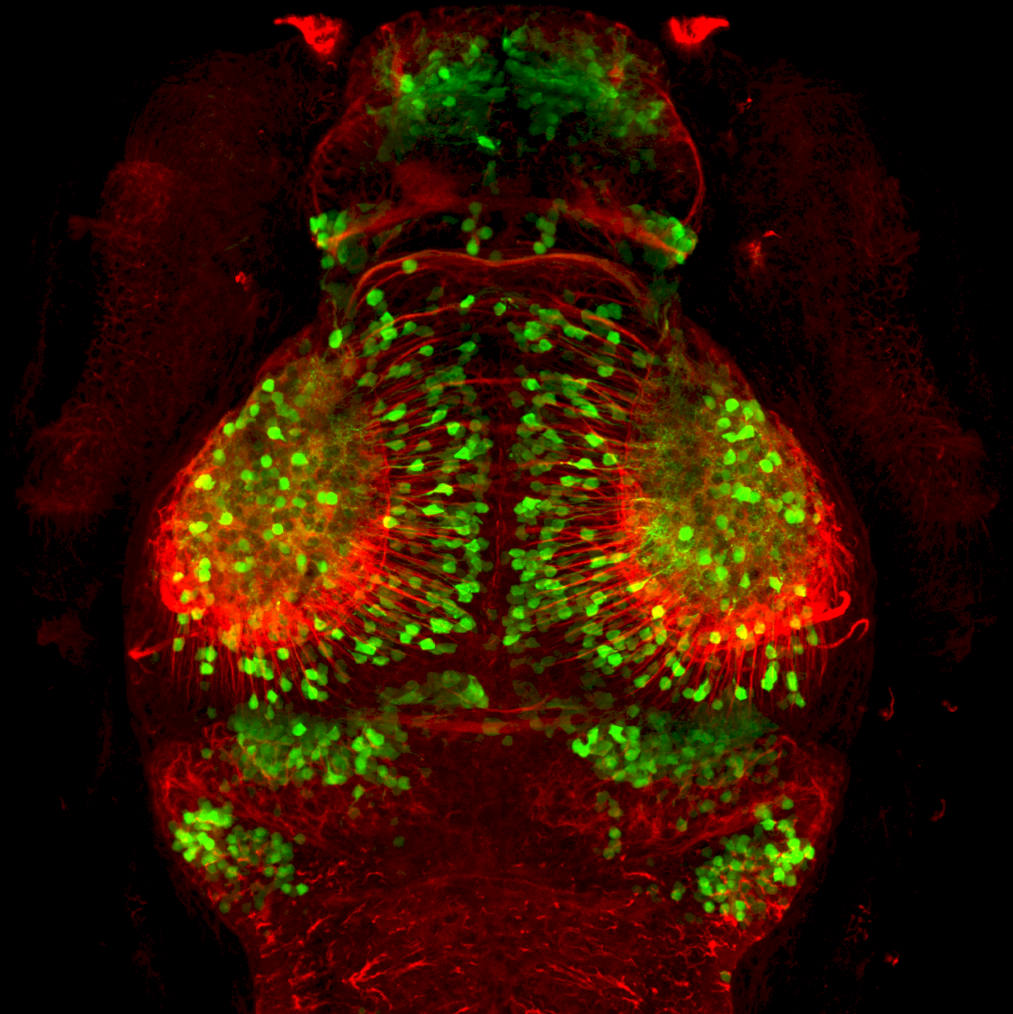
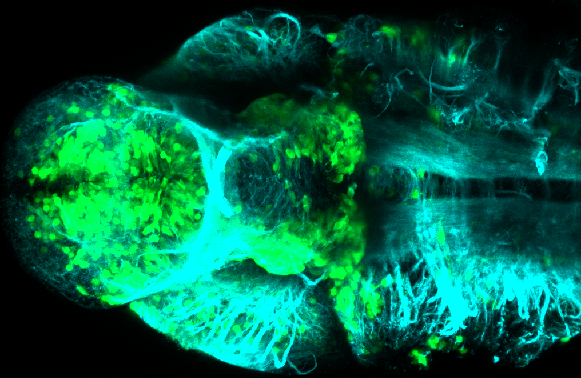
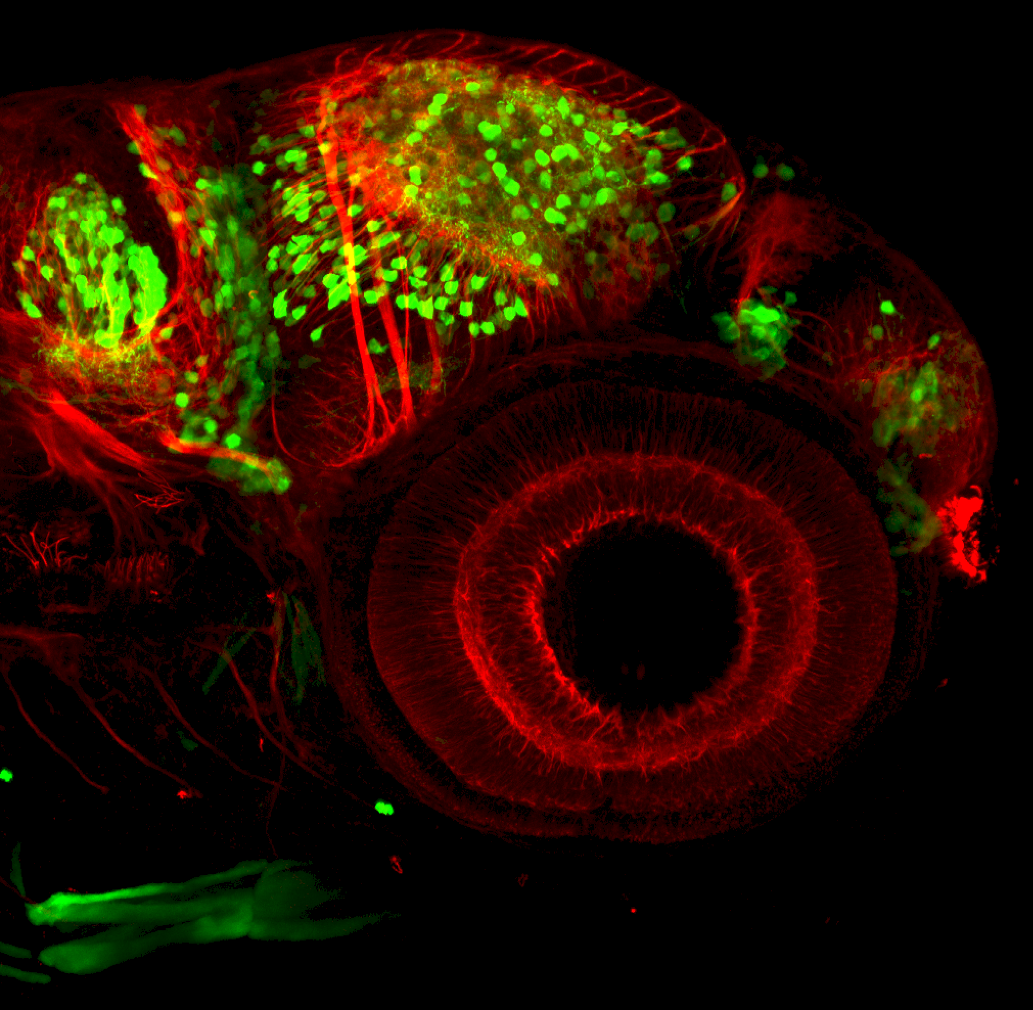
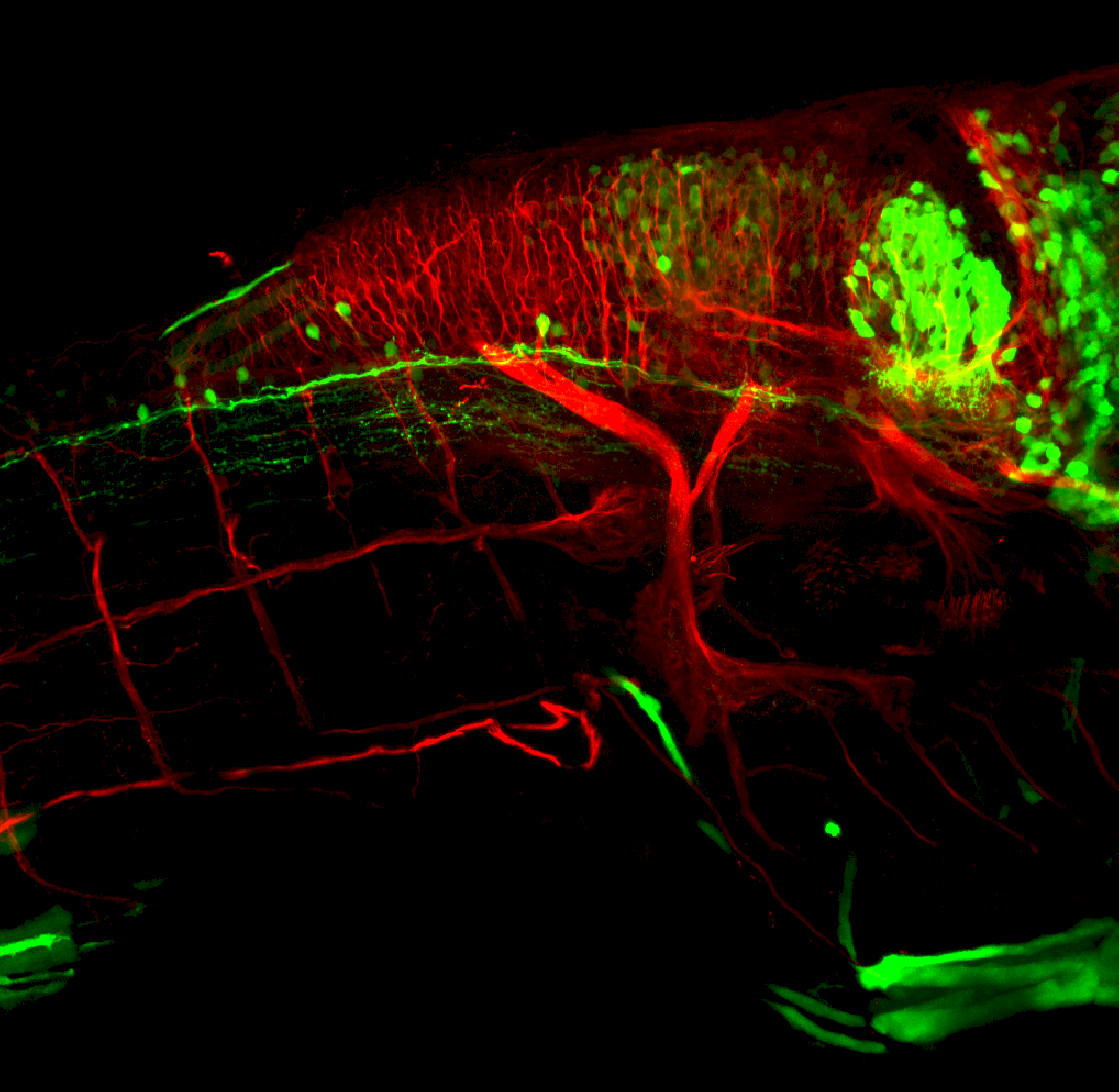
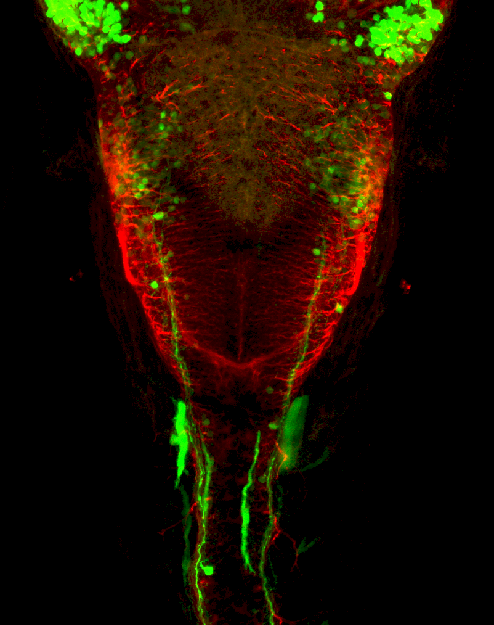
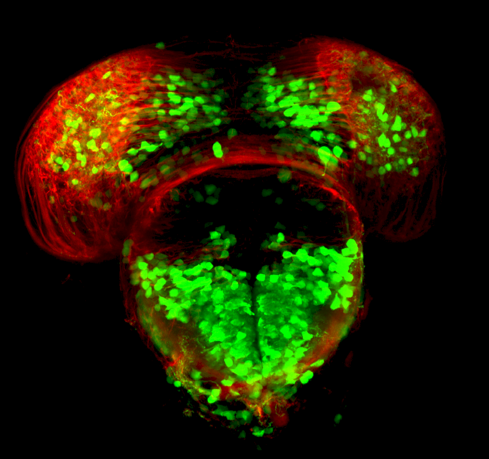
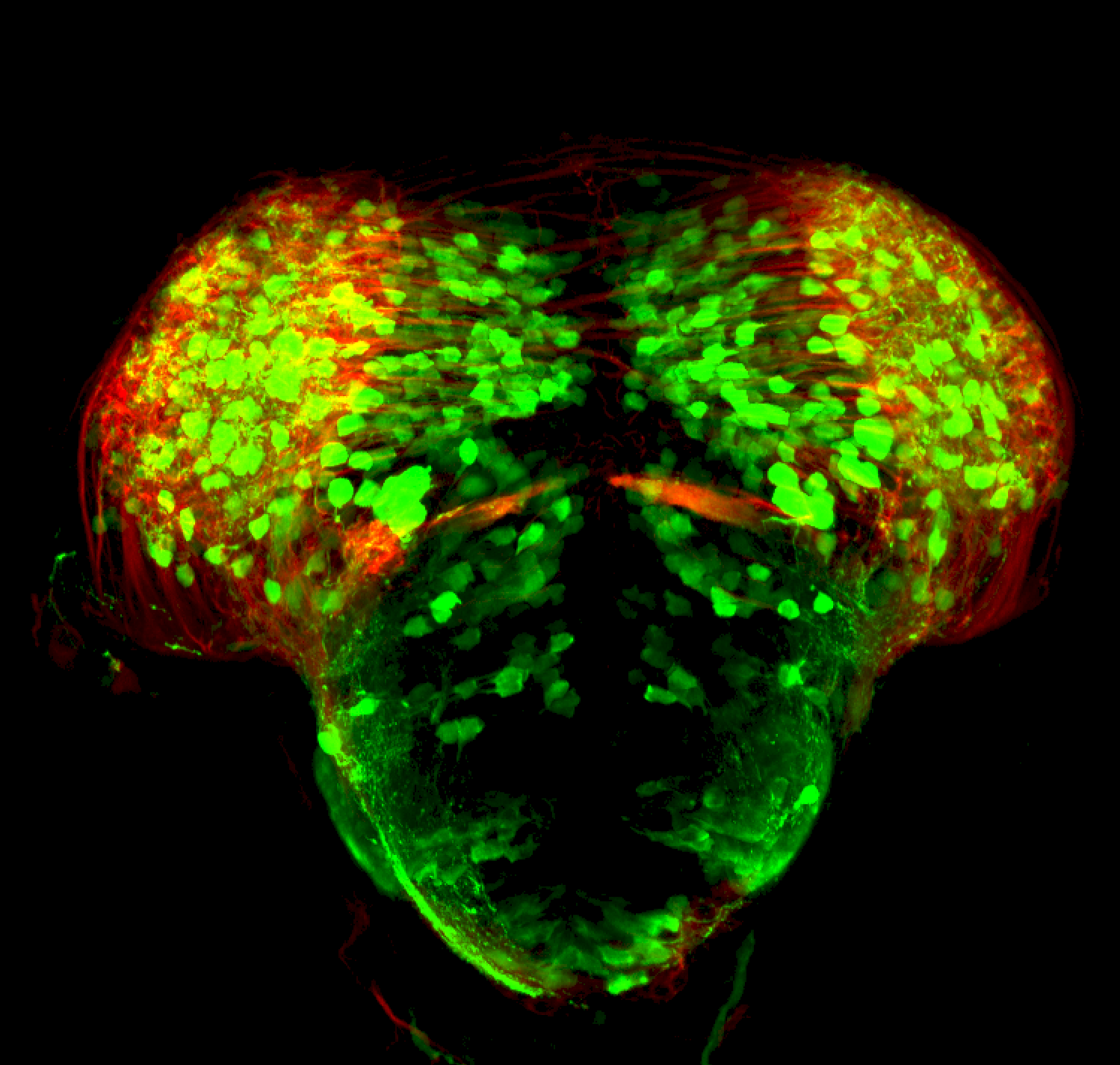
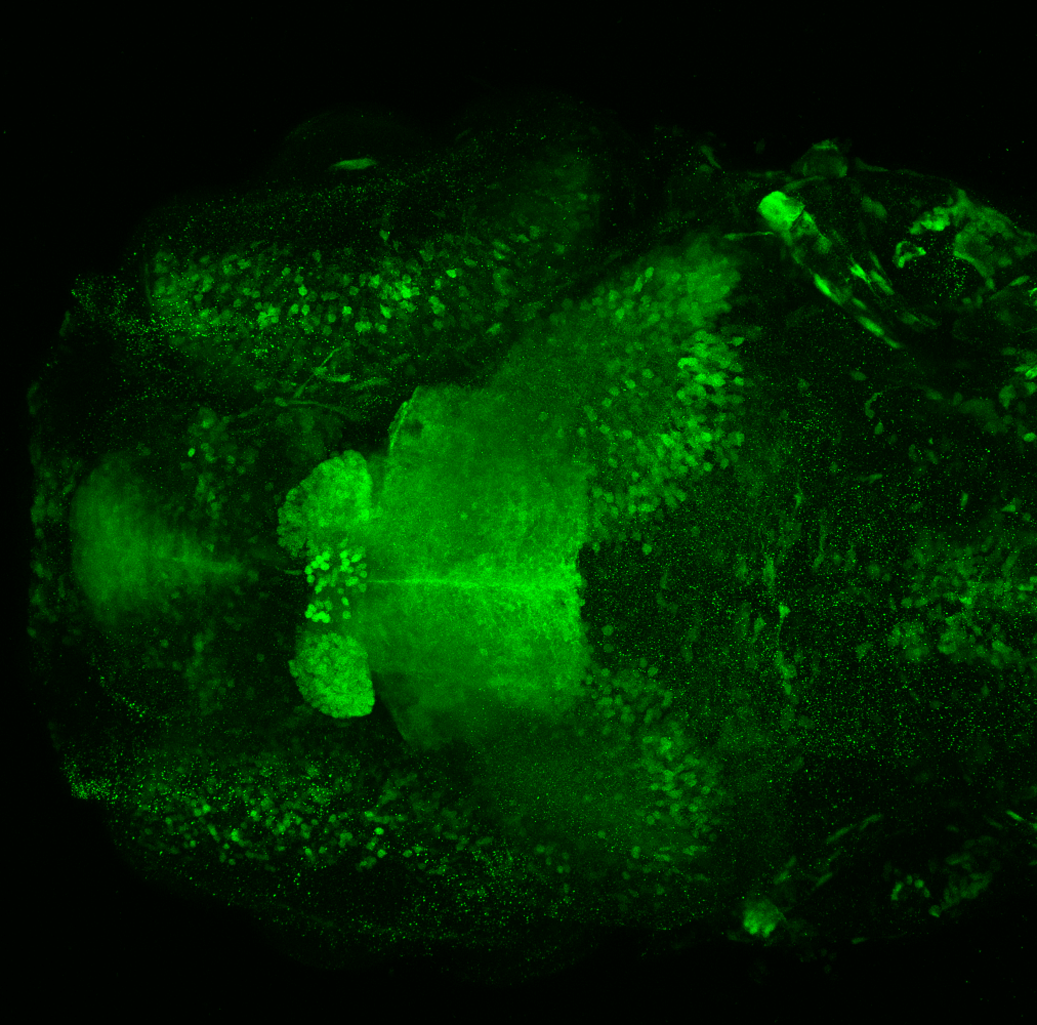
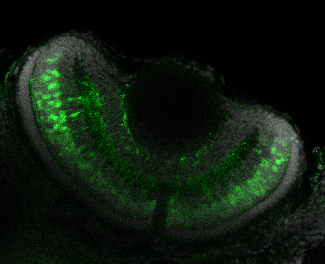
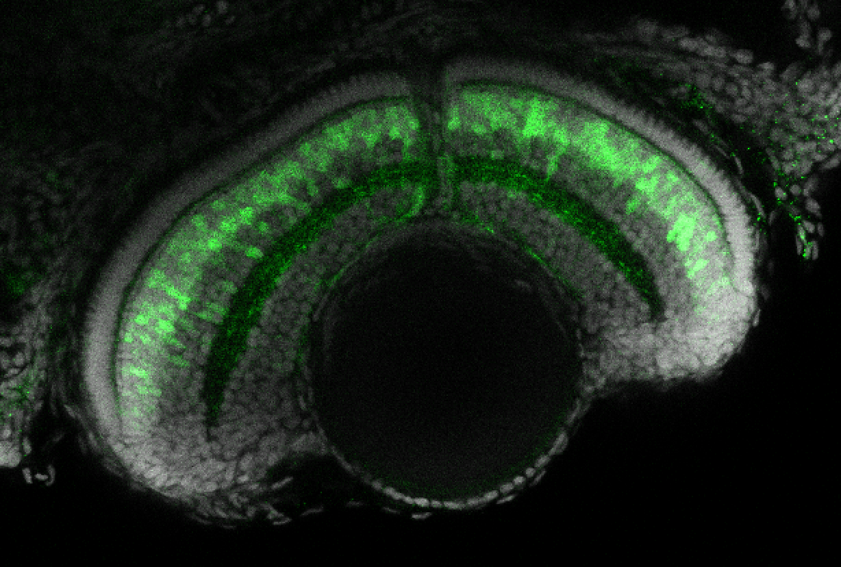
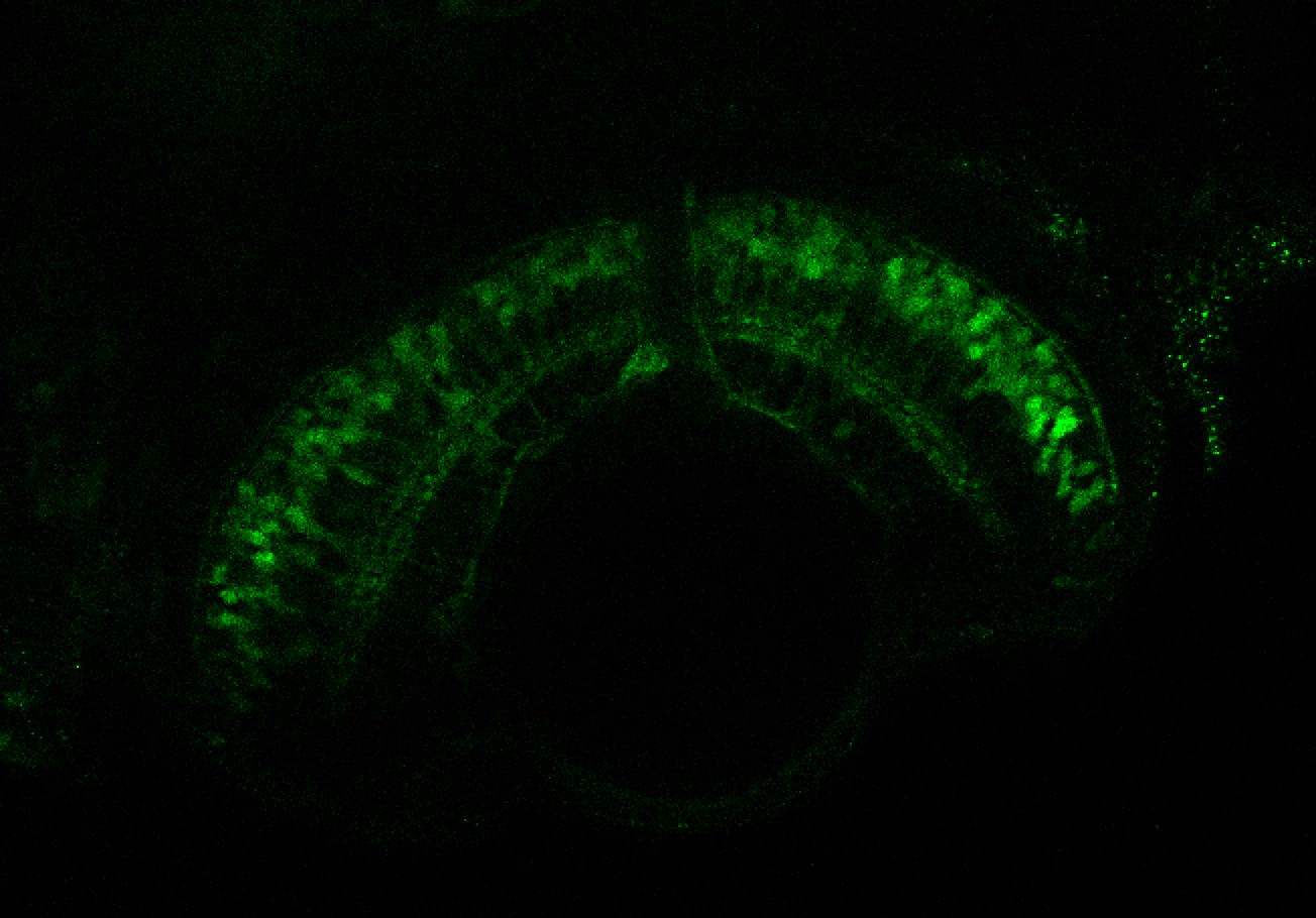
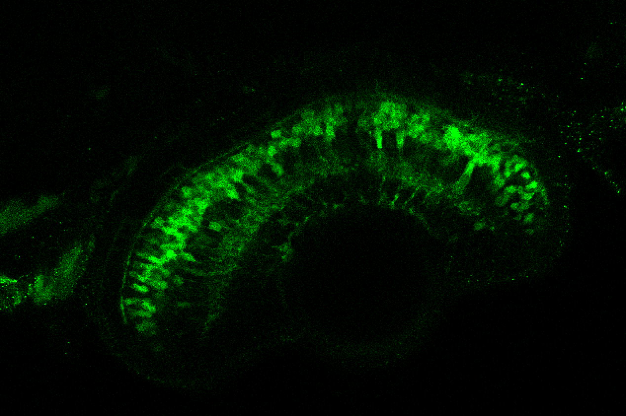
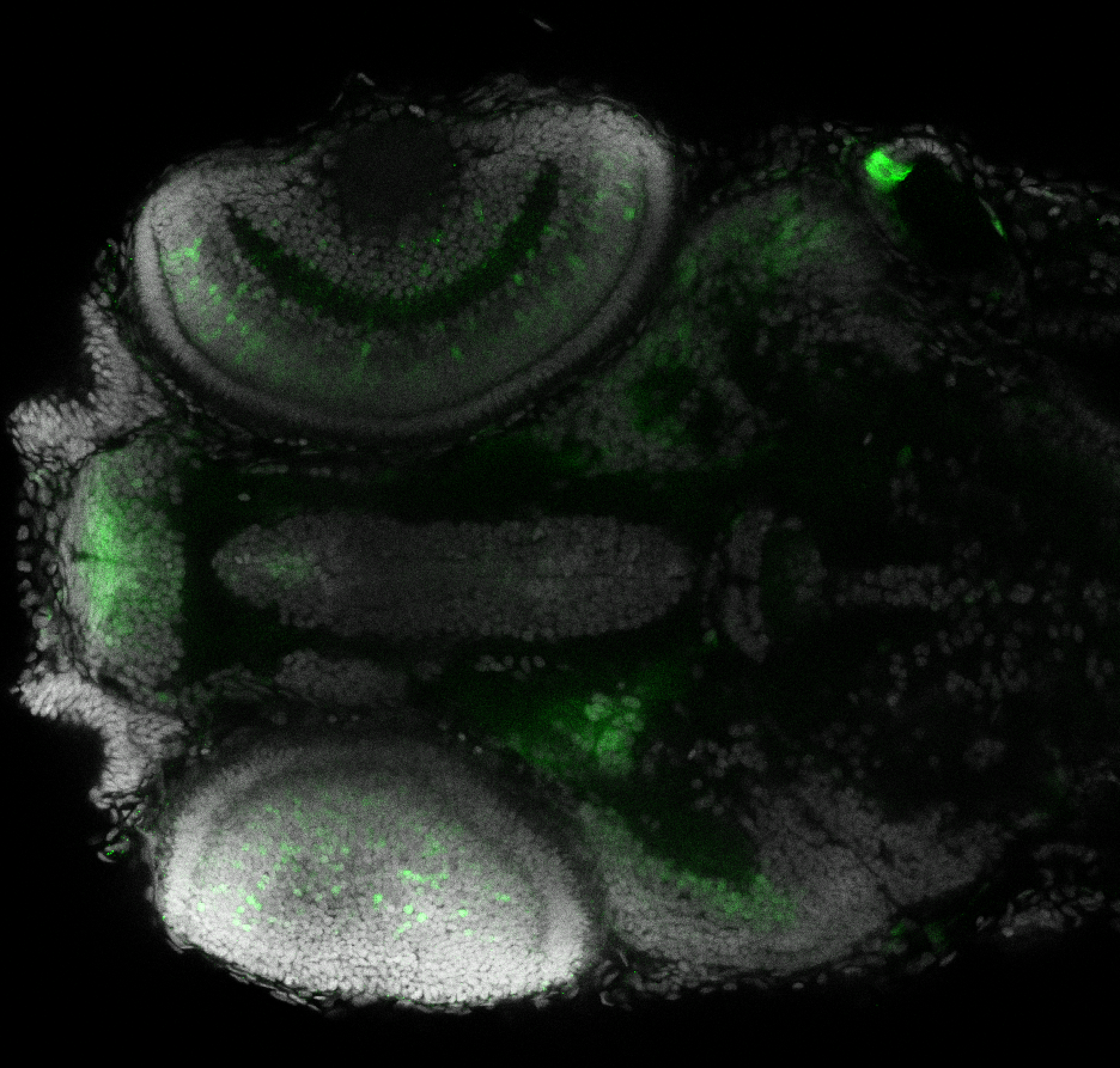
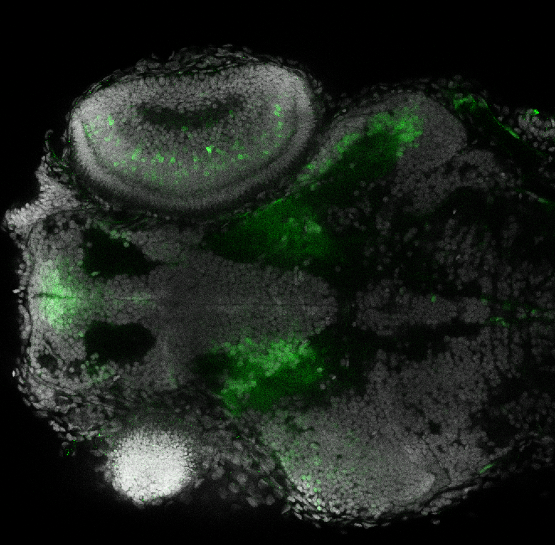
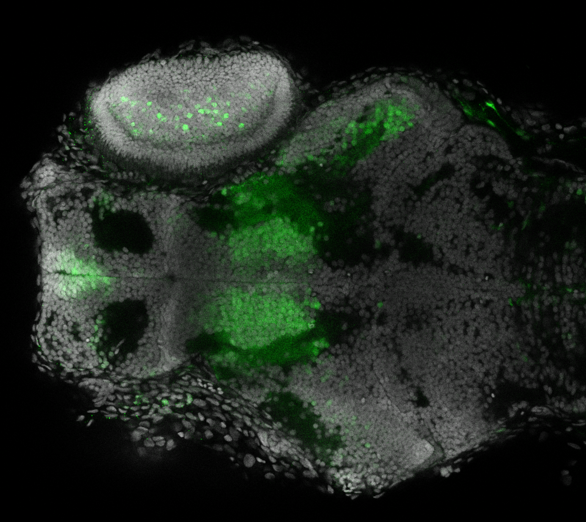
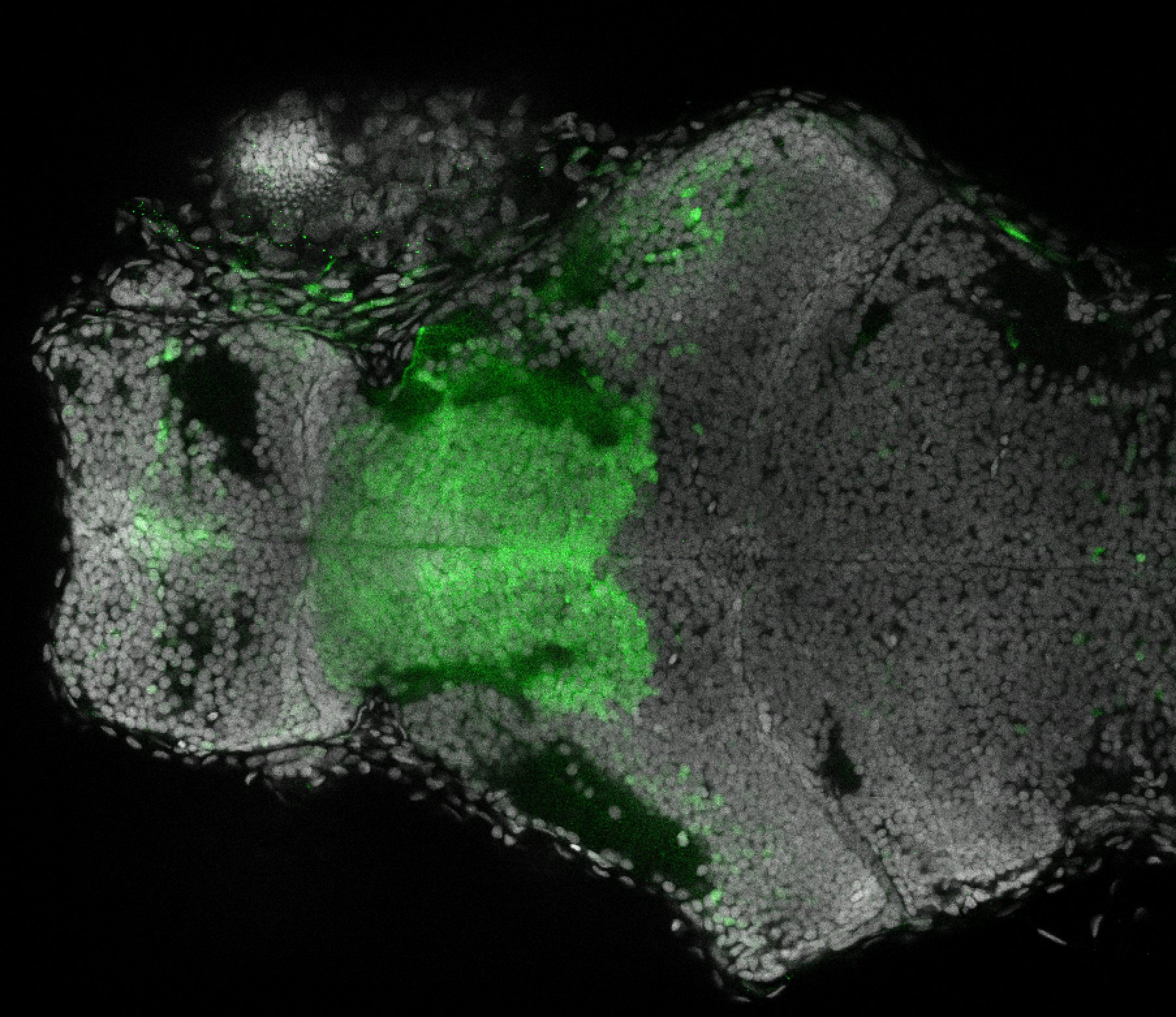
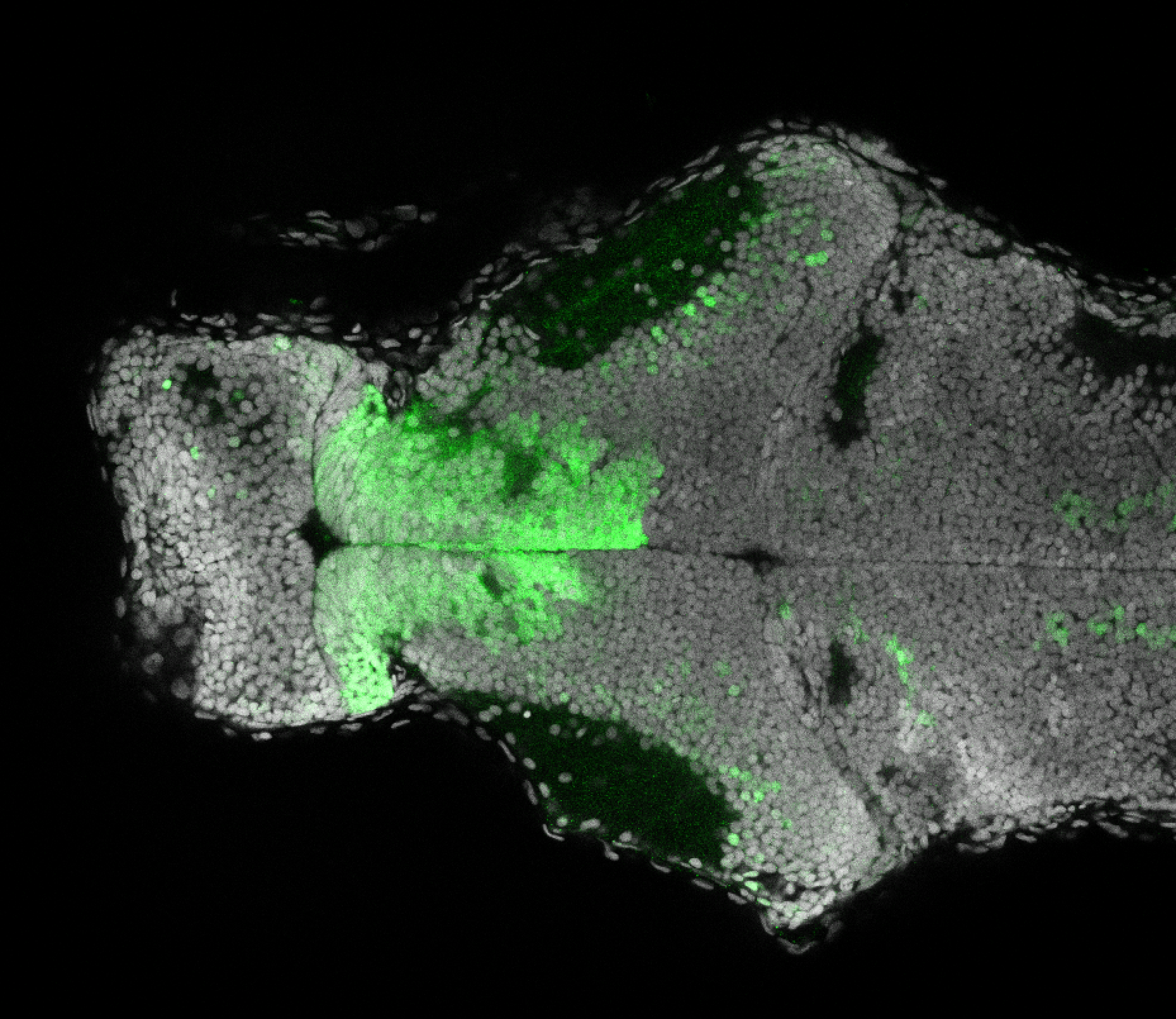
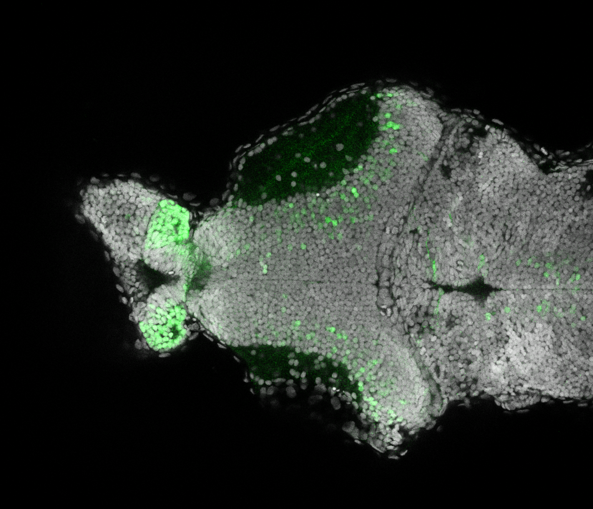
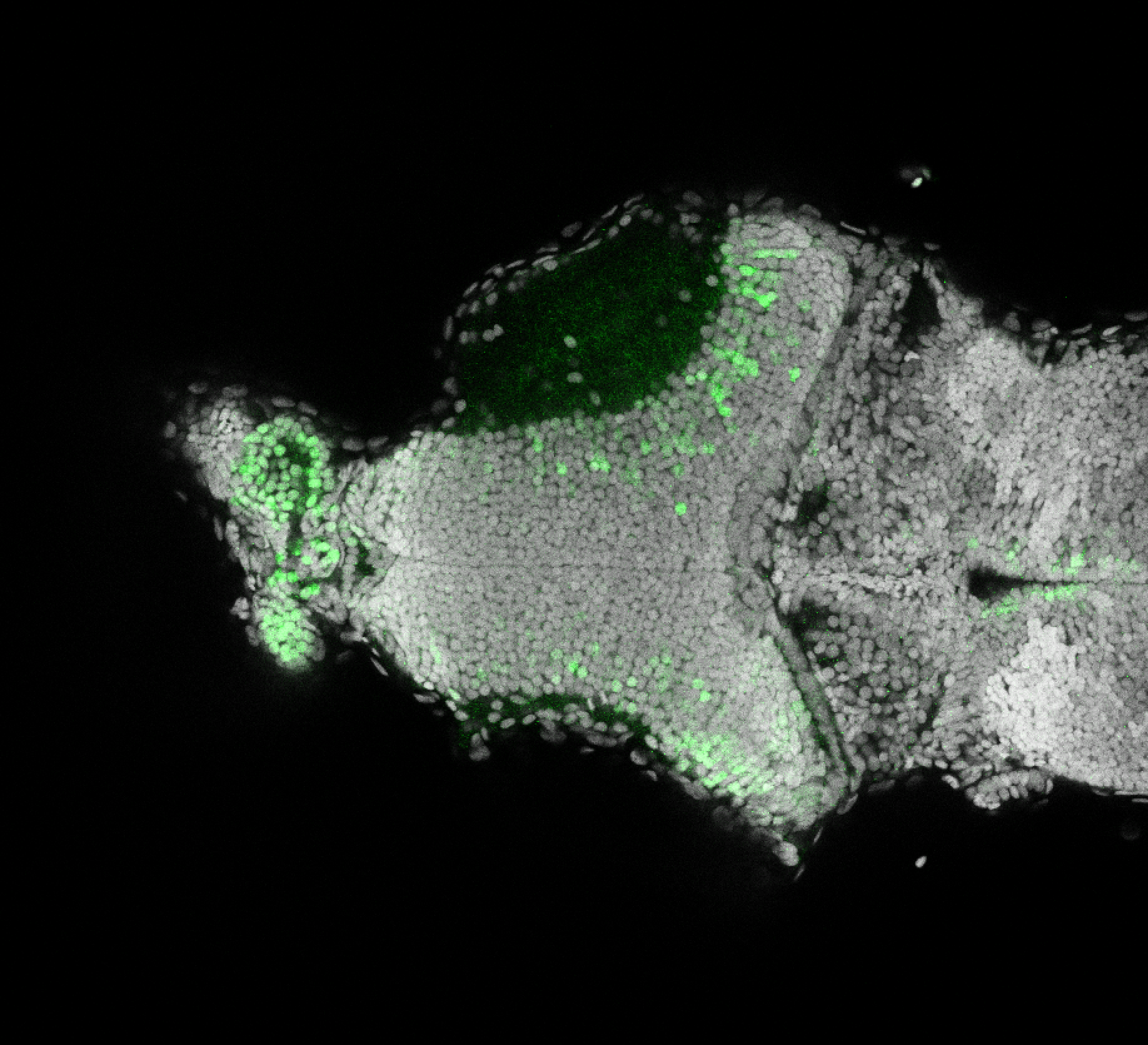
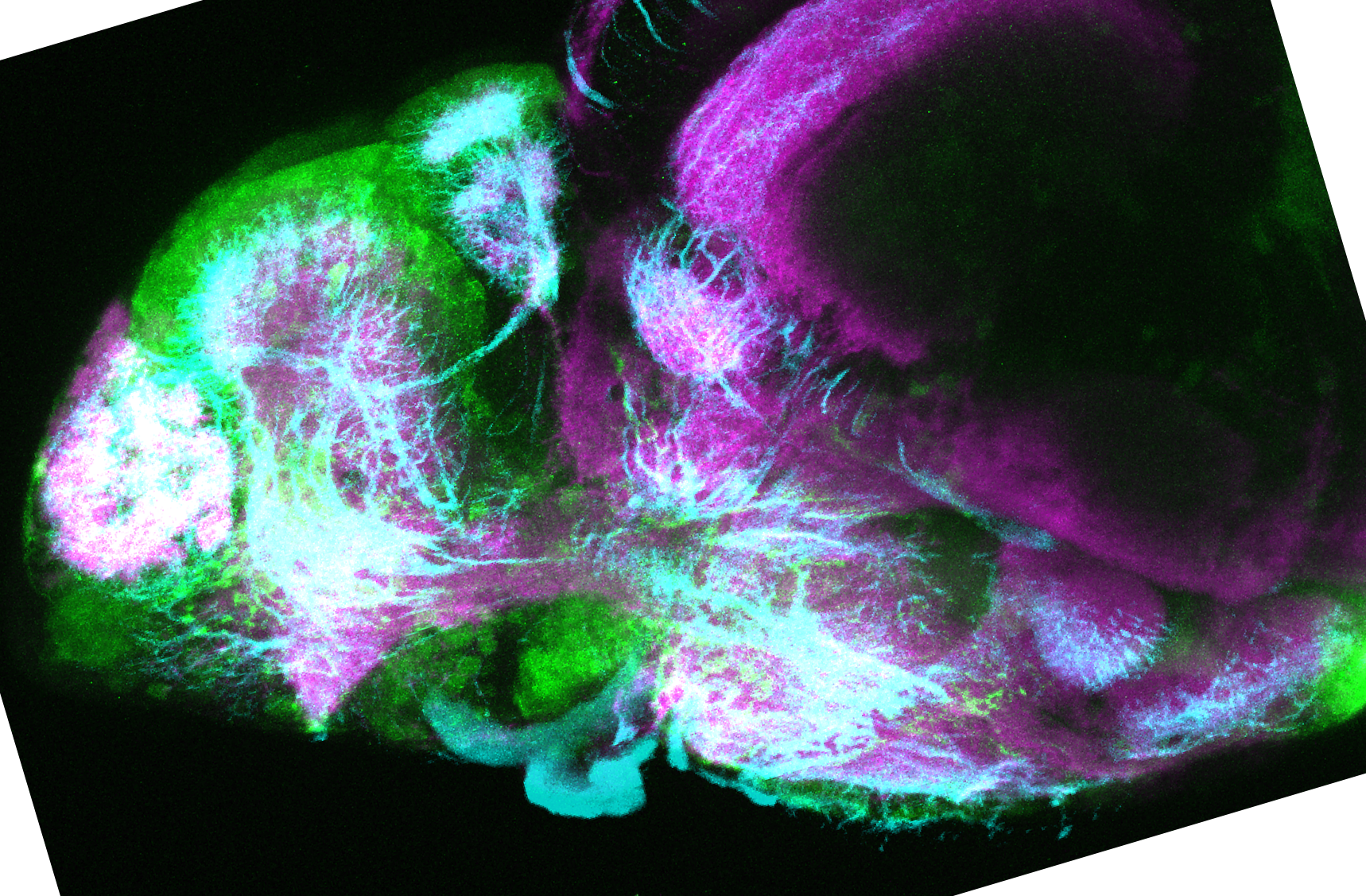
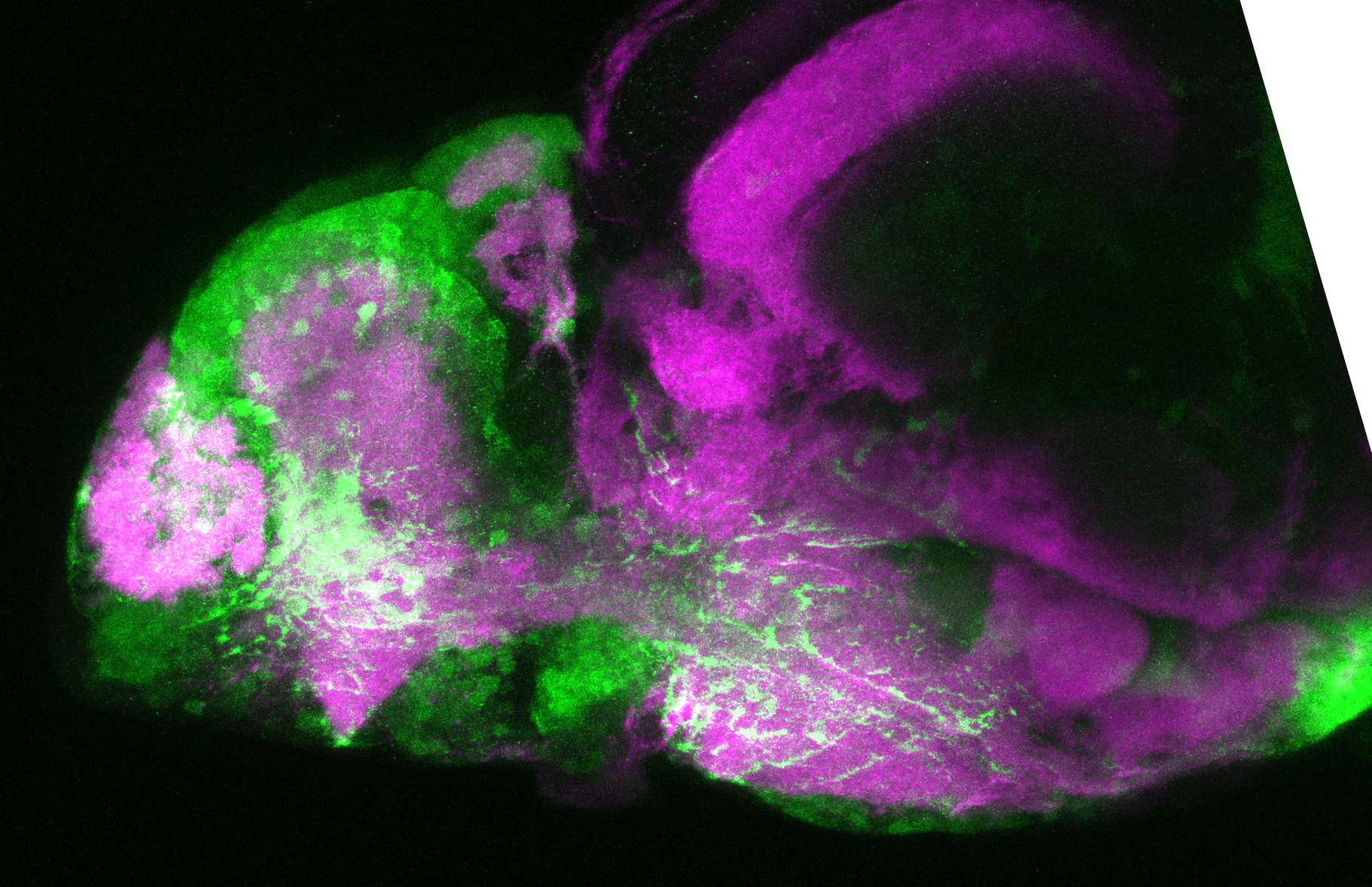
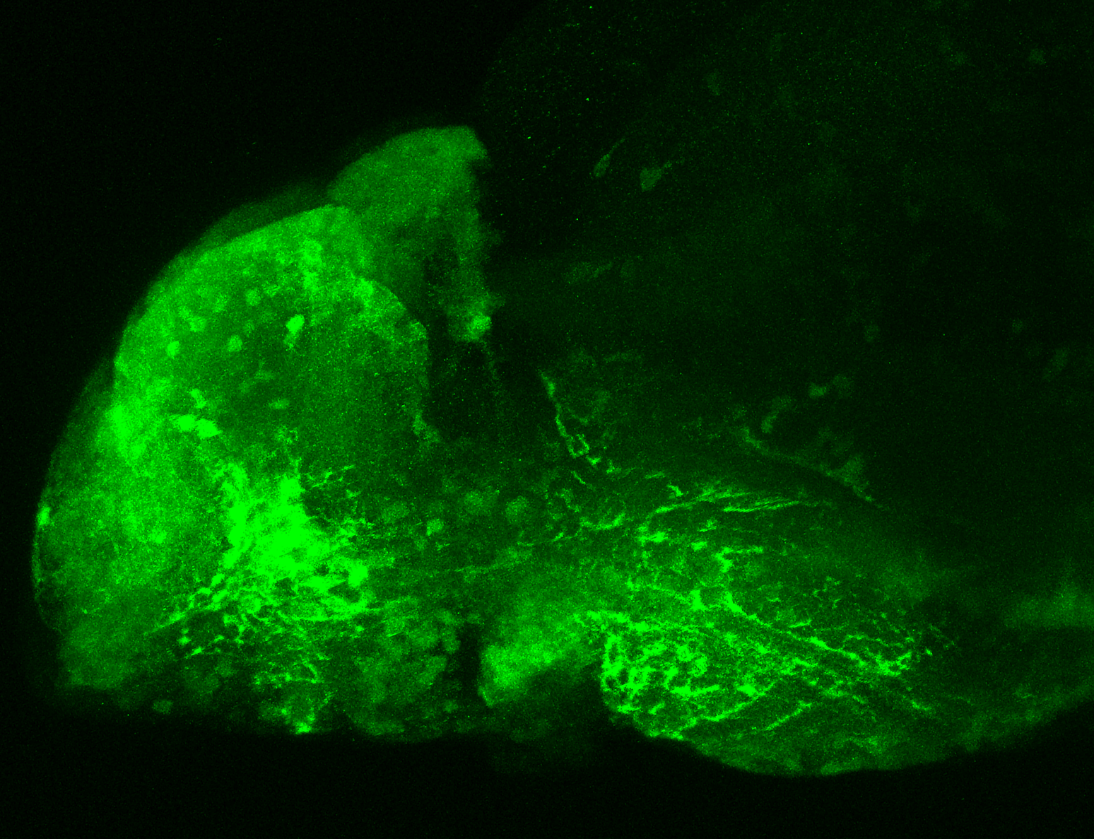
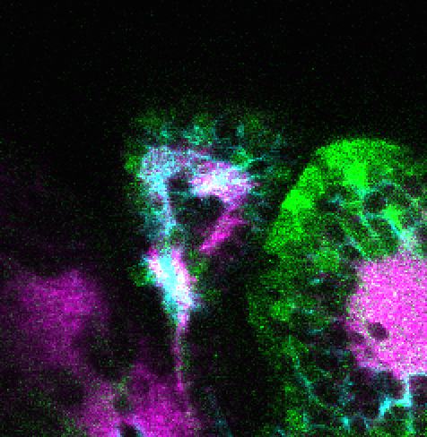
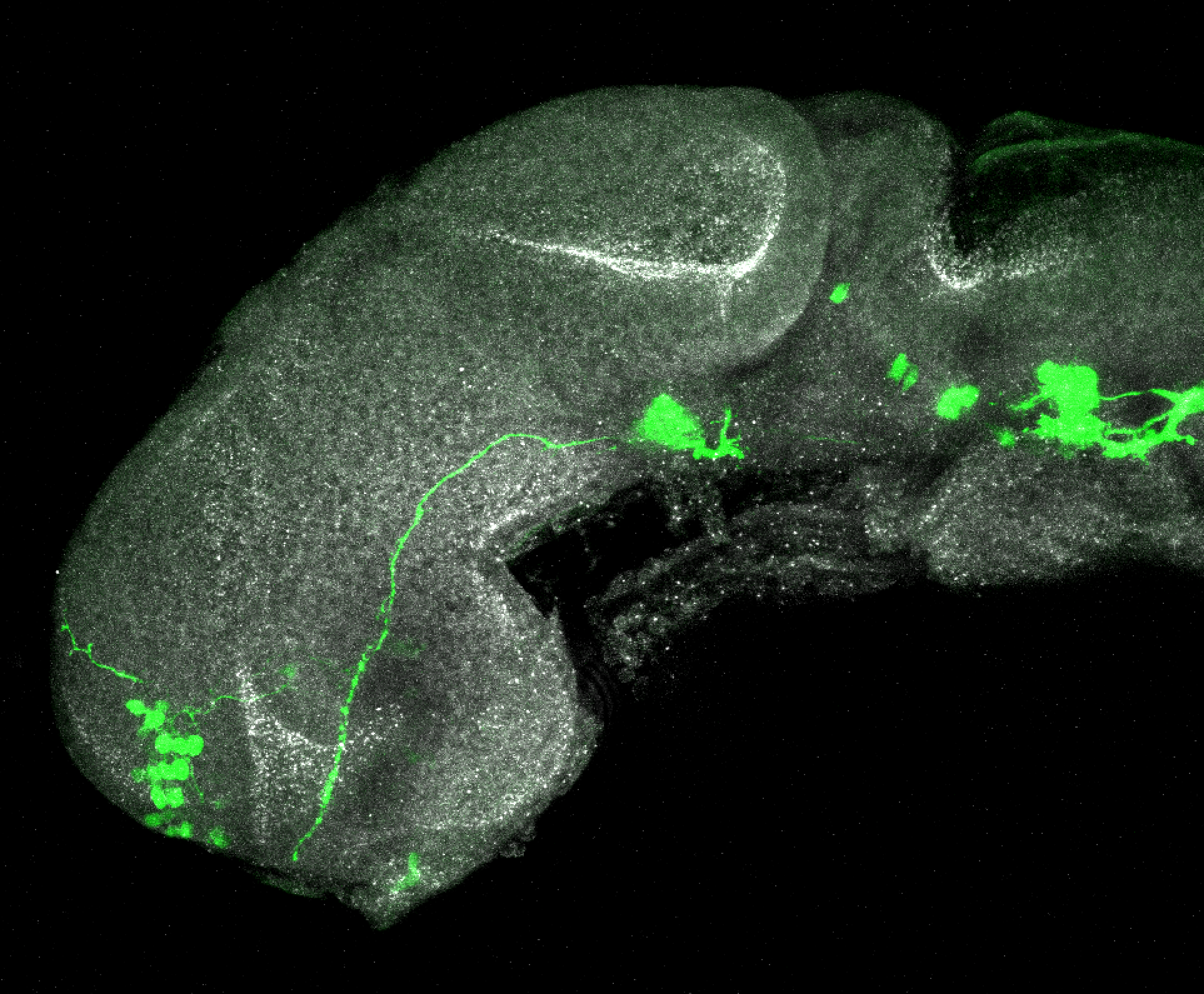
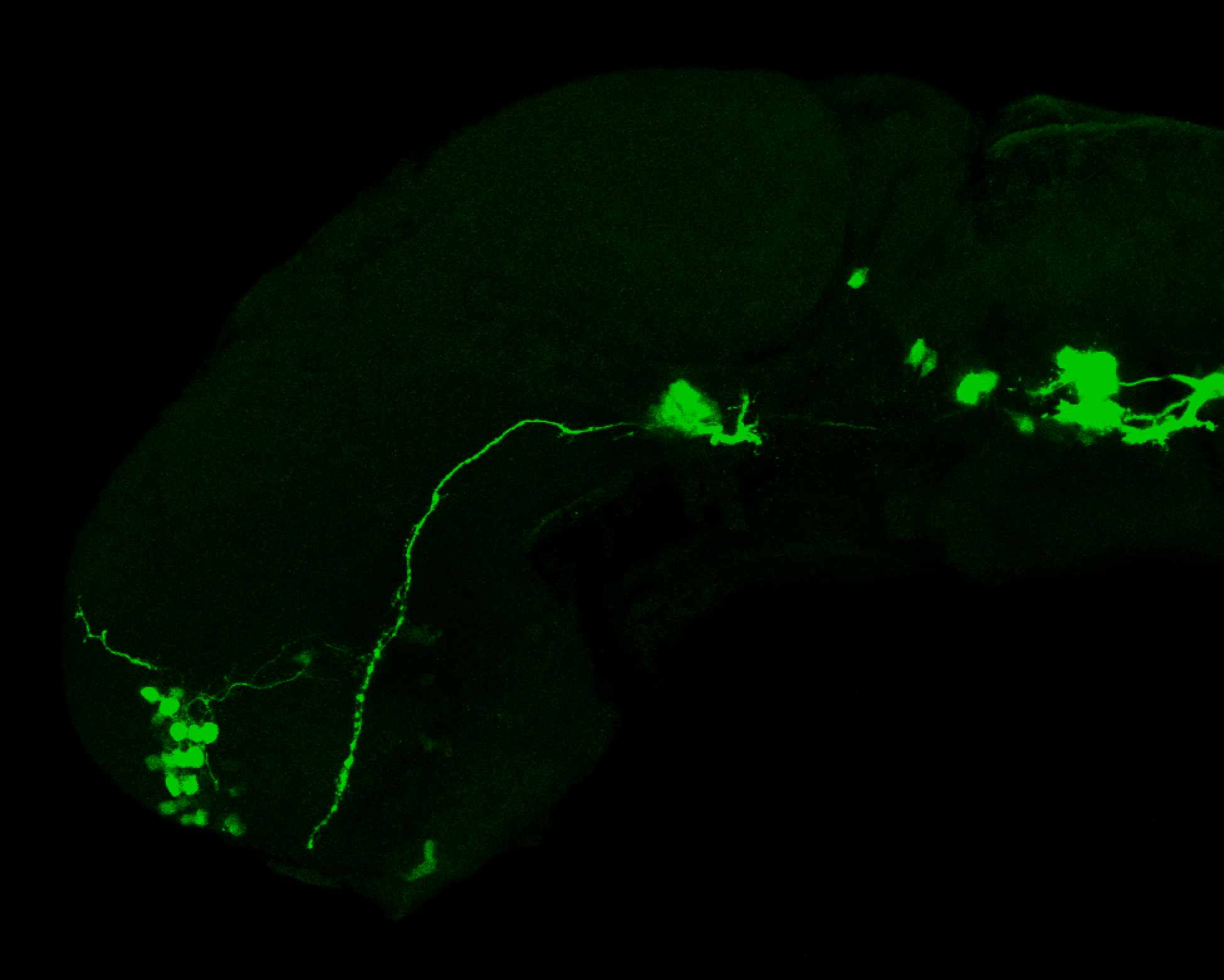
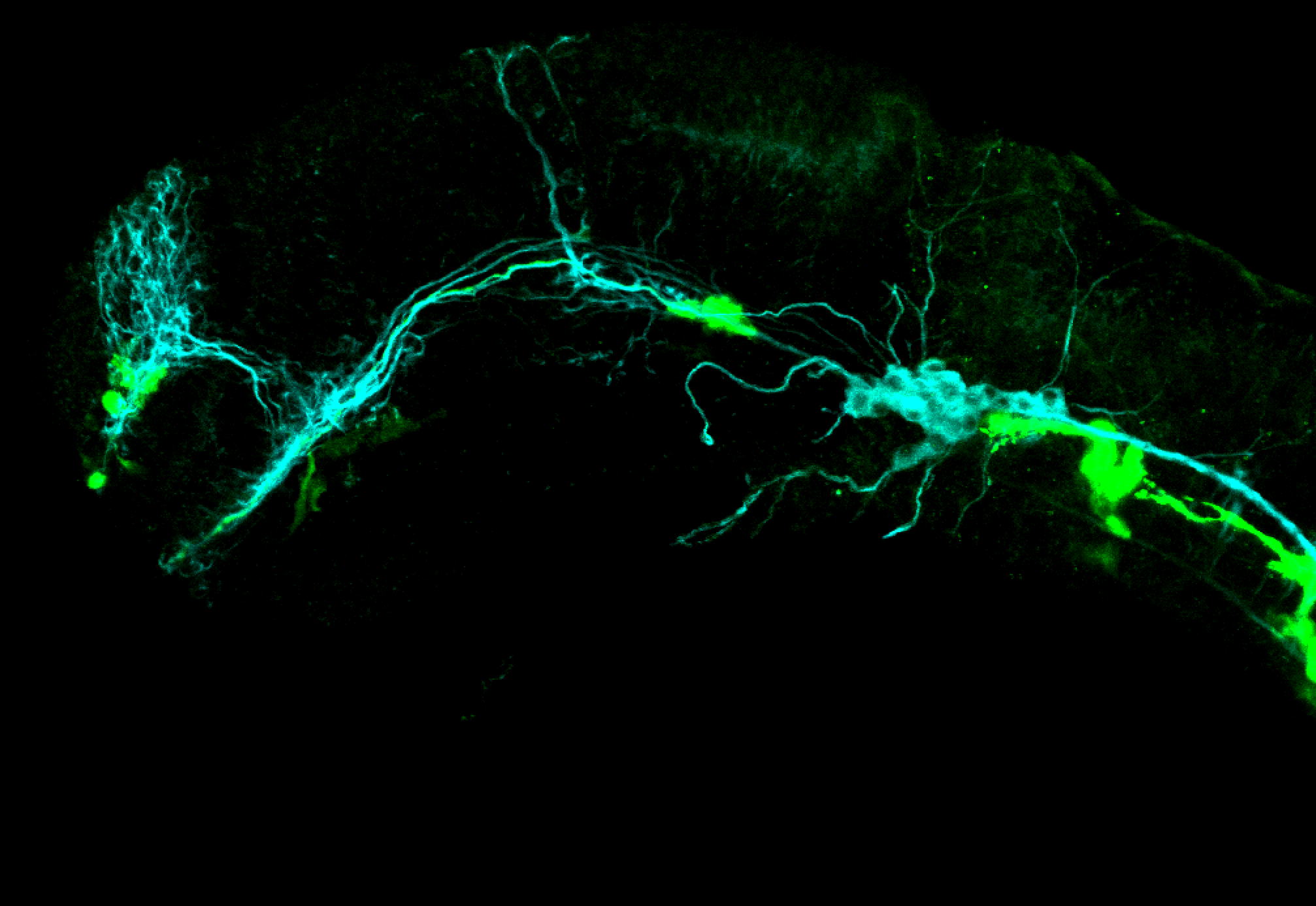
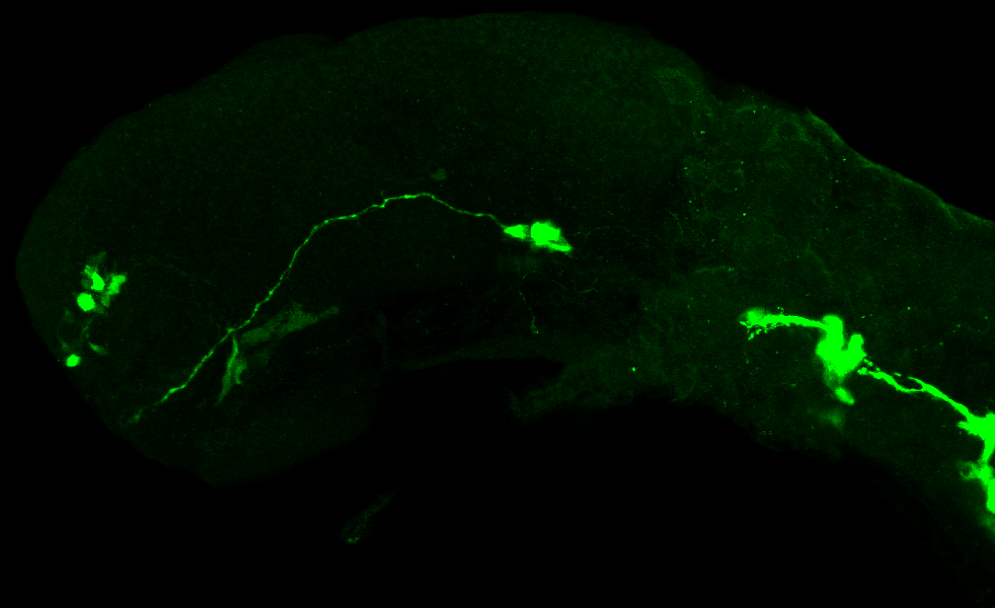
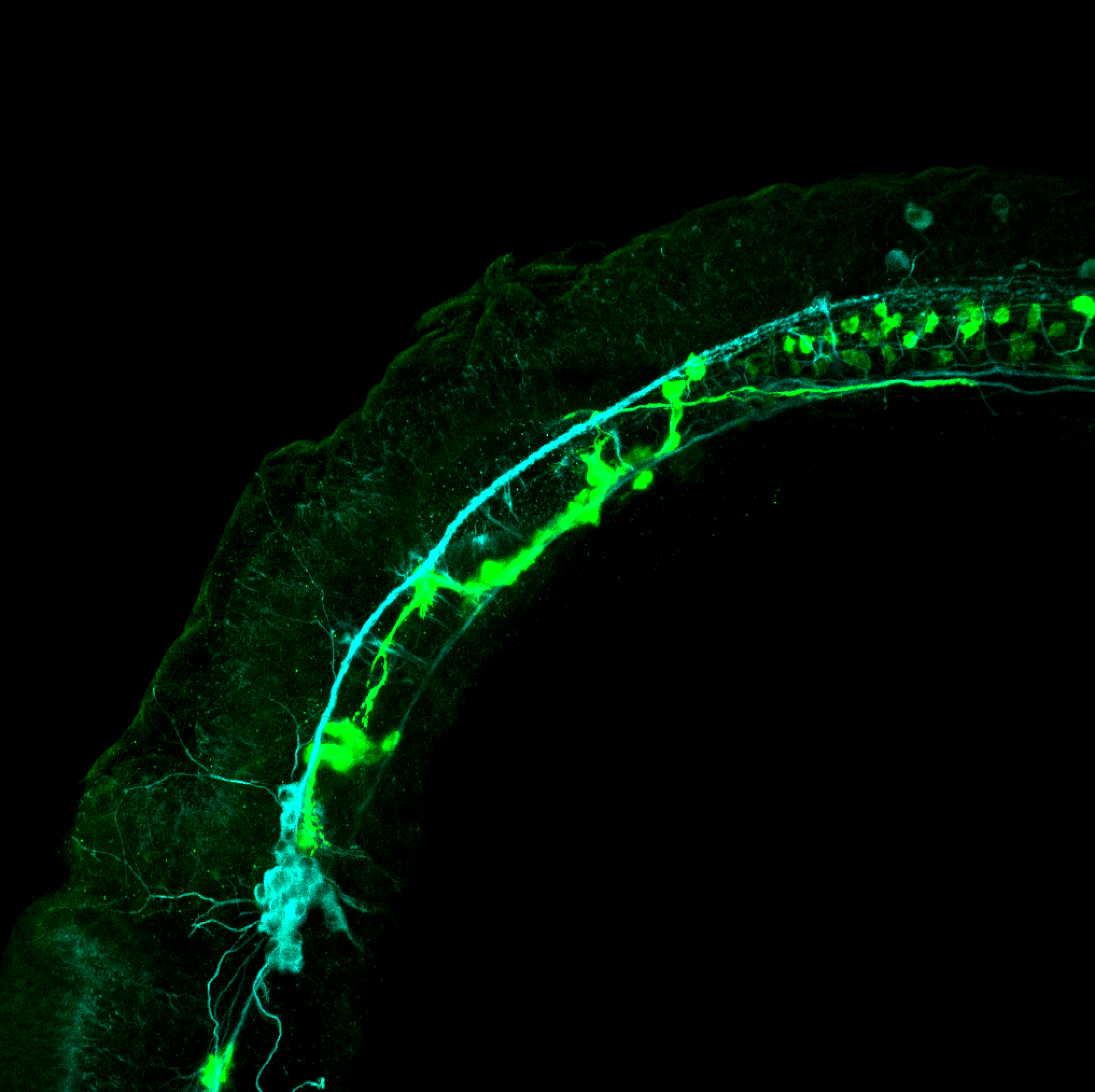
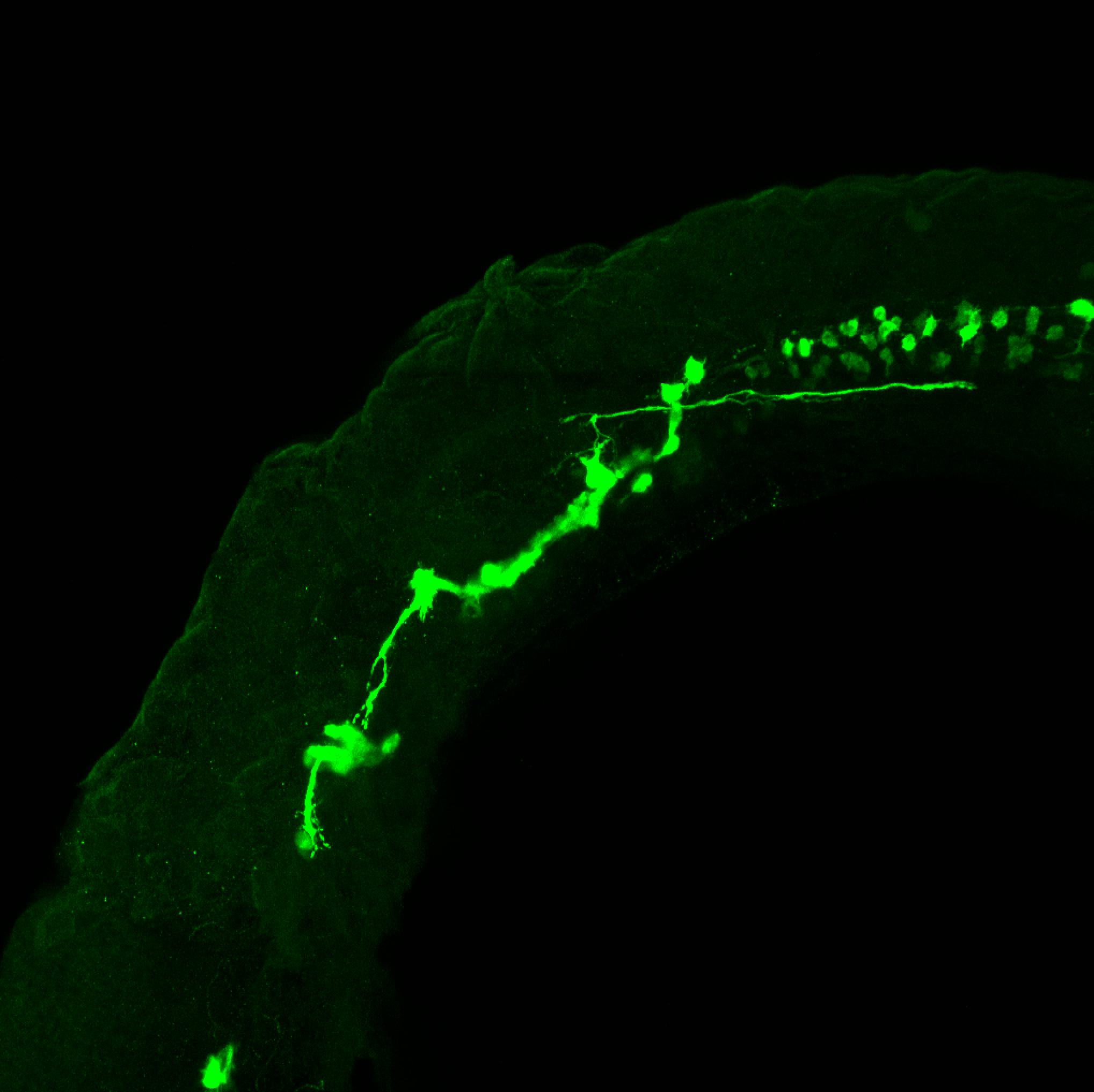
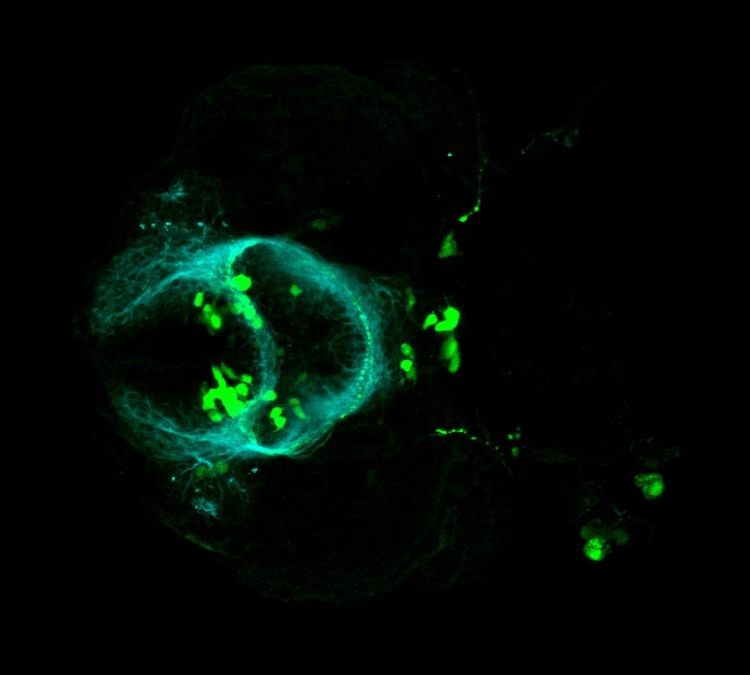
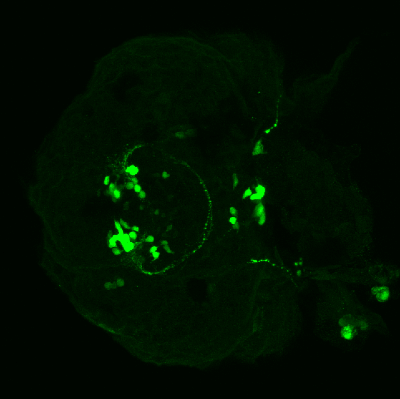
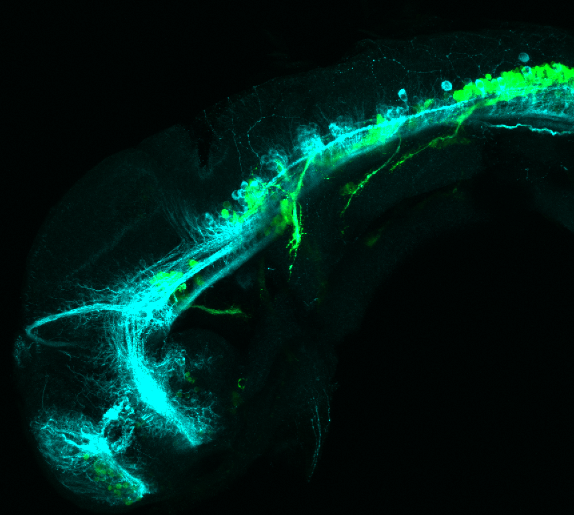
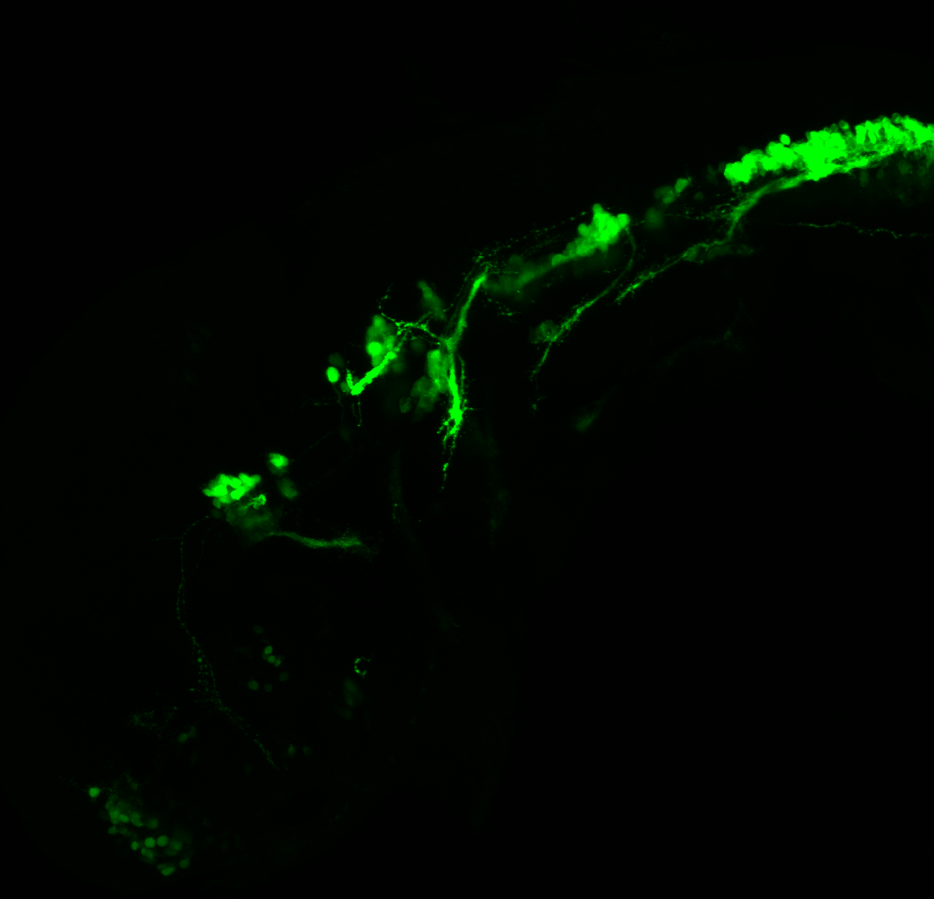
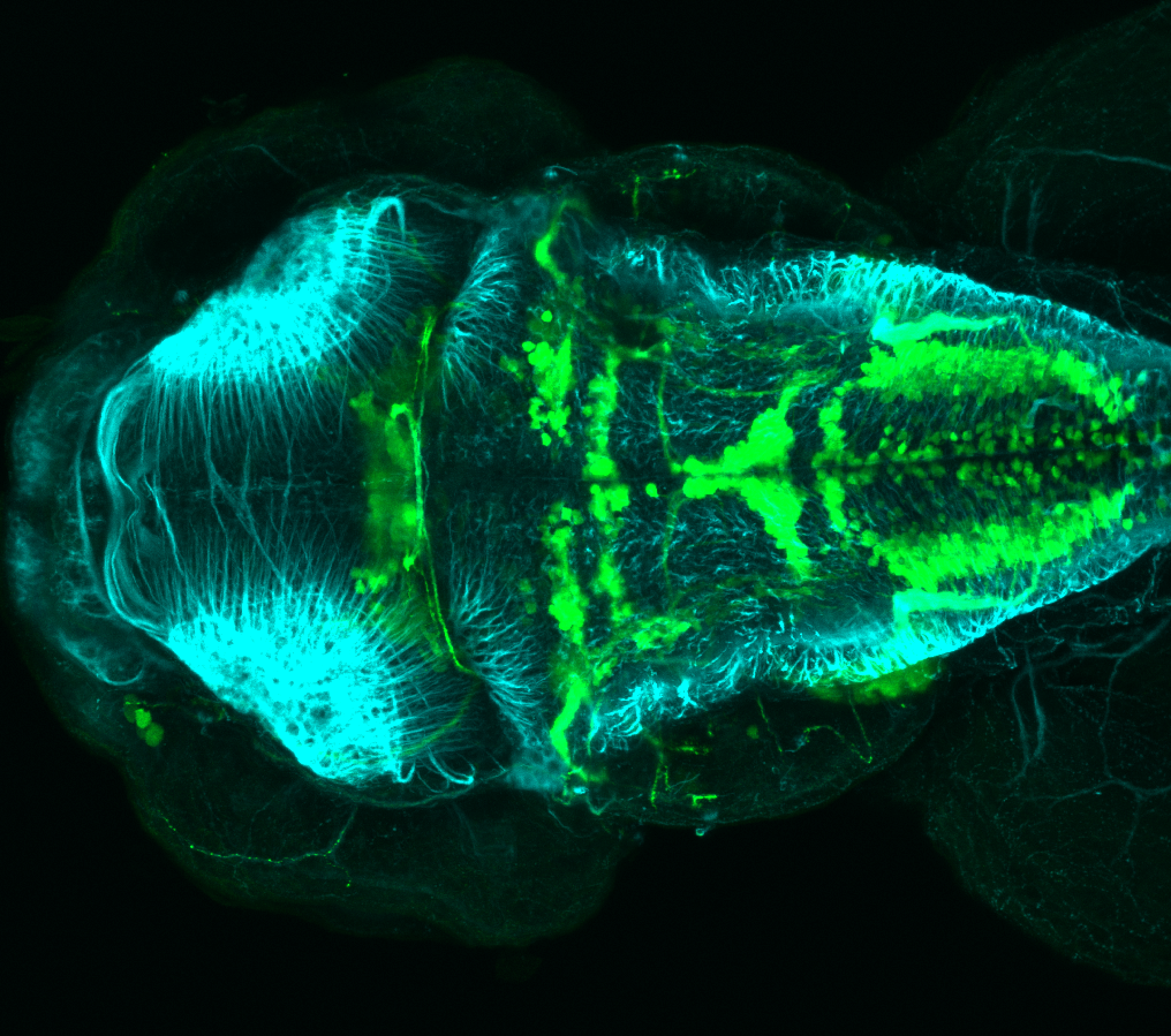
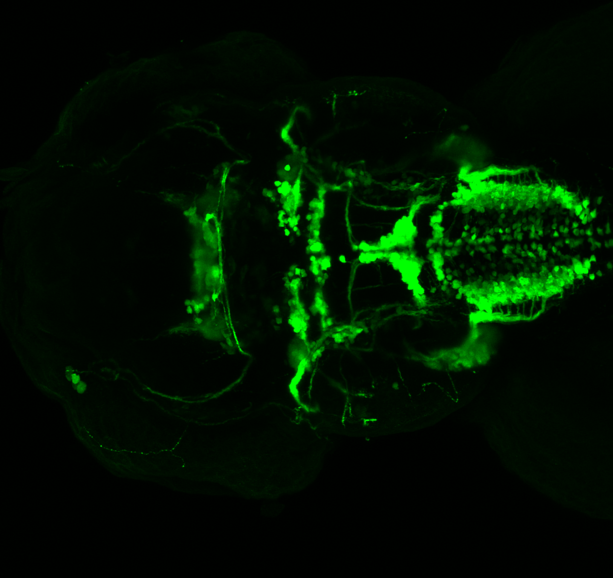
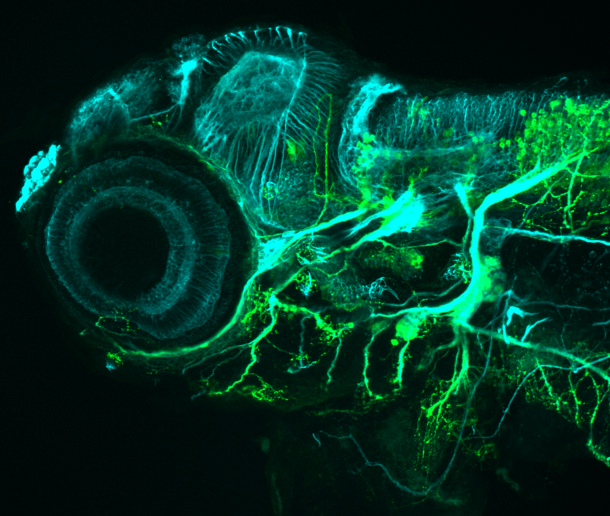
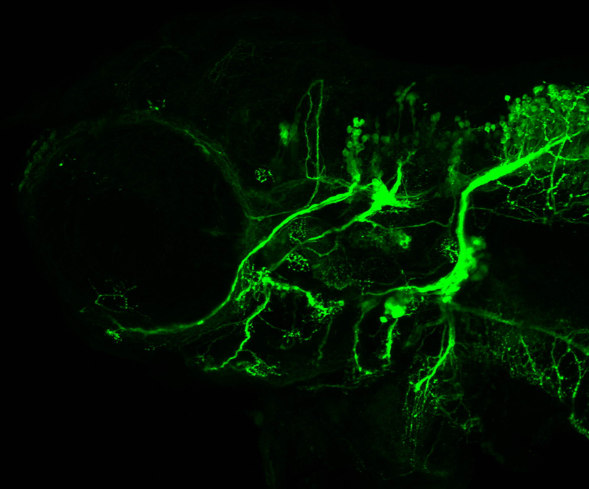
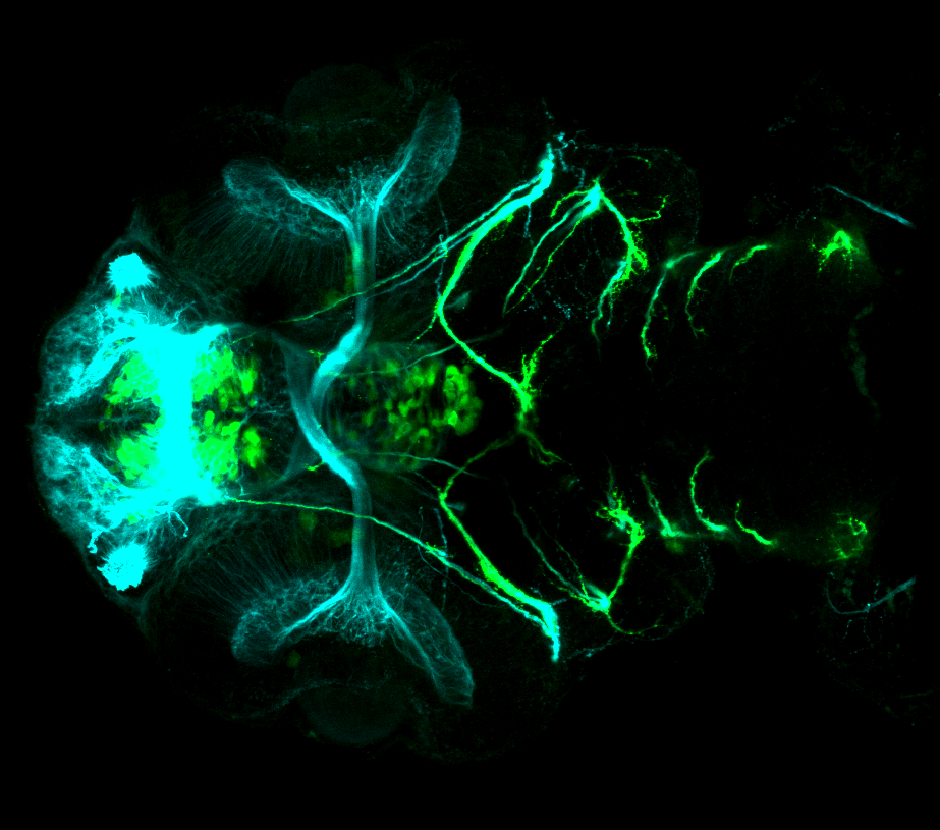
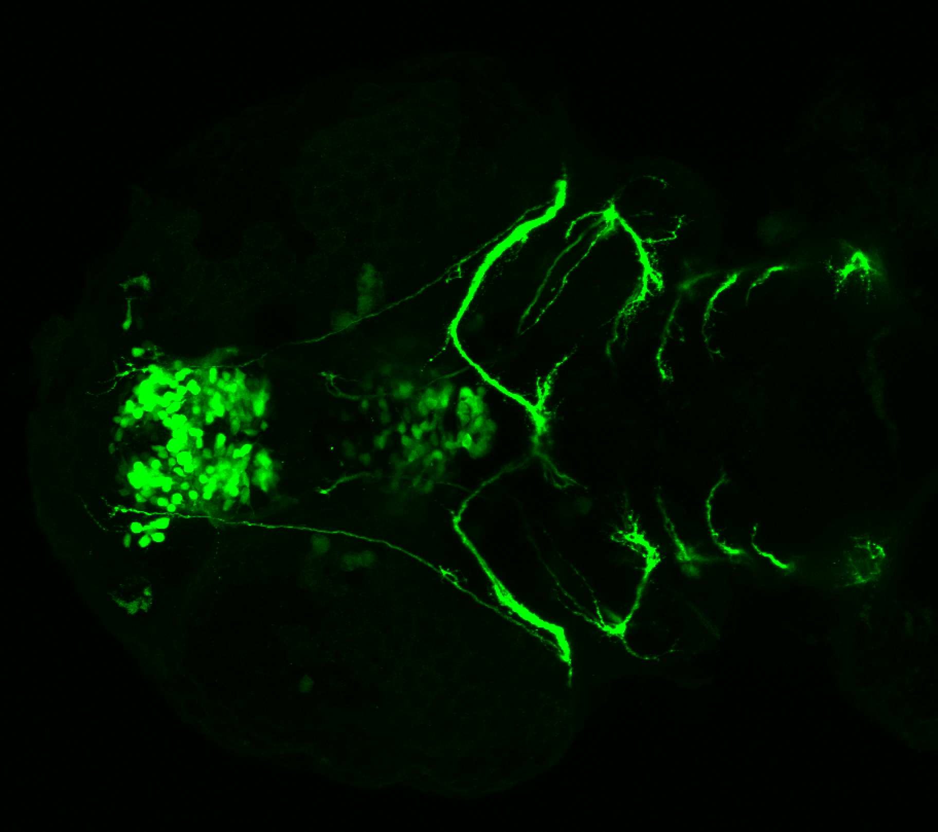
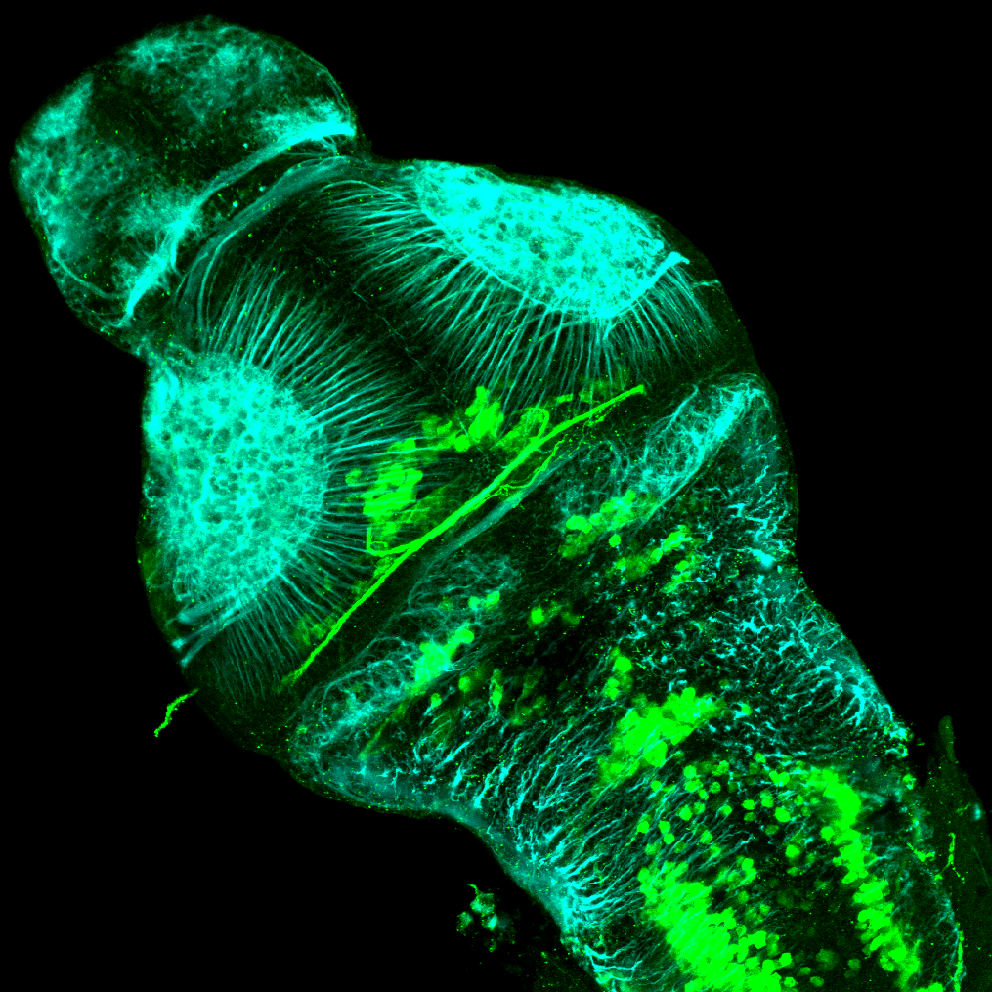
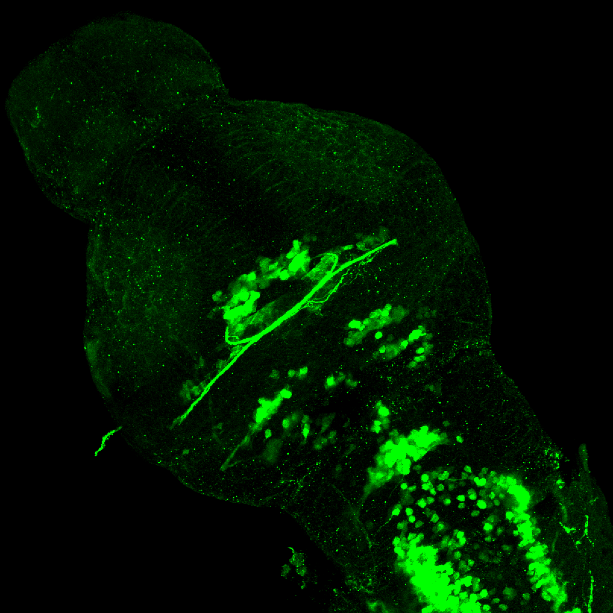
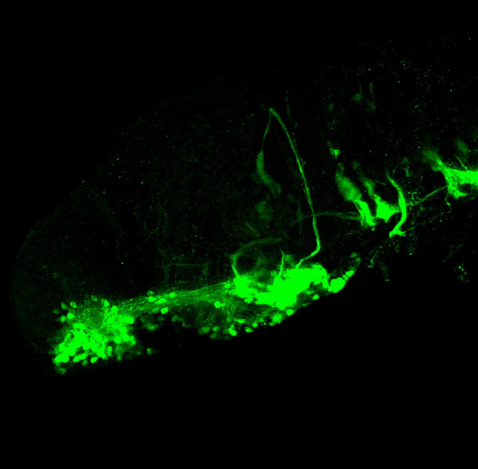
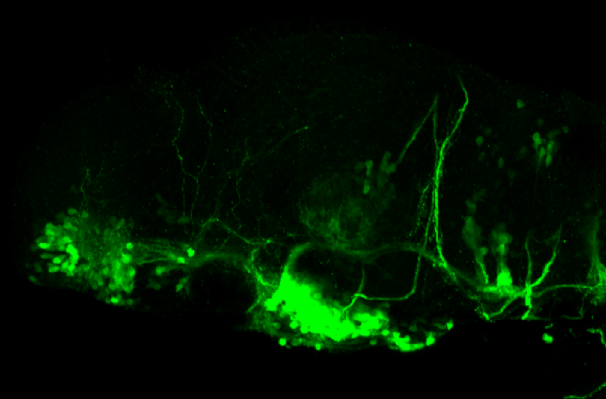
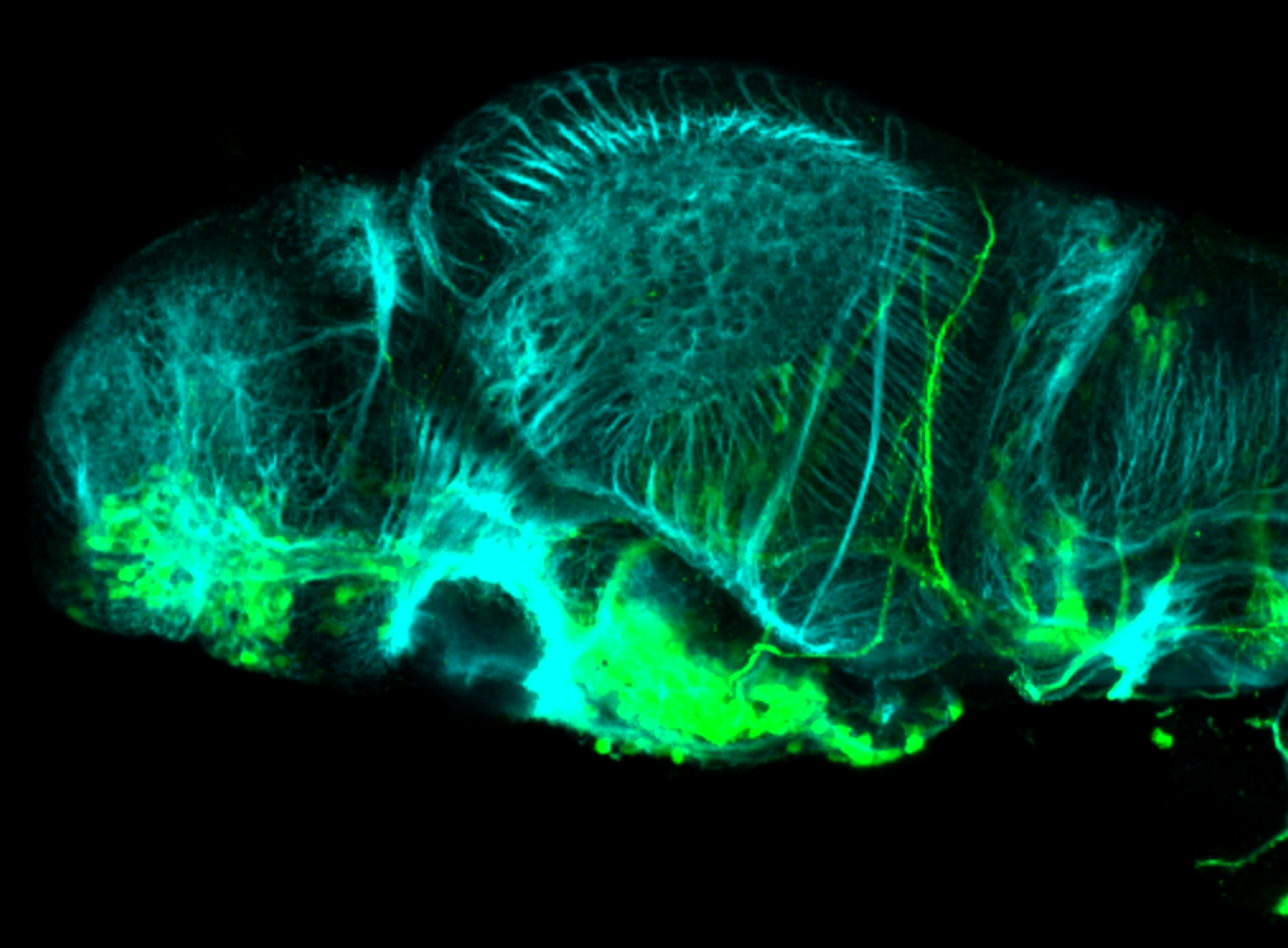
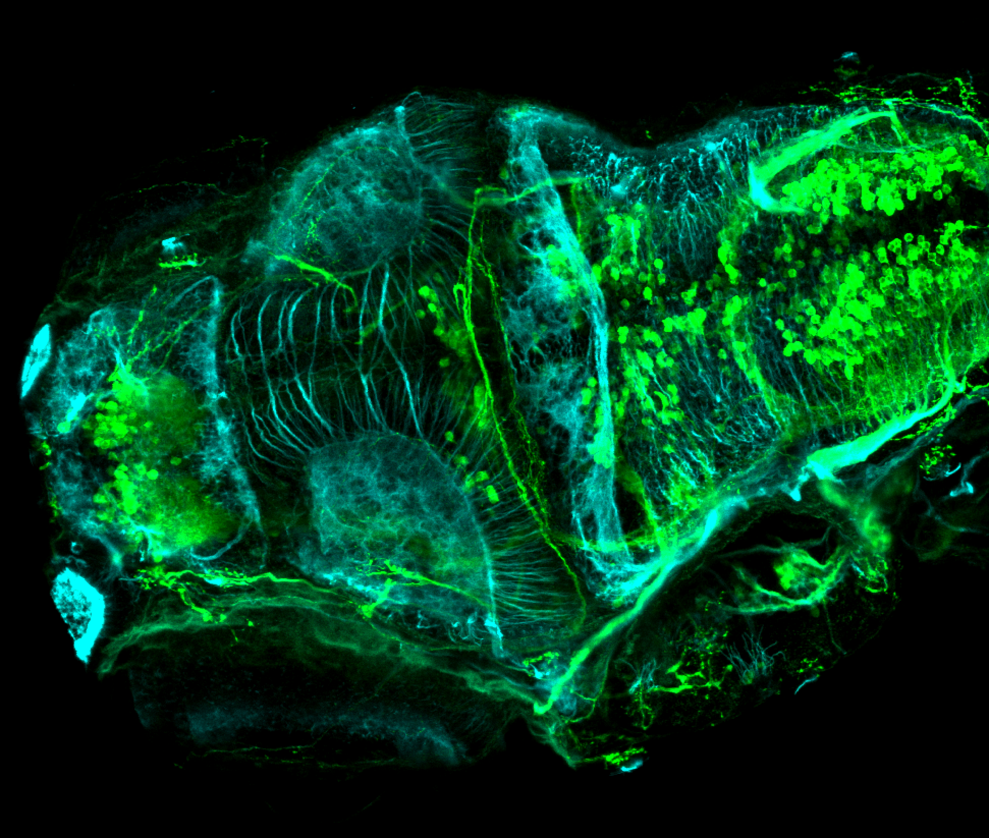
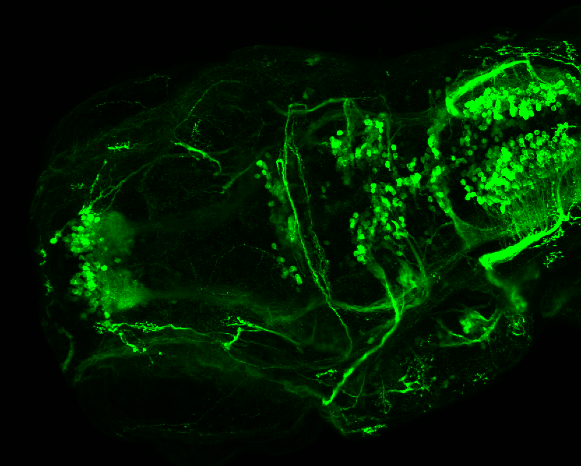
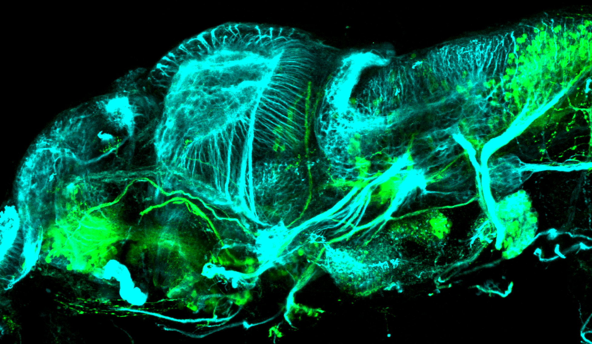
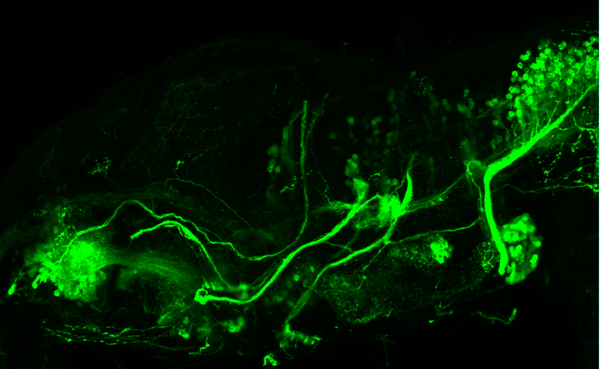
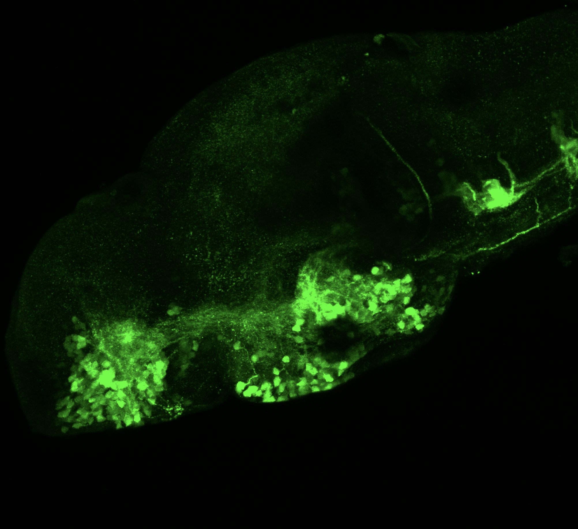
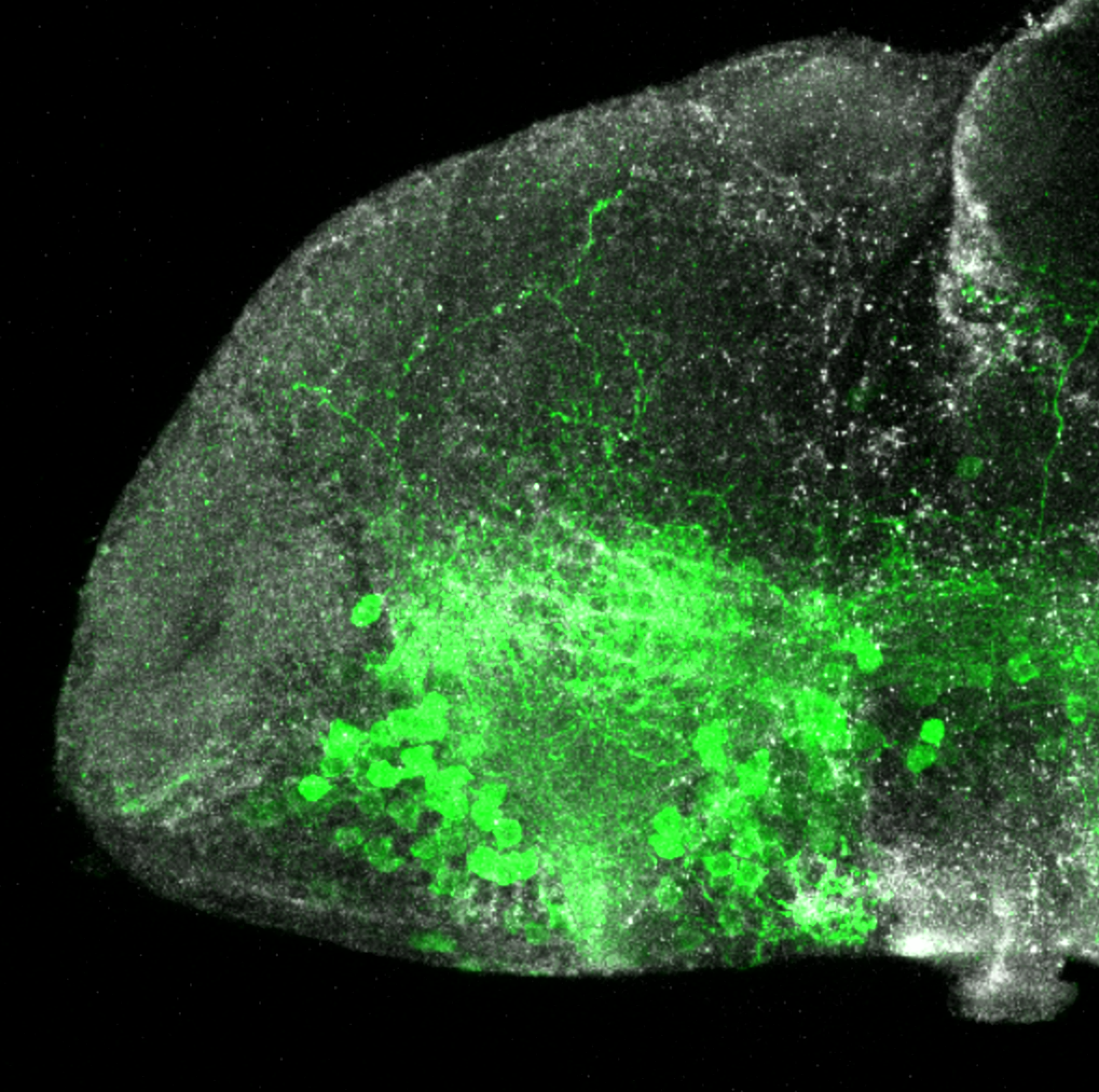
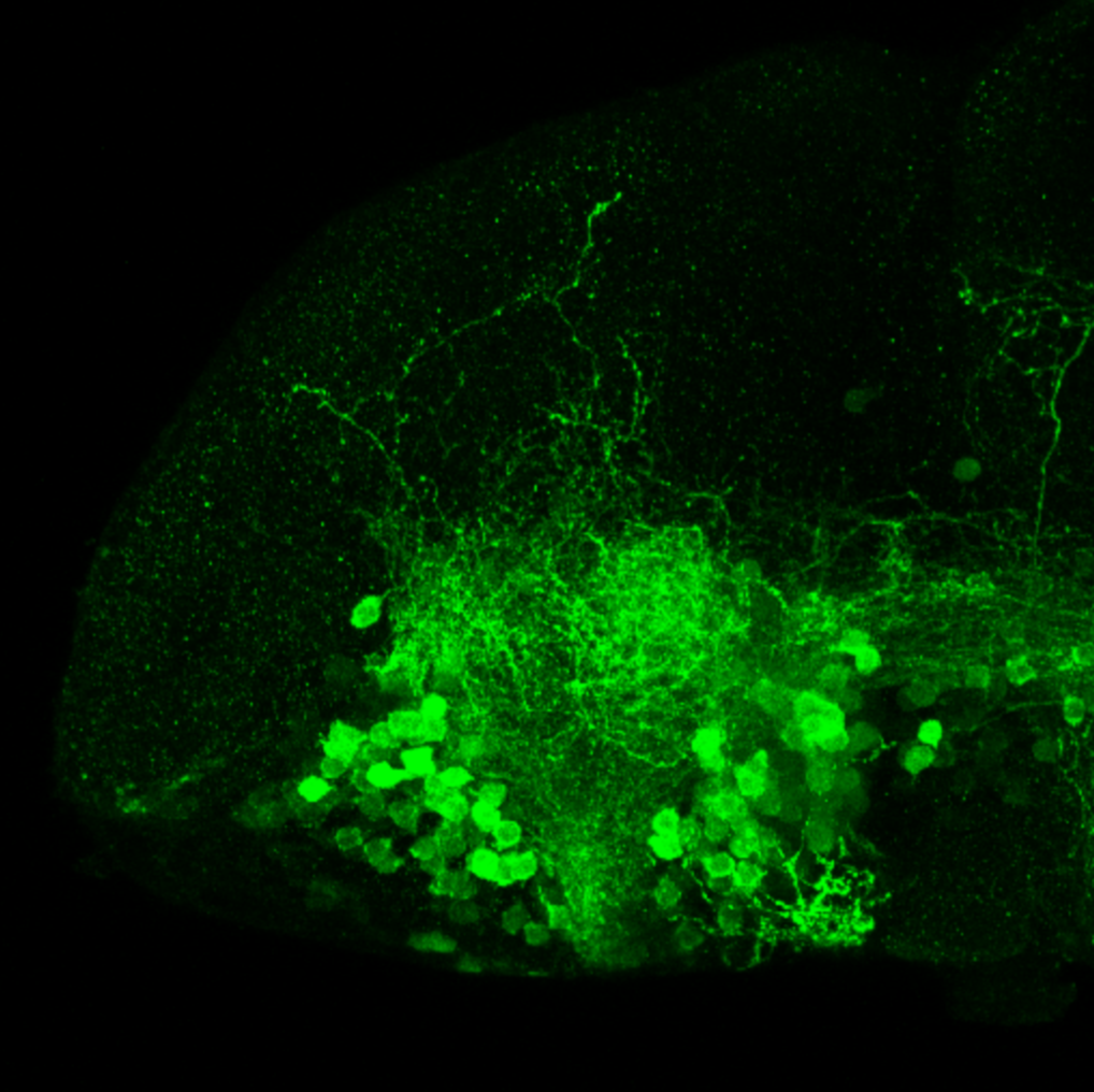
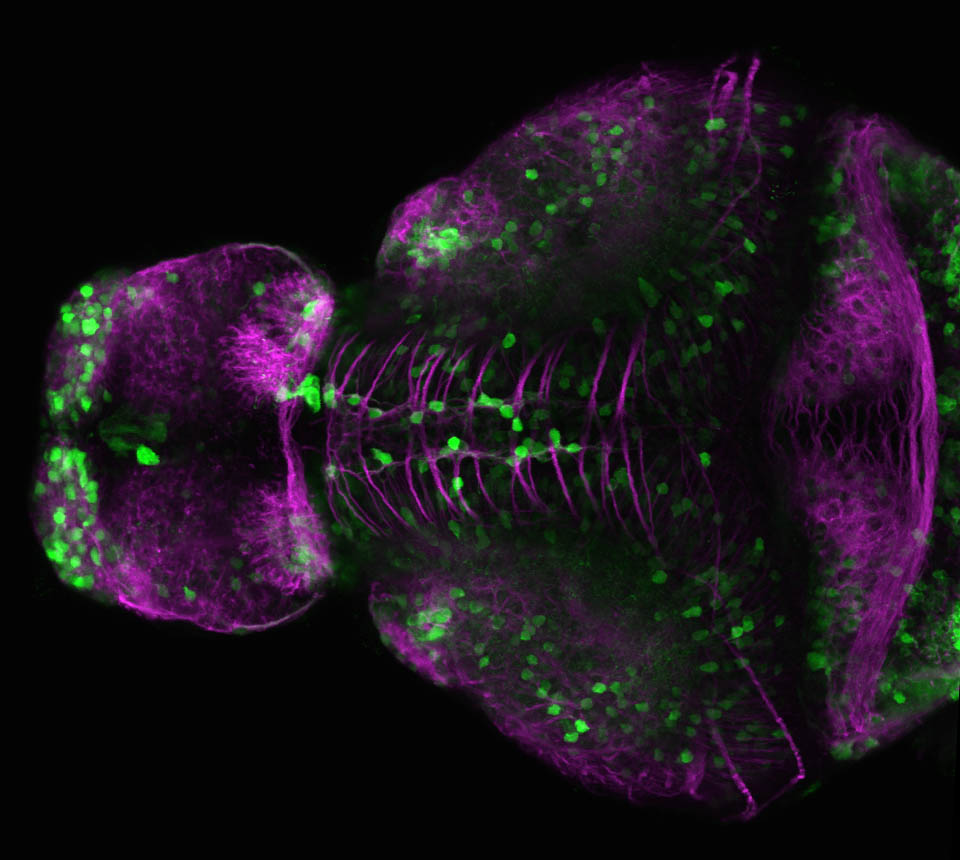
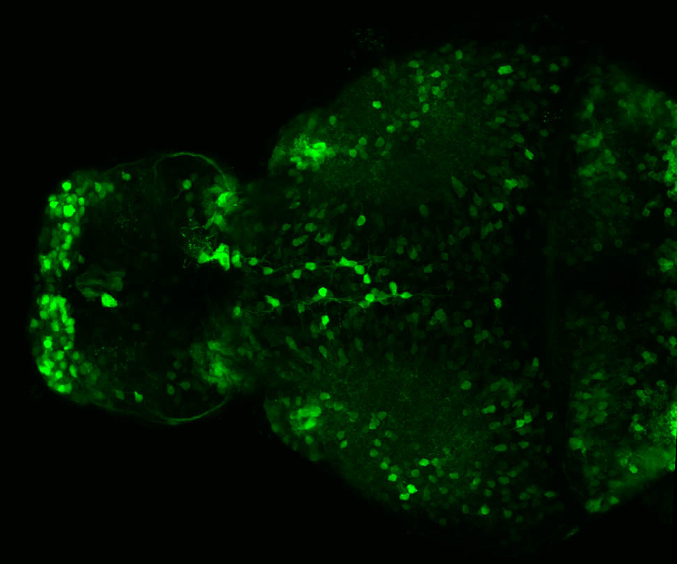
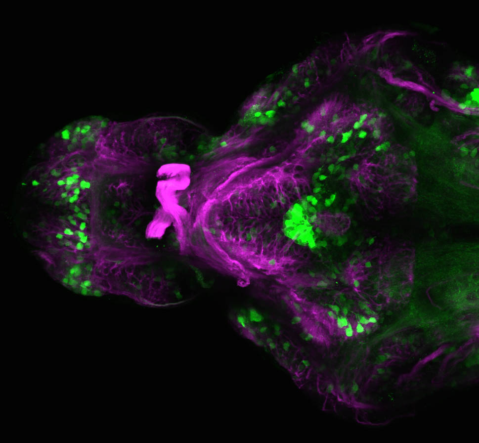
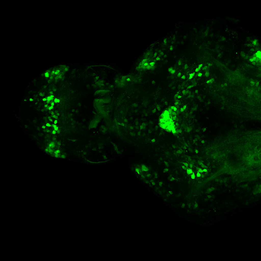
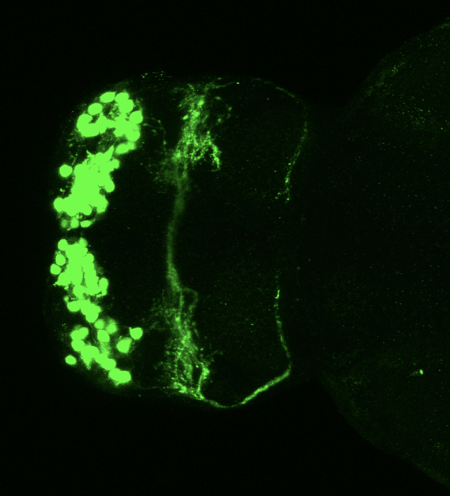
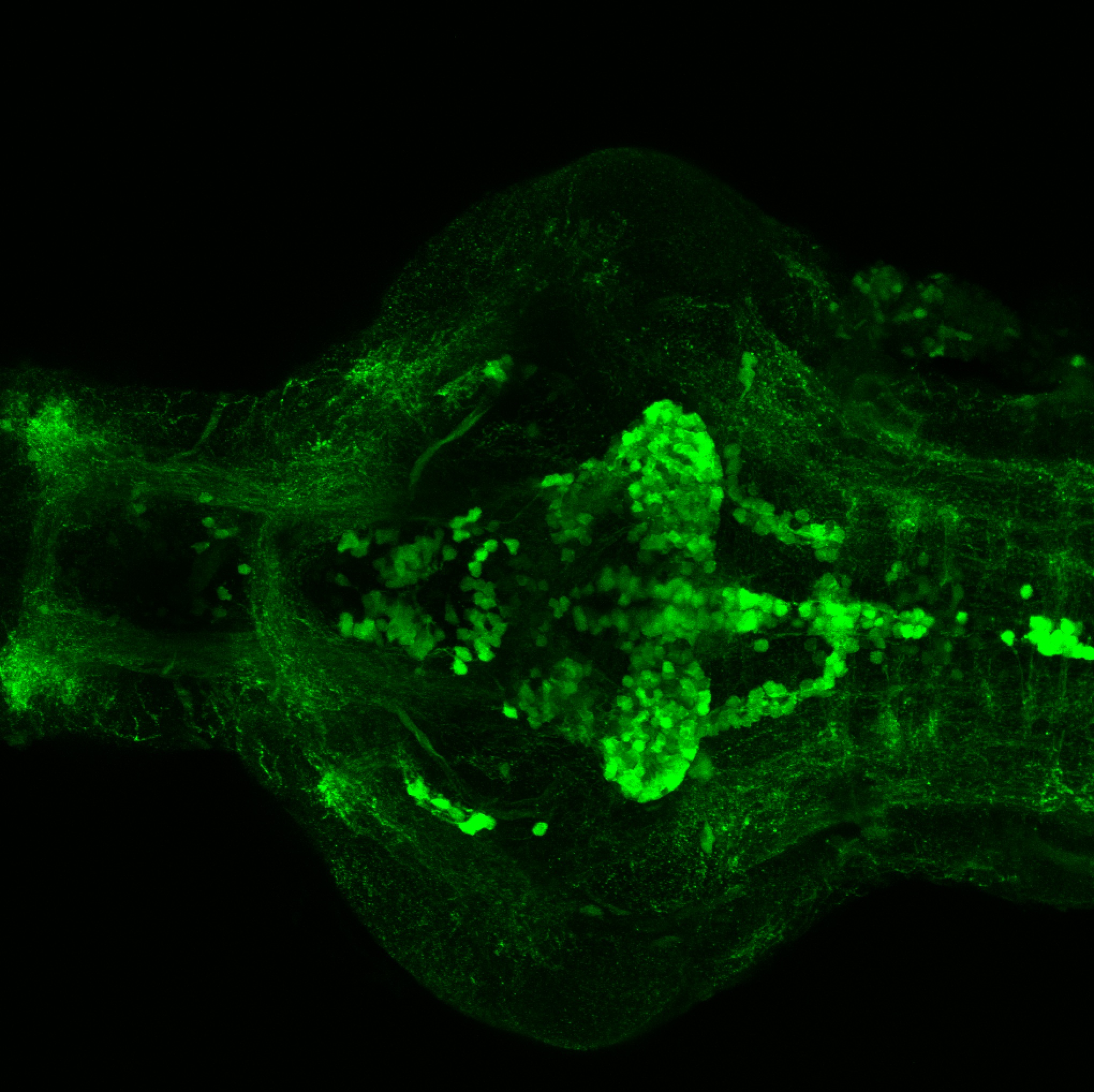
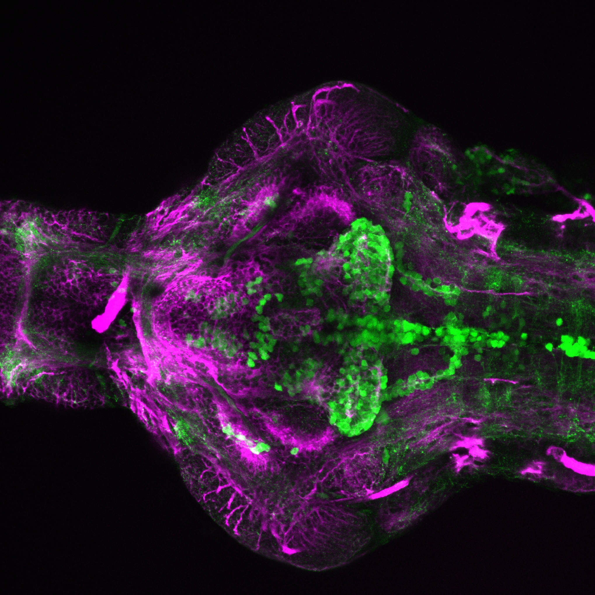
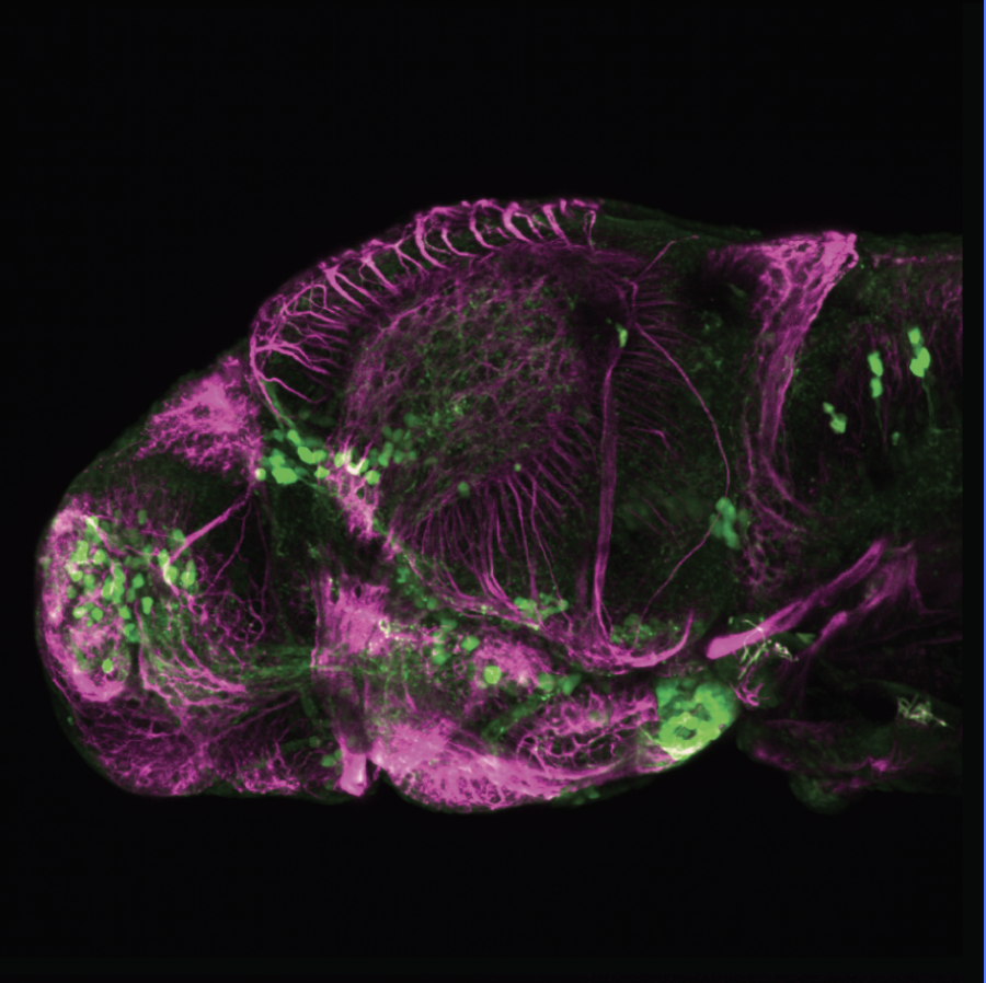
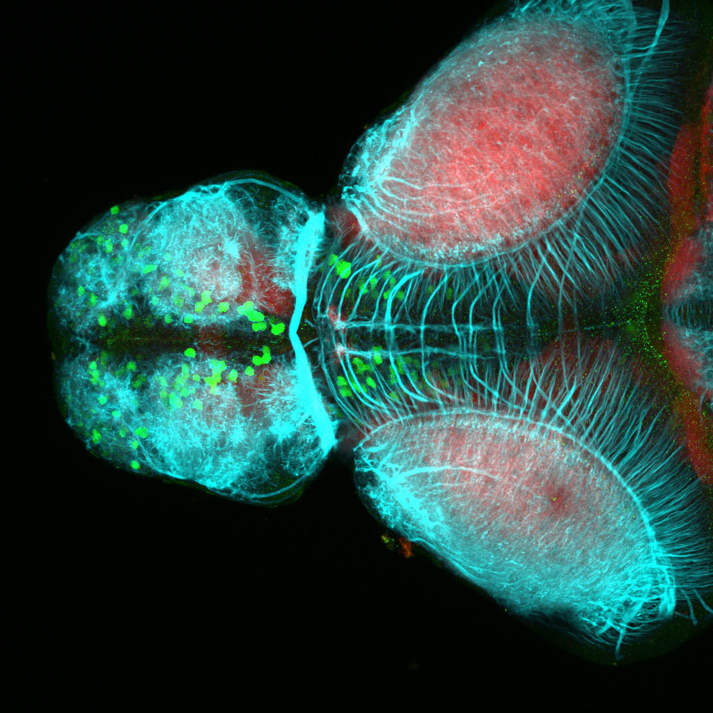
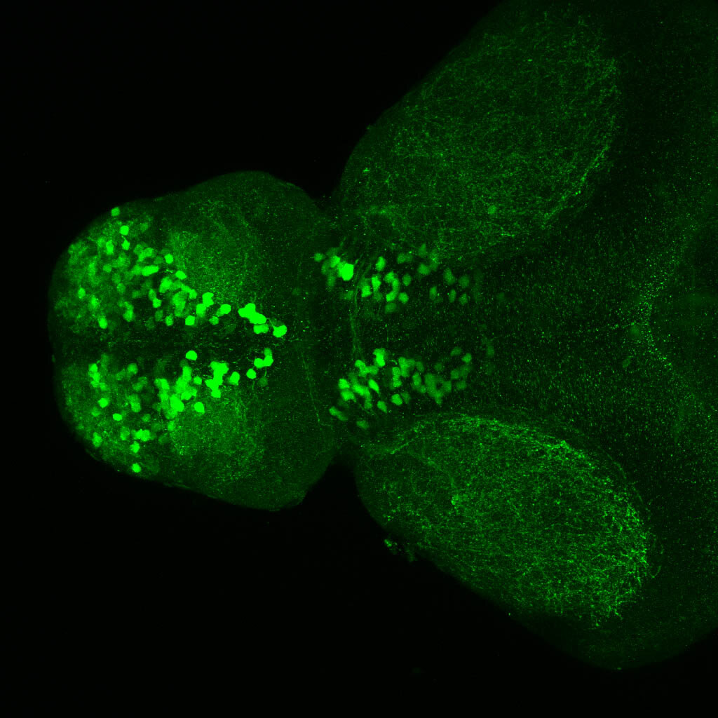
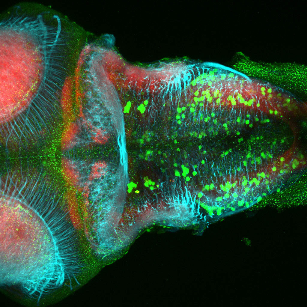
Tg(-8.0cldnb:LY-EGFP)

This transgenic line was created by the Gilmore lab “ Eight kilobases of sequence directly upstream of the Claudin B start codon were amplified from BAC zK241F11 by using the Expand Long Template PCR System (Roche). The resultant fragment was cloned into a vector containing lynEGFPpA (Koster and Fraser, 2001) flanked by sites for I-SceI, and the resultant construct was injected into one-cell zebrafish embryos by following the meganuclease transgenesis protocol (Thermes et al., 2002).”(Haas & Gilmore, 2006).

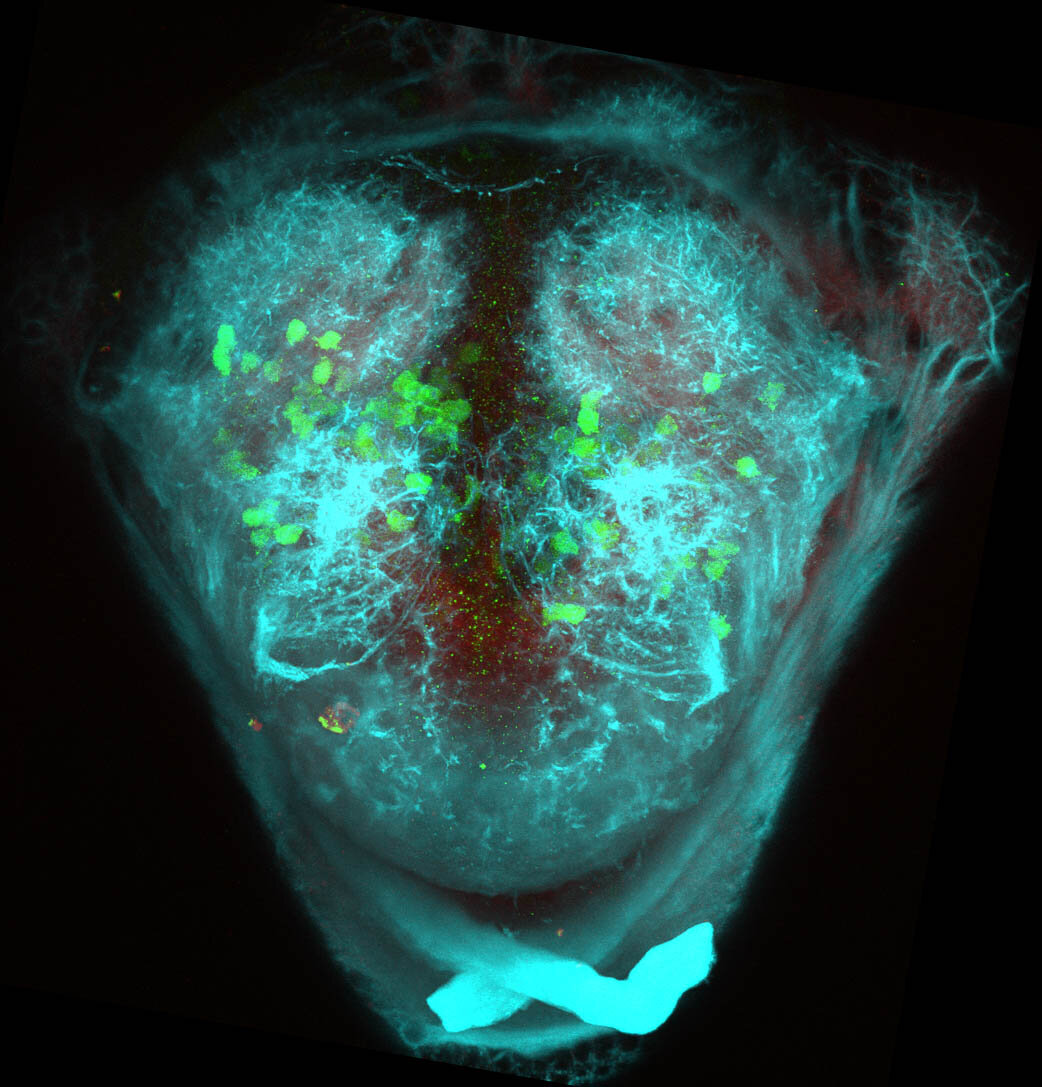
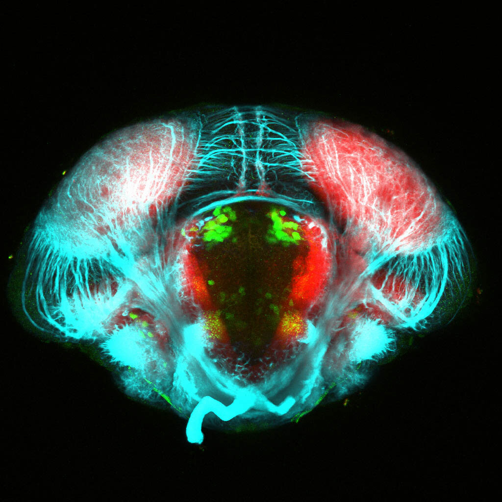
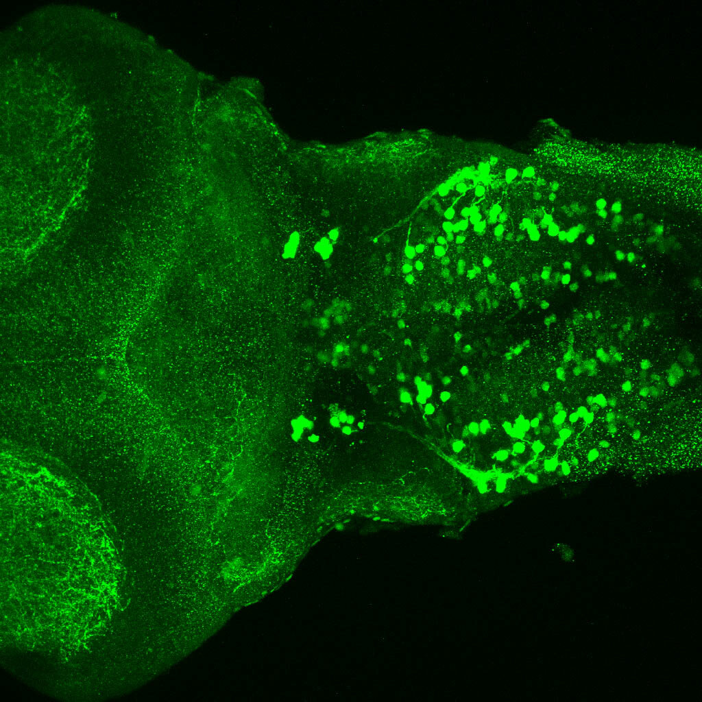
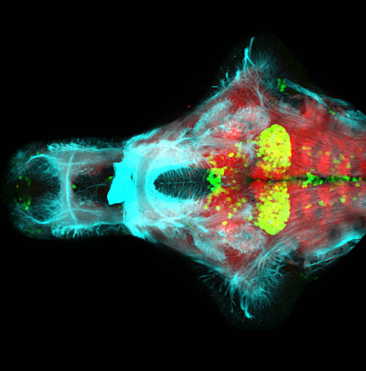
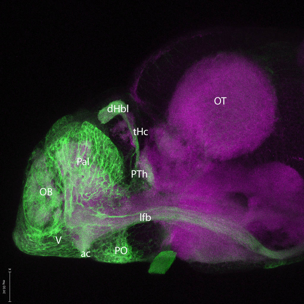
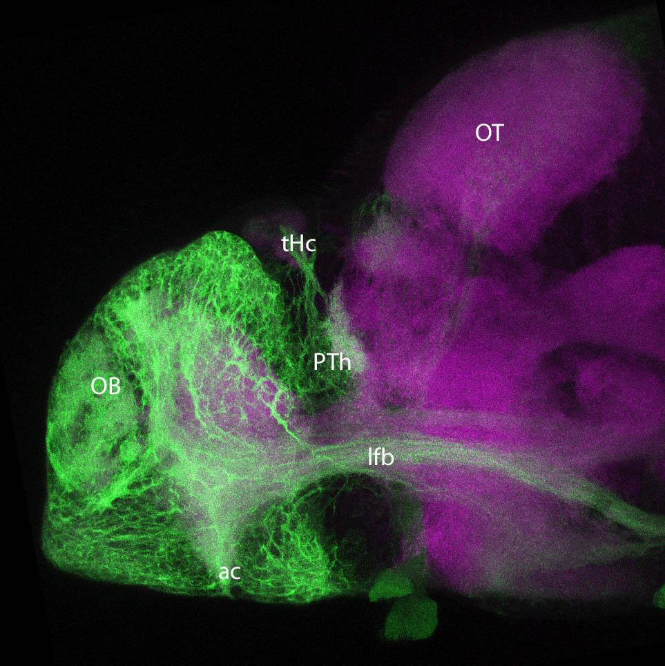
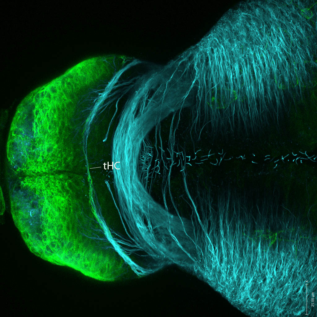
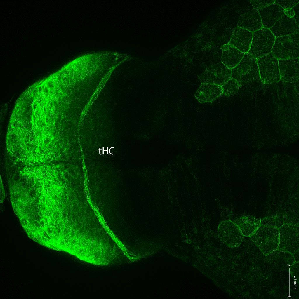
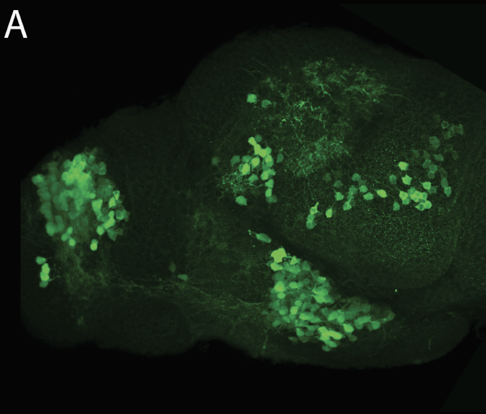
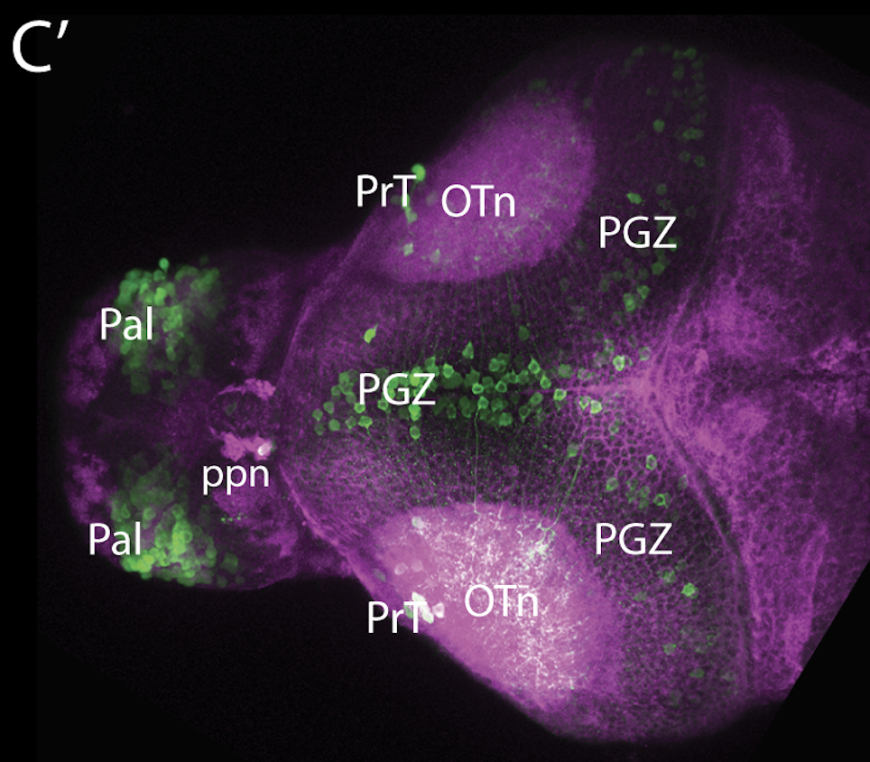
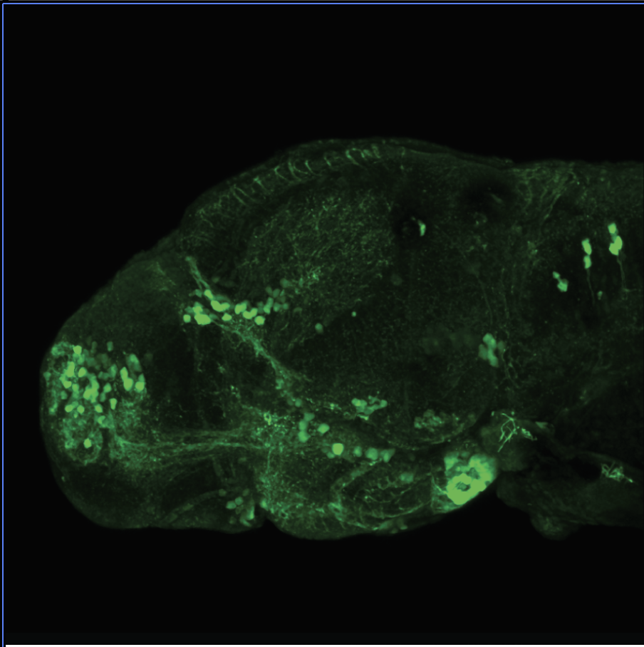
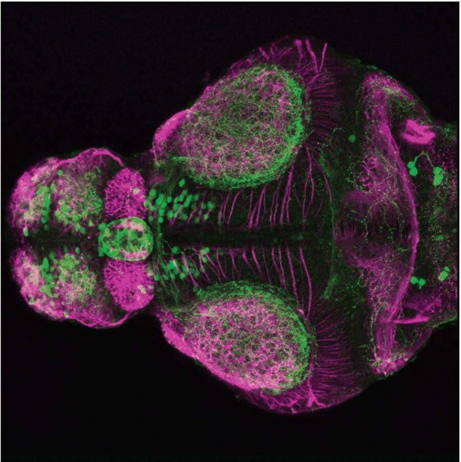
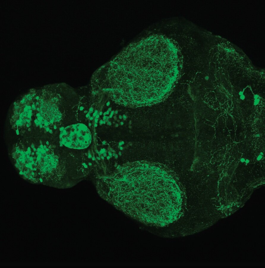
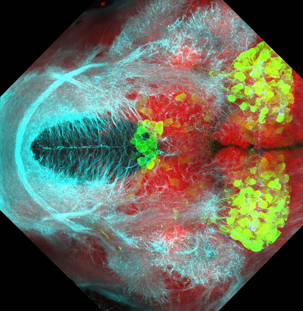
The Gilmore lab wanted to label the lateral line and neuromasts of the lateral line system, one allele of this transgenic also had EGFP expression in the nasal retina and telencephalon and has been used by other labs to study eye and telencephalic morphogenesis.


External Links:

 ZFIN

Lab or Origin: Gilmore Lab


Expressed in: 

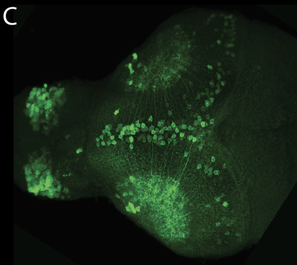
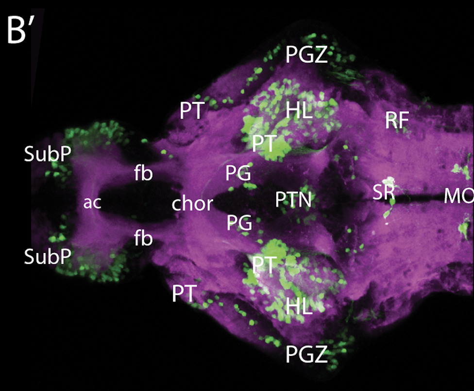
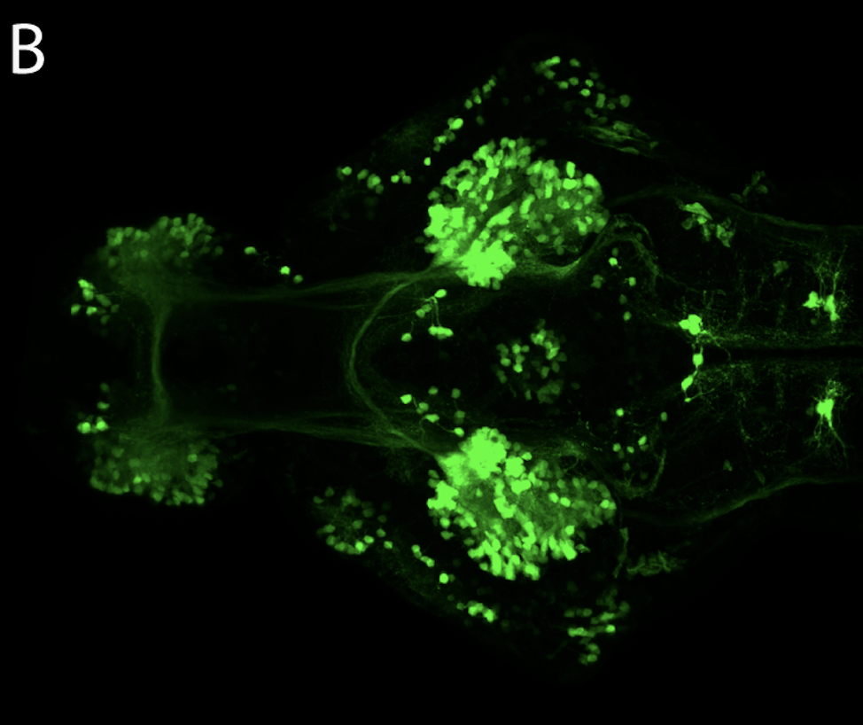
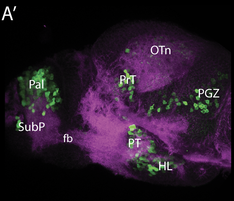
neuromasts, lateral line, olfactory epithelium, olfactory bulb, , pallium, subpallium, tract of the habenula commissure, nasal retina.


Key Publications

 Haas, P., and Gilmour, D. (2006)
Chemokine signaling mediates self-organizing tissue migration in the zebrafish lateral line.
Developmental Cell. 10(5):673-680.

Folgueira, M., Bayley, P., Navratilova, P., Becker, T.S., Wilson, S.W., and Clarke, J.D. (2012)
Morphogenesis underlying the development of the everted teleost telencephalon.
Neural Development. 7(1):32.

Valdivia, L.E., Young, R.M., Hawkins, T.A., Stickney, H.L., Cavodeassi, F., Schwarz, Q., Pullin, L.M., Villegas, R., Moro, E., Argenton, F., Allende, M.L., and Wilson, S.W. (2011)
Lef1-dependent Wnt/β-catenin signalling drives the proliferative engine that maintains tissue homeostasis during lateral line development.
Development (Cambridge, England). 138(18):3931-3941.

Valentin, G., Haas, P., and Gilmour, D. (2007)
The chemokine SDF1a coordinates tissue migration through the spatially restricted activation of Cxcr7 and Cxcr4b.
Current biology : CB. 17(12):1026-1031.

Picker, A., Cavodeassi, F., Machate, A., Bernauer, S., Hans, S., Abe, G., Kawakami, K., Wilson, S.W., and Brand, M. (2009)
Dynamic coupling of pattern formation and morphogenesis in the developing vertebrate retina.
PLoS Biology. 7(10):e1000214.


Tg(1.4dlx5a-dlx6a:GFP)ot1

Tg(1.4dlx5a-dlx6a:GFP)ot1

About

Tg(1.4dlx5a-dlx6a:GFP)ot1 larvae express GFP in subpallial neurons γ-aminobutyric acid (GABA)-expressing neurons. Several other brain regions also show GFP expression in this transgenic line such as the optic tectum and cerebellum.
 

 Dlx homeobox genes play essential roles in the differentiation, migration and survival of subpallial precursor cells that will later give rise to diverse subtypes of γ-aminobutyric acid (GABA)-expressing neurons. They also participate in the regulation of the Gad genes encoding the enzymes necessary for GABA synthesis (Yu et al., 2011).


Mouse over the different areas of GFP expression in the interactive images below to see the name of the brain area. 


More images

External Links:

 ZFIN

Lab of Origin: Mark Ekker Lab


Expressed in: 

olfactory bulb, subpallium, pallium, preoptic area, prethalamus, posterior tuberculum, hypothalamus, optic tectum, cerebellum.


Key Publications

Zerucha, T., Stuhmer, T., Hatch, G., Park, B.K., Long, Q., Yu, G., Gambarotta, A., Schultz, J.R., Rubenstein, J.L., and Ekker, M. (2000) 
A highly conserved enhancer in the Dlx5/Dlx6 intergenic region is the site of cross-regulatory interactions between dlx genes in the embryonic forebrain. 
The Journal of neuroscience : the official journal of the Society for Neuroscience. 20(2):709-721.

Yu, M., Xi, Y., Pollack, J., Debiais-Thibaud, M., Macdonald, R.B., and Ekker, M. (2011) 
Activity of dlx5a/dlx6a regulatory elements during zebrafish GABAergic neuron development. 
Int. J. Dev. Neurosci.. 29(7):681-91.

Et(fos:Gal4-VP16)s1026t

Et(fos:Gal4-VP16)s1026t

About

 This transgenic originates from Herwig Baier’s laboratory and is one of many enhancer trap Gal4 lines created by them. It labels prethalamus, thalamus and posterior tubercular regions. It also has retinal, habenular, pineal, optic tectum and subpallial expression.


External Links:

 ZFIN

Lab or Origin:


Expressed in: 

 subpallium, habenulae, pineal, emminentia thalami, prethalamus, thalamus, posterior tuberculum, retina, optic tectum, hypothalamus, preglomerular complex.


Key Publications

 Scott, E.K., and Baier, H. (2009) The cellular architecture of the larval zebrafish tectum, as revealed by gal4 enhancer trap lines. Frontiers in neural circuits. 3:13.

Et(fos:Gal4-VP16)s1137t

Et(fos:Gal4-VP16)s1137t

About

 This transgenic originates from Herwig Baier’s laboratory and is one of many enhancer trap Gal4 lines created by them. It shows dense expression of kaede in the telencephalon and throughout all the habenula subnuclei. The insertion of this transgene is currently unmapped.


External Links:

 ZFIN

Lab or Origin: Baier Laboratory


Expressed in: 

 telencephalon, posterior tuberculum, habenula, hypothalamus


Key Publications

Mason, L., Scott, E.K., Staub, W., Finger-Baier, K., and Baier, H. (2009) Expression patterns from GAL4 enhancer trap screen. ZFIN Direct Data Submission. . (http://zfin.org).

Scott, E.K., and Baier, H. (2009) The cellular architecture of the larval zebrafish tectum, as revealed by gal4 enhancer trap lines. Frontiers in neural circuits. 3:13. 

Tg(gata2:eGFP)bi105

Tg(gata2:eGFP)bi105

About

The Tg(gata2:eGFP)bi105 enhancer trap transgenic line was generated as part of an enhancer trap screen by the Becker Lab using the Tol2-transposase system. It has GFP expression throughout the pallium, in a small subpallial nucleus, parapineal organ, optic tectum, pretectum, posterior tuberculum and lateral hypothalamus. In the hindbrain, several cell bodies are labelled in the superior raphe and areas of the reticular formation and caudally in areas of the medulla oblongata


External Links:

 ZFIN

Lab of Origin: Tom Becker Lab


Transgene expressed in: 

Key Publications


Folgueira, M., Bayley, P., Navratilova, P., Becker, T.S., Wilson, S.W., and Clarke, J.D. (2012)
Morphogenesis underlying the development of the everted teleost telencephalon.
Neural Development. 7(1):32


Ragvin, A., Moro, E., Fredman, D., Navratilova, P., Drivenes, O., Engström, P.G., Alonso, M.E., Mustienes, E.D., Gomez Skarmeta, J.L., Tavares, M.J., Casares, F., Manzanares, M., van Heyningen, V., Molven, A., Njølstad, P.R., Argenton, F., Lenhard, B., and Becker, T.S. (2010)
Long-range gene regulation links genomic type 2 diabetes and obesity risk regions to HHEX, SOX4, and IRX3.
Proceedings of the National Academy of Sciences of the United States of America. 107(2):775-780

Turner, K.J., Hawkins, T.A., Yáñez, J., Anadón, R., Wilson, S.W., Folgueira, M. (2016)
Afferent Connectivity of the Zebrafish Habenulae.
Frontiers in neural circuits. 10:30


Tg(isl1:GFP)rw0

Tg(isl1:GFP)rw0

About

 This transgenic line expresses GFP in cranial motor neurons. It was made using a construct that fuses Islet-1 promotor/enhancer sequences to GFP.

isl1 cranial nerves expanded-01.png

isl1 cranial nerves expanded-02.png

More images of this transgenic

External Links:

 ZFIN

Lab or Origin: Okomoto Lab


Expressed in: 

cranial motor neurons, hindbrain, subpallium, preoptic area. 


Key Publications

Higashijima, S., Hotta, Y., and Okamoto, H. (2000)
Visualization of cranial motor neurons in live transgenic zebrafish expressing green fluorescent protein under the control of the islet-1 promoter/enhancer.
The Journal of neuroscience. 20(1):206-218

Suli, A., Mortimer, N., Shepherd, I., and Chien, C.B. (2006)
Netrin/DCC signaling controls contralateral dendrites of octavolateralis efferent neurons.
The Journal of neuroscience. 26(51):13328-13337.

Schoppik, D., Bianco, I.H., Prober, D.A., Douglass, A.D., Robson, D.N., Li, J.M.B., Greenwood, J.S.F., Soucy, E., Engert, F., Schier, A.F. (2017)
Gaze-stabilizing central vestibular neurons project asymmetrically to extraocular motoneuron pools. The Journal of neuroscience. 37(47):11353-11365.

Rebman, J.K., Kirchoff, K.E., Walsh, G.S. (2016)
Cadherin-2 Is Required Cell Autonomously for Collective Migration of Facial Branchiomotor Neurons.
PLoS One. 11:e0164433.

Barsh, G.R., Isabella, A.J., Moens, C.B. (2017)
Vagus Motor Neuron Topographic Map Determined by Parallel Mechanisms of hox5 Expression and Time of Axon Initiation.
Current biology : CB. 27(24):3812-3825.e3.

Cox, J.A., Lamora, A., Johnson, S.L., and Voigt, M.M. (2011)
Diverse mechanisms for assembly of branchiomeric nerves.
Developmental Biology. 357(2):305-17.

Et(krt4:EGFP)sqet11

Et(krt4:EGFP)sqet11

About

This enhancer trap construct carries the EGFP gene controlled by a partial epithelial promoter from the keratin8 gene.


External Links:

 ZFIN

Lab or Origin: Korzh Lab


Expressed in: 

 olfactory bulb, subpllium, parapineal, torus longitudinlis, optic tectum, posterior tuberal region, hypothalamus, pituitary


Key Publications

 Parinov, S., Kondrichin, I., Korzh, V., and Emelyanov, A. (2004) Tol2 transposon-mediated enhancer trap to identify developmentally regulated zebrafish genes in vivo. Developmental dynamics : an official publication of the American Association of Anatomists. 231(2):449-459.

pku2Et/ ETvmat2:GFP

pku2Et/ ETvmat2:GFP

About

 Synonyms: Et(gata2a:EGFP)pku2, Et(gata2a:GFP)zf81, pku2Et, ETvmat2:GFP

The ETvmat2:GFP transgenic zebrafish line was identified from a large scale enhancer trap screen (unpublished data (Wen et al., 2008)) using a Tol2 vector containing a 249 bp zebrafish gata2 minimal promoter linked to a GFP reporter gene. (Wen et al., 2008). The enhancer trap vector was inserted in the second intron of vmat2 gene with the transcription direction of the GFP reporter opposite to that of the vmat2 gene (Wen et al., 2008).

This enhancer trap line recapitulates the expression pattern of vmat2 and labels most monaminergic neurons in the zebrafish. Vesicular monoamine transporter 2 (Vmat2) is a monoamine transporter VMAT2 is a membrane protein that transports monoamines, including neurotransmitters such as dopamine, norepinephrine, serotonin, and histamine from the cytosol into synaptic vesicles.


External Links:

 ZFIN

Lab or Origin: PKU Zebrafish Functional Genomics Group


Expressed in: 

 monoaminergic neurons, olfactory bulb, telencephalon, pretectum, pineal, posterior tuberculum, torus longitudinalis, raphe, ventral lateral group of serotinergic neurons in hindbrain(VL) , hypothalamus, locus coerulus.


Key Publications

Wen, L., Wei, W., Gu, W., Huang, P., Ren, X., Zhang, Z., Zhu, Z., Lin, S., and Zhang, B. (2008)
Visualization of monoaminergic neurons and neurotoxicity of MPTP in live transgenic zebrafish. Developmental Biology. 314(1):84-92. 

Farrar, M.J., Kolkman, K.E., Fetcho, J.R. (2018)
Features of the structure, development and activity of the Zebrafish Noradrenergic System explored in new CRISPR transgenic lines.
The Journal of comparative neurology. 526(15):2493-2508.

Tg(slc6a3:EGFP)ot80

Tg(slc6a3:EGFP)ot80

About

The Tg(slc6a3:EGFP)ot80 transgenic line expresses GFP under the control of
cis-regulatory elements of the dopamine transporter (DAT) gene slc6a3.


External Links:

 ZFIN

Lab of Origin: Mark Ekker Lab


Transgene expressed in: 

olfactory bulb, subpallium, preoptic region, pretectum, posterior tuberculum, hypothalamus, optic tectum, cerebellum

Key Publications


Xi Y1, Yu M, Godoy R, Hatch G, Poitras L, Ekker M.
Transgenic zebrafish expressing green fluorescent protein in dopaminergic neurons of the ventral diencephalon.
Dev Dyn. 2011 Nov;240(11):2539-47. doi: 10.1002/dvdy.22742. Epub 2011 Sep 19.


Tg(slc17a6b: DsRed)nns9Tg

Tg(slc17a6b: DsRed)nns9Tg

About

 Slc17a6b is a vesicular glutamate transporter that that mediates the uptake of the excitatory neurotransmitter glutamate into vesicles in the presynaptic terminals of excitatory neurons. This BAC transgenic line from the Yoshihara lab drives the expression of DSRed in glutamatergic neurons. Expression can be seen in many neurons throughout the brain. Strong expression in the dorsal and ventral habenular subnuclei, olfactory bulbs, the pallium and optic tectum


External Links:

 ZFIN

Lab or Origin: Yoshihara Lab


Expressed in: 

 glutamatergic neurons, olfactory epithelium, olfactory bulb, pallium, subpallium, dorsal habenula, ventral habenula, preoptic area, prethalamus, pretectum (AF9), optic tectum, trigeminal sensory ganglion.


Key Publications

 
Miyasaka, N., Morimoto, K., Tsubokawa, T., Higashijima, S., Okamoto, H., and Yoshihara, Y. (2009)
From the olfactory bulb to higher brain centers: genetic visualization of secondary olfactory pathways in zebrafish.
The Journal of neuroscience. 29(15):4756-4767.

Kani, S., Bae, Y.K., Shimizu, T., Tanabe, K., Satou, C., Parsons, M.J., Scott, E., Higashijima, S.I., and Hibi, M. (2010) Proneural gene-linked neurogenesis in zebrafish cerebellum.
Developmental Biology. 343(1-2):1-17.

Tg5(Hsa.SOX3-gata2a:EGFP)bi85Tg

Tg5(Hsa.SOX3-gata2a:EGFP)bi85Tg

About


The construct used to create this enhancer trap line contains the highly conserved noncoding element  "hs7" from the regulatory region of the human SOX3 gene.


External Links:

 ZFIN

Lab or Origin: Tom Becker Lab


Expressed in: 

 olfactory bulb, subpallium, habenula, AF9 pretectum, optic tectum, cerebellum.


Key Publications

 Navratilova, P., Fredman, D., Hawkins, T.A., Turner, K., Lenhard, B., and Becker, T.S. (2009) Systematic human/zebrafish comparative identification of cis-regulatory activity around vertebrate developmental transcription factor genes. Developmental Biology. 327(2):526-540.


	io005Tg/Tg(Xleomes:GFP)io005

io005Tg/Tg(Xleomes:GFP)io005

About

 Synonyms: Tg(Xleomes:GFP)io005, tg(Xeom:GFP)

The tg(Xeom:GFP) transgenic lines were made in the Mione lab and used to study the dorsal to ventral migration of the paraseptal neurons in the subpallium.

“GFP-expressing cells appear around 28 hpf in the telencephalon of tg(Xeom:GFP) transgenic embryos. Paired groups of GFP-expressing cells appear in the lateral region of the telencephalon more or less at the level of the olfactory placode, at the telencephalic/diencephalic border and in the midbrain tegmentum.
We describe the migration of the telencephalic group: these cells originate from the corresponding ventricular zone, they first move towards the lateral side and then proceed rapidly towards the ventral telencephalon. Most of the GFP+ cells will congregate at the level of the septal area just rostral to the anterior commissure The 3 cell groups are interconnected through the anterior commissure and the middle forebrain bundle.” (Mione et al., 2008).

“Telencephalic Xeom:GFP- expressing cells probably correspond to the eomes/tbr1- expressing cells located in the ventral telencephalon of zebrafish [Mione et al., 2001] and of many other vertebrates [Brox et al., 2004; Puelles et al., 2000]. The observation that these cells originate from a dorsal telencephalic position and undergo an extensive migration towards the ventral telencephalon and diencephalon, accompanying the growth of the MOT and MFB, suggests that they may play pioneer roles on the formation of these major axon tracts. “(Mione et al., 2008).


External Links:

 ZFIN

Lab or Origin: Mione Lab


Expressed in: 

 septal region, subpallium, entopeduncular nucleus, thalamic emminence, midbrain tegmentum, optic tectum, cerebellum.


Key Publications

Mione, M., Baldessari, D., Deflorian, G., Nappo, G., and Santoriello, C. (2008)
How neuronal migration contributes to the morphogenesis of the CNS: insights from the zebrafish. Developmental neuroscience. 30(1-3):65-81.

Mione M, Shanmugalingam S, Kimelman D, Griffin K (2001):
Overlapping expression of zebrafish T-brain-1 and eomesodermin during forebrain development.
Mech Dev 100: 93–97.

Puelles L, Rubenstein JL (2003)
Forebrain gene expression domains and the evolving prosomeric model.
Trends Neurosci 26:469–476.

Puelles L, Kuwana E, Puelles E, Bulfone A, Shi- mamura K, Keleher J, Smiga S, Rubenstein JL (2000)
Pallial and subpallial derivatives in the embryonic chick and mouse telencephalon, traced by the expression of the genes Dlx-2, Emx-1, Nkx-2.1, Pax-6, and Tbr-1.
J Comp Neurol 424:409–438.