Viewing entries in
transgenic line

Tg44(FRT-Xla.Actc1:DsRed-GAB-FRT,LOXP-Hsa.IRX3-LOXP-gata2a:EGFP-5HS4)upo73Tg

Tg44(FRT-Xla.Actc1:DsRed-GAB-FRT,LOXP-Hsa.IRX3-LOXP-gata2a:EGFP-5HS4)upo73Tg

About

 Construct contains the highly conserved noncoding sequence C99 from the human IRXB cluster, containing the genes IRX3, IRX5, and IRX6.  

TG44 is an enhancer trap line from the Skarmeta Lab. Generated as part of a study looking at the transcriptional regulatory properties of highly-conserved noncoding elements on chromosome 16.
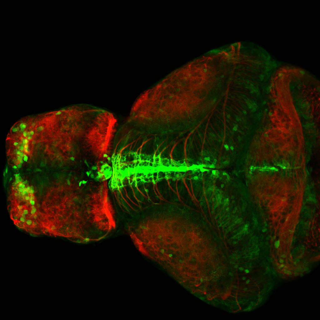
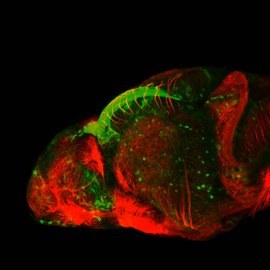
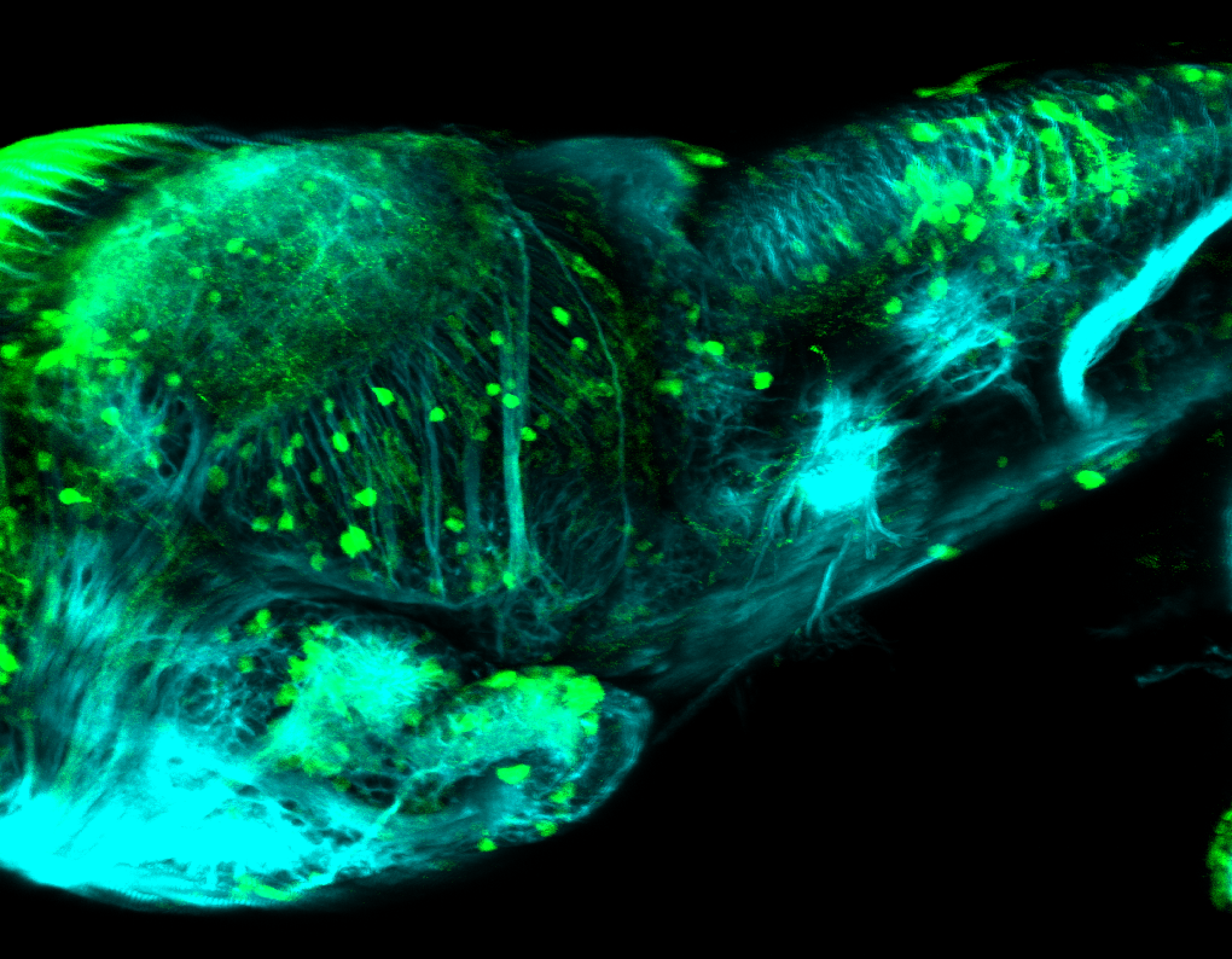
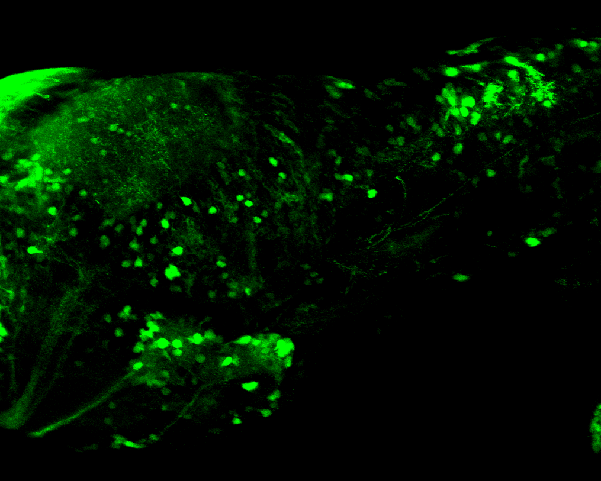
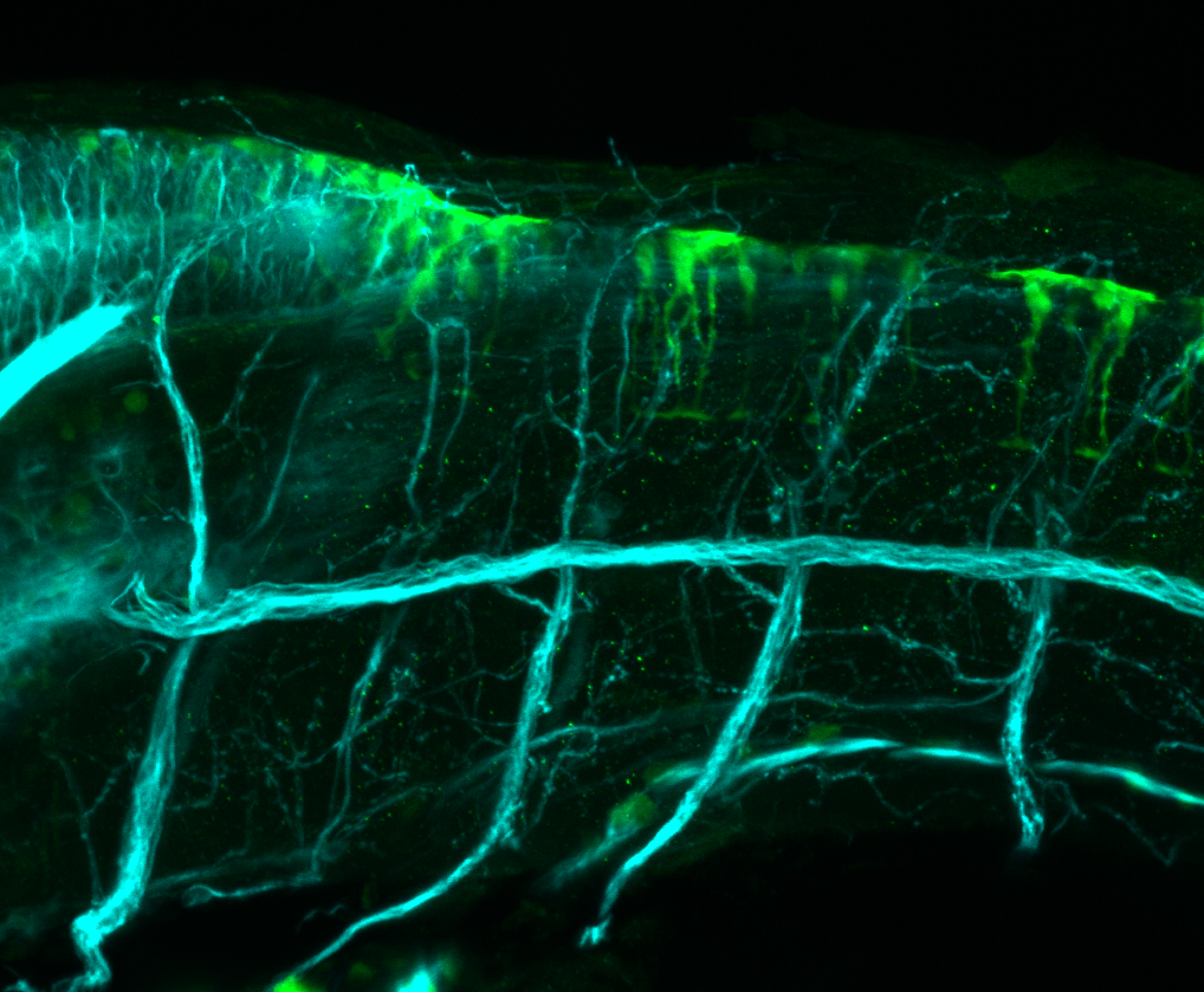
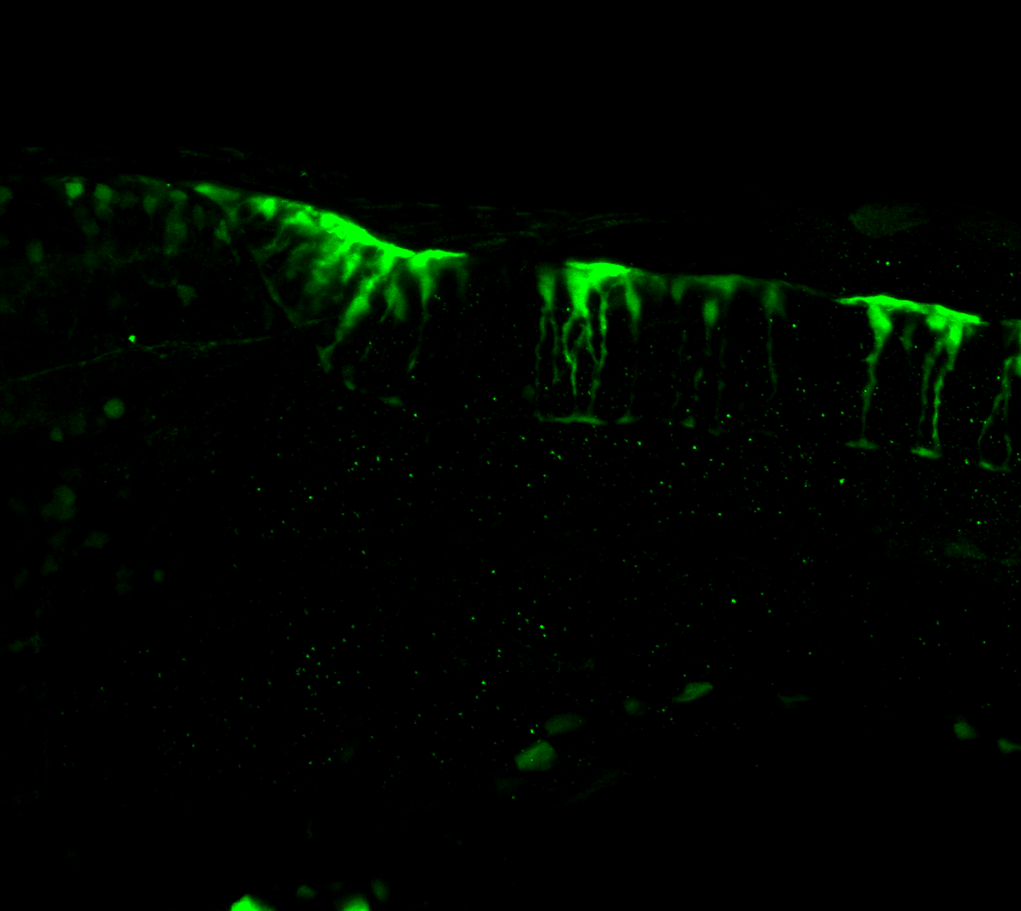
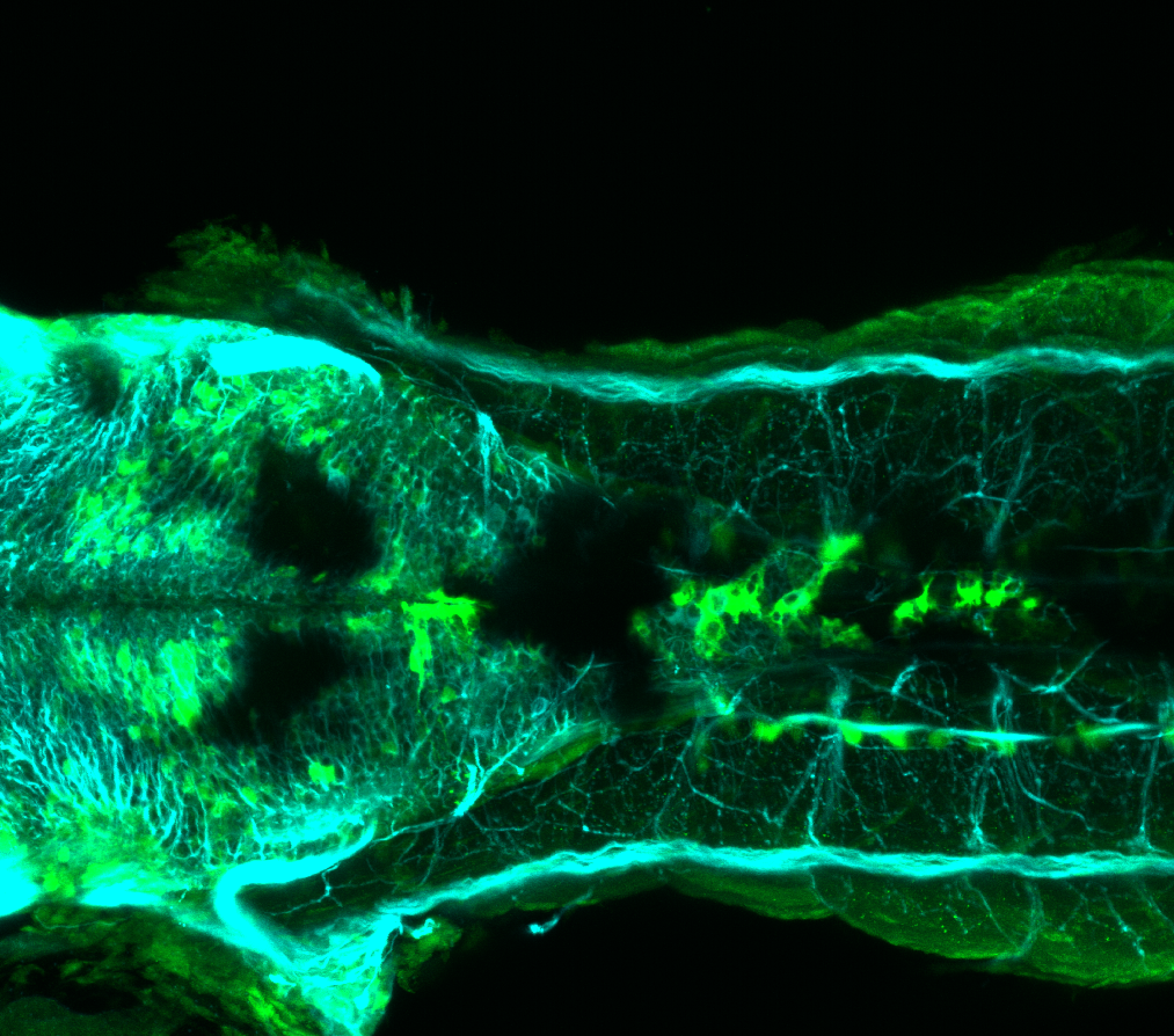
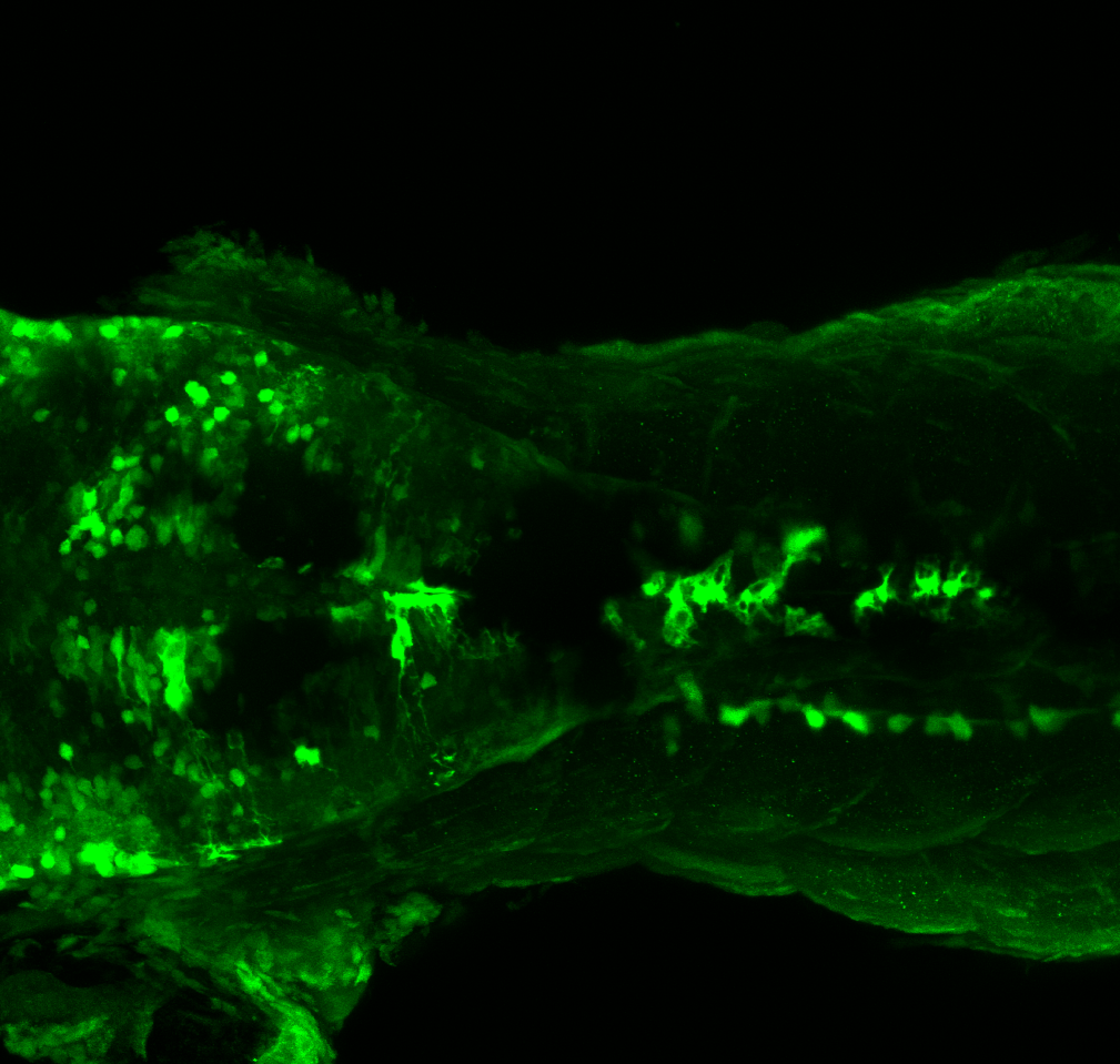
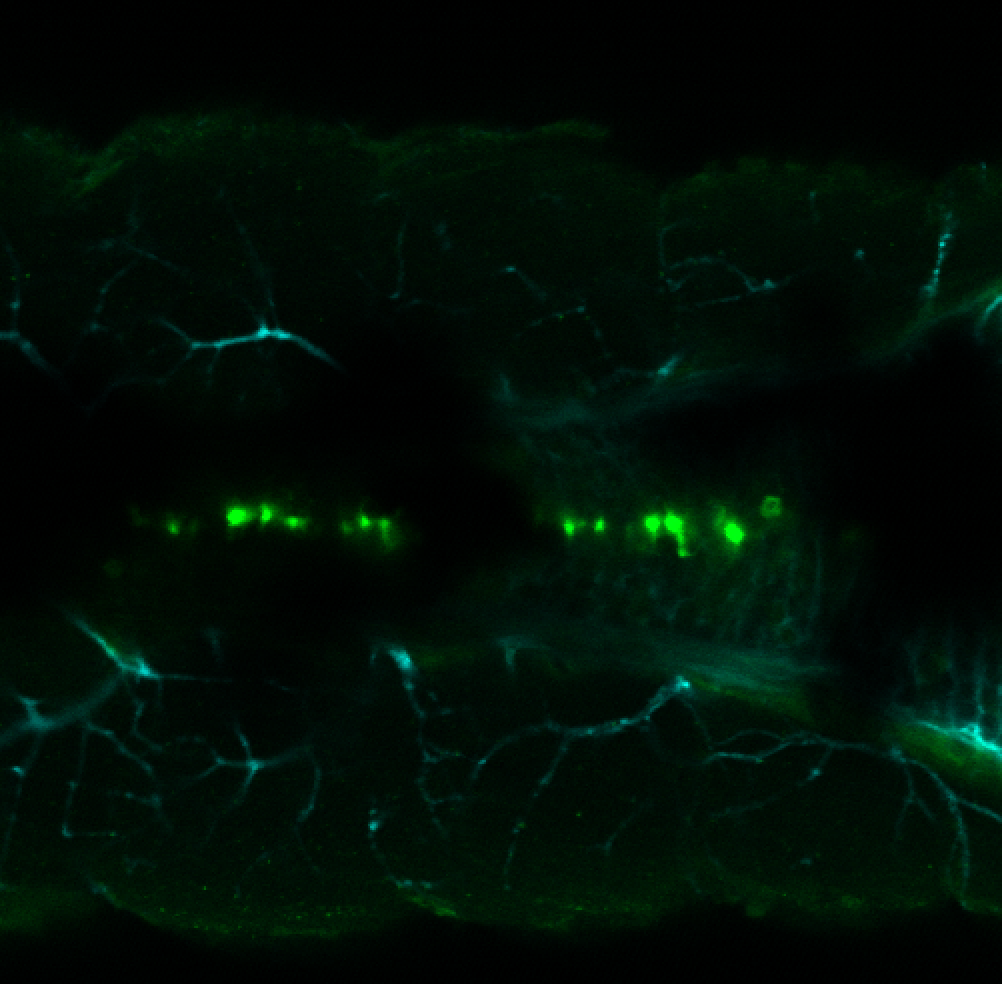
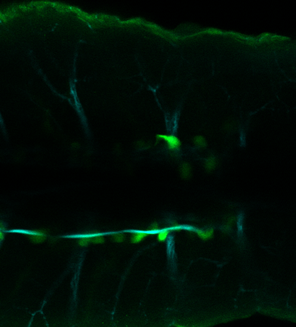
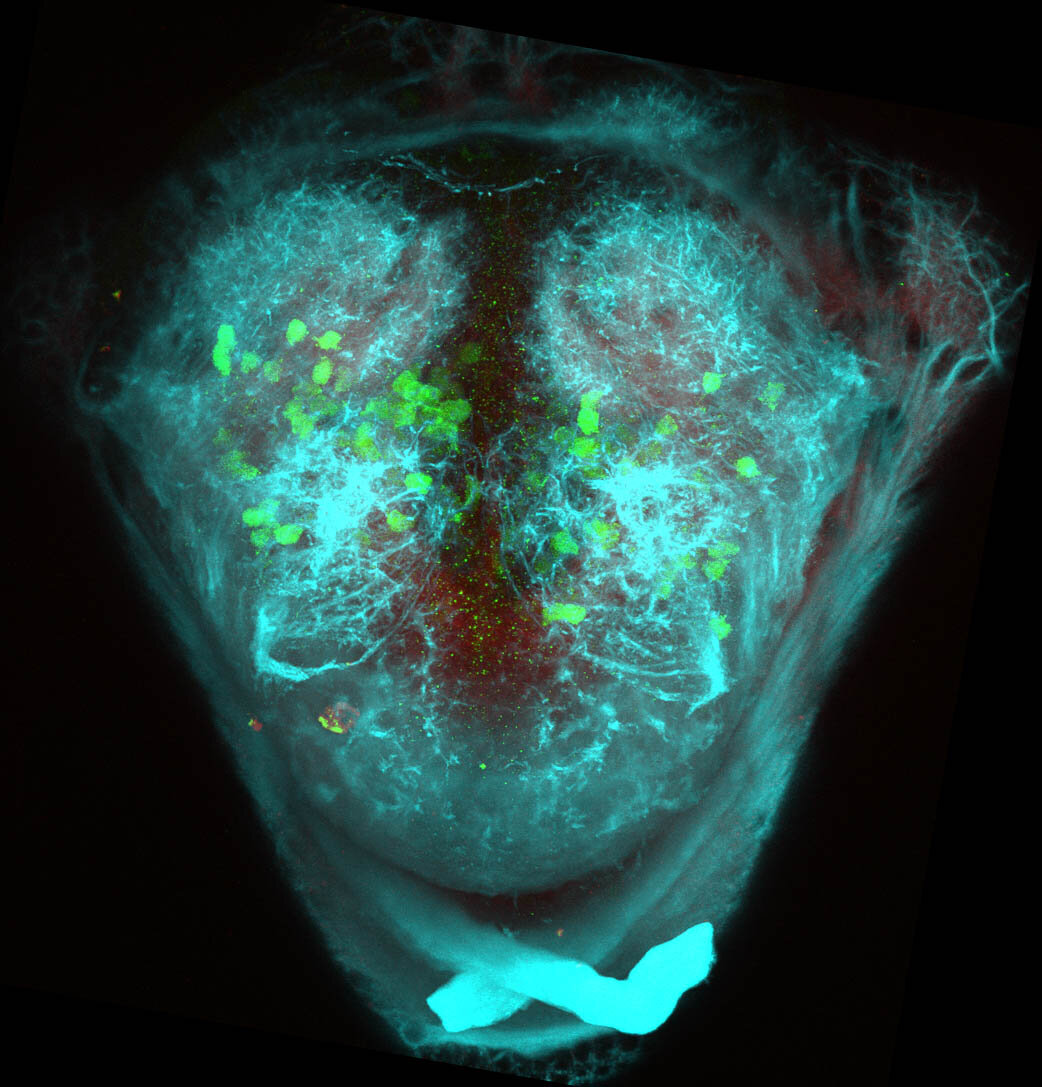
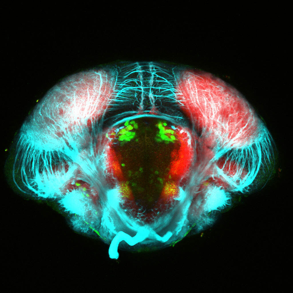
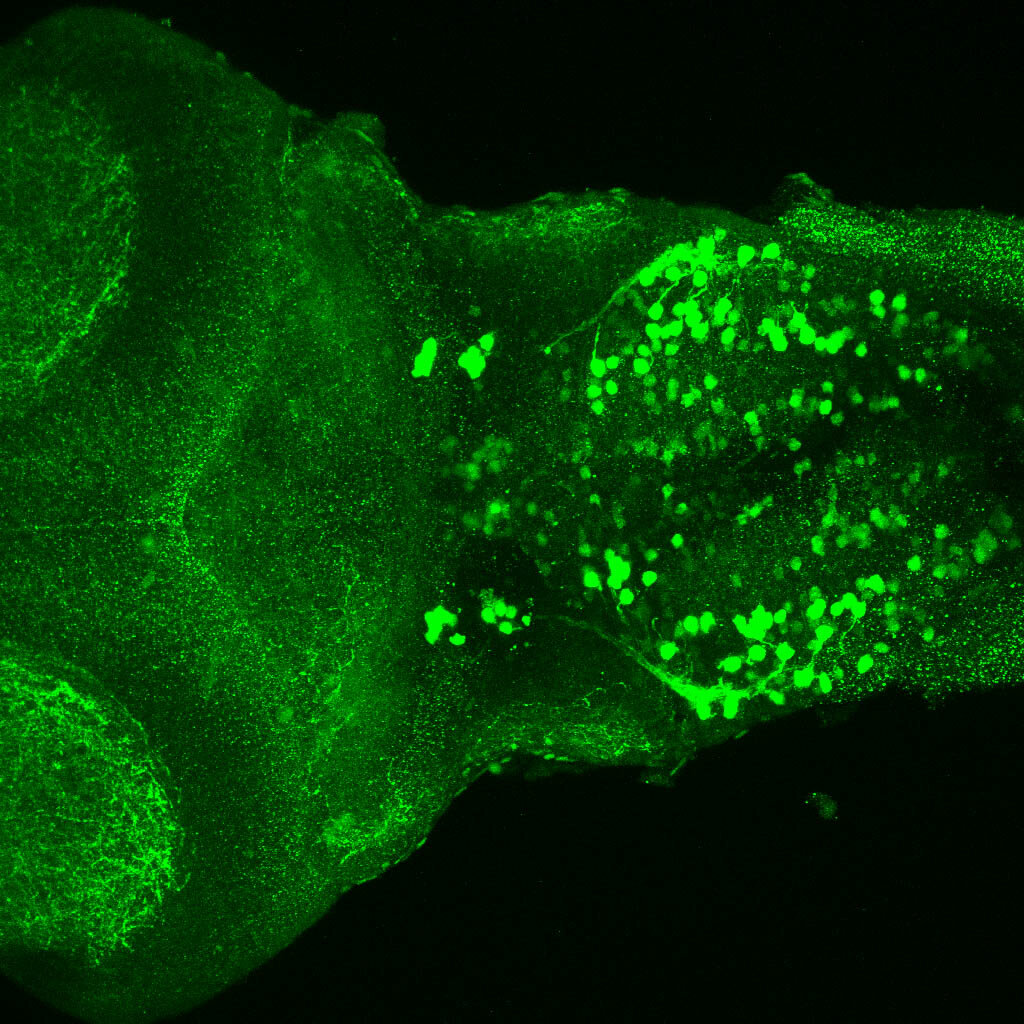
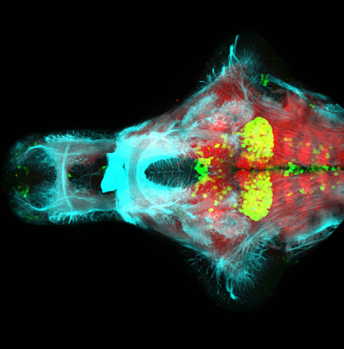
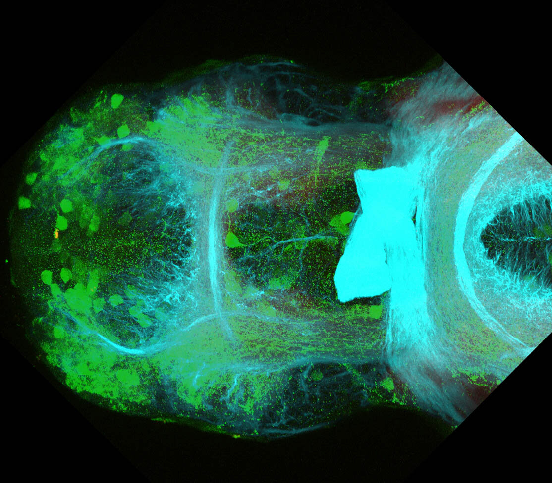
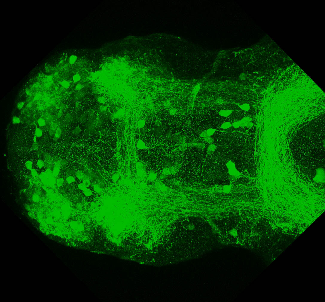
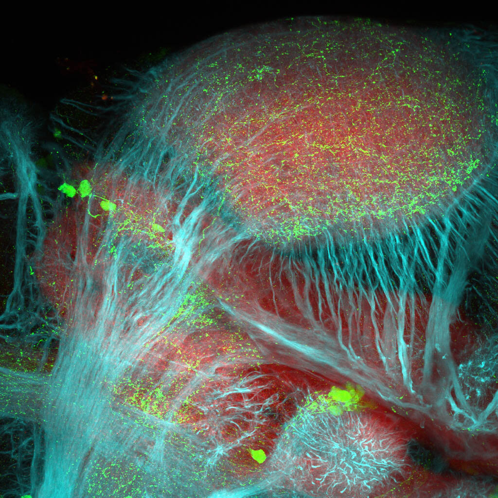
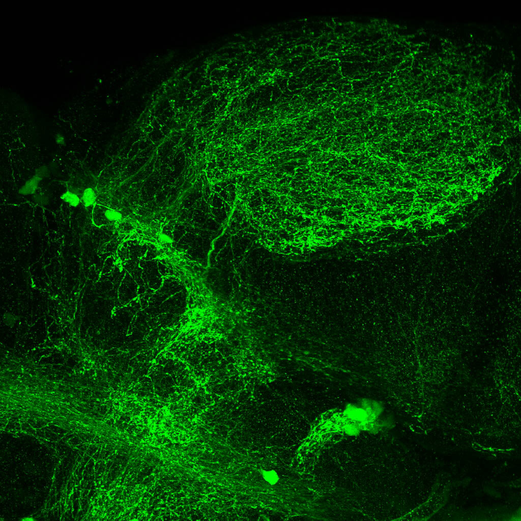
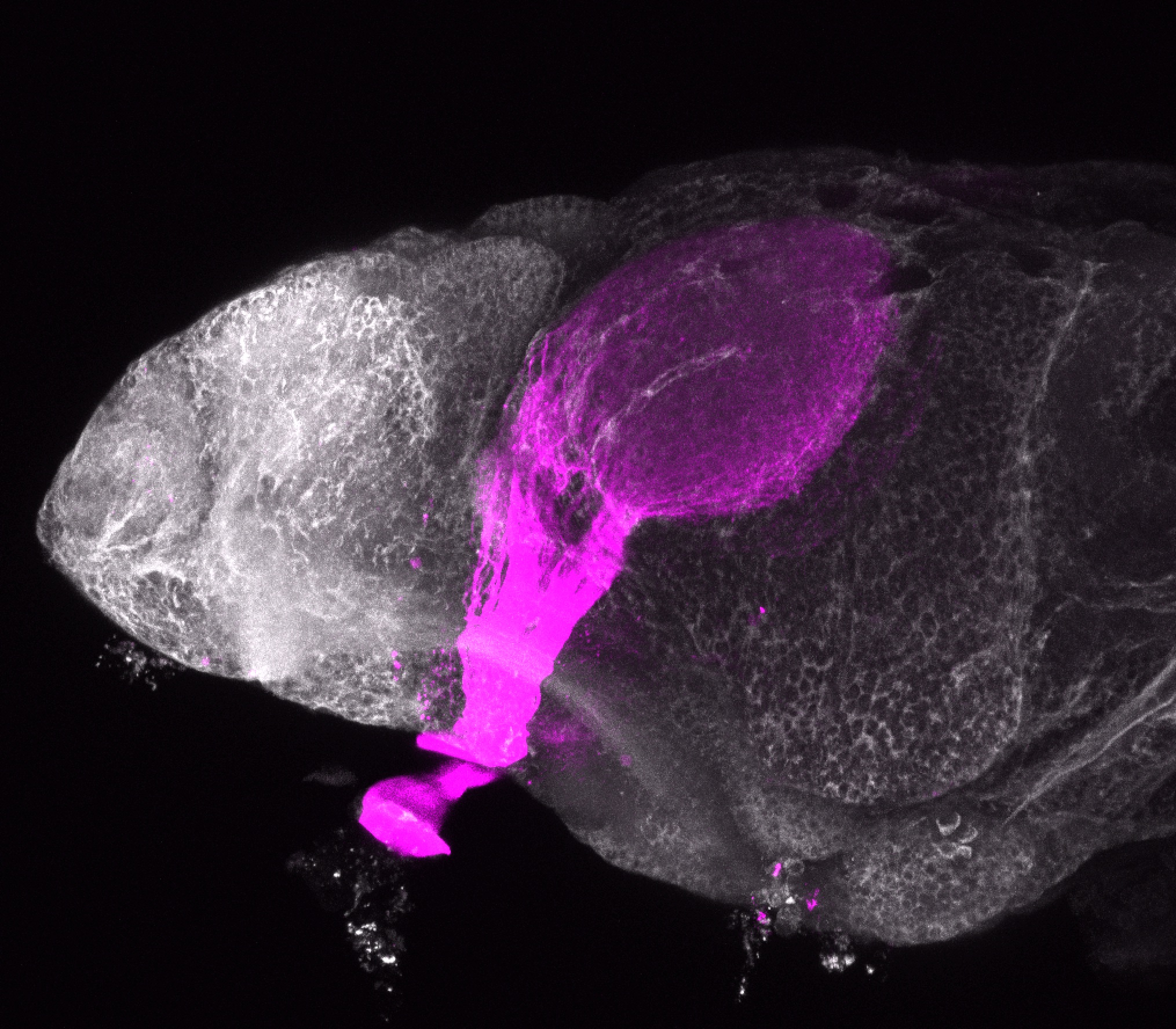
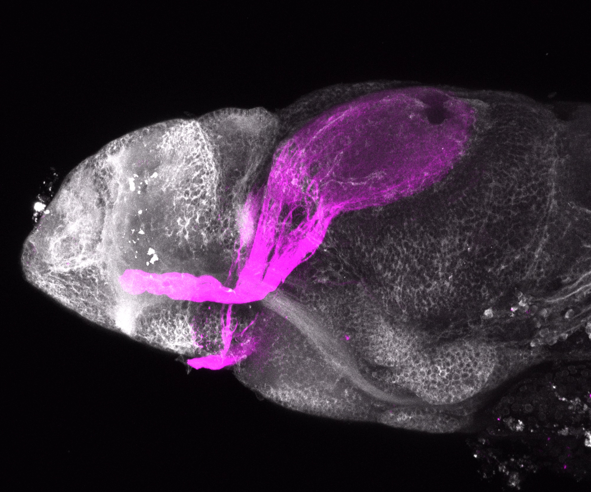
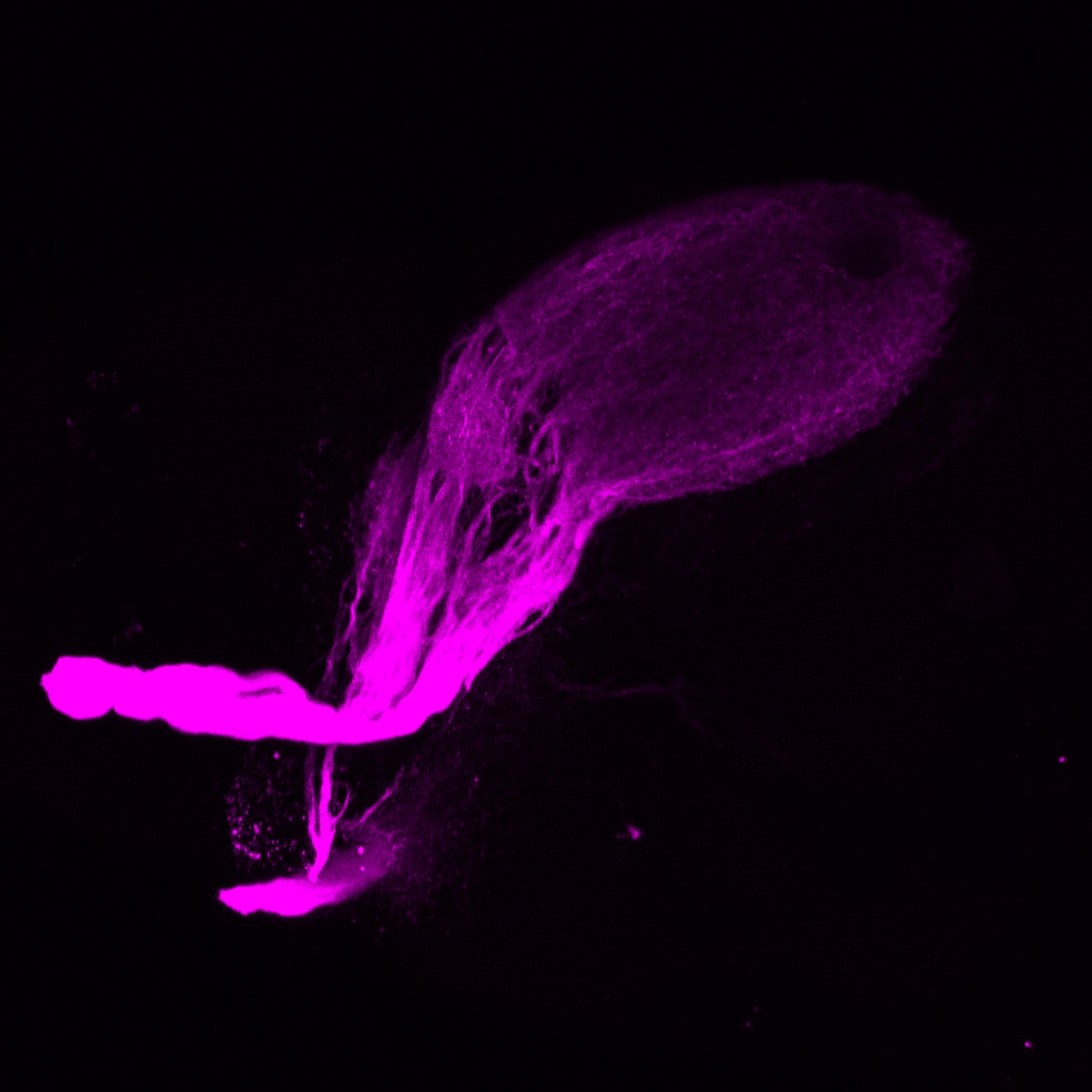
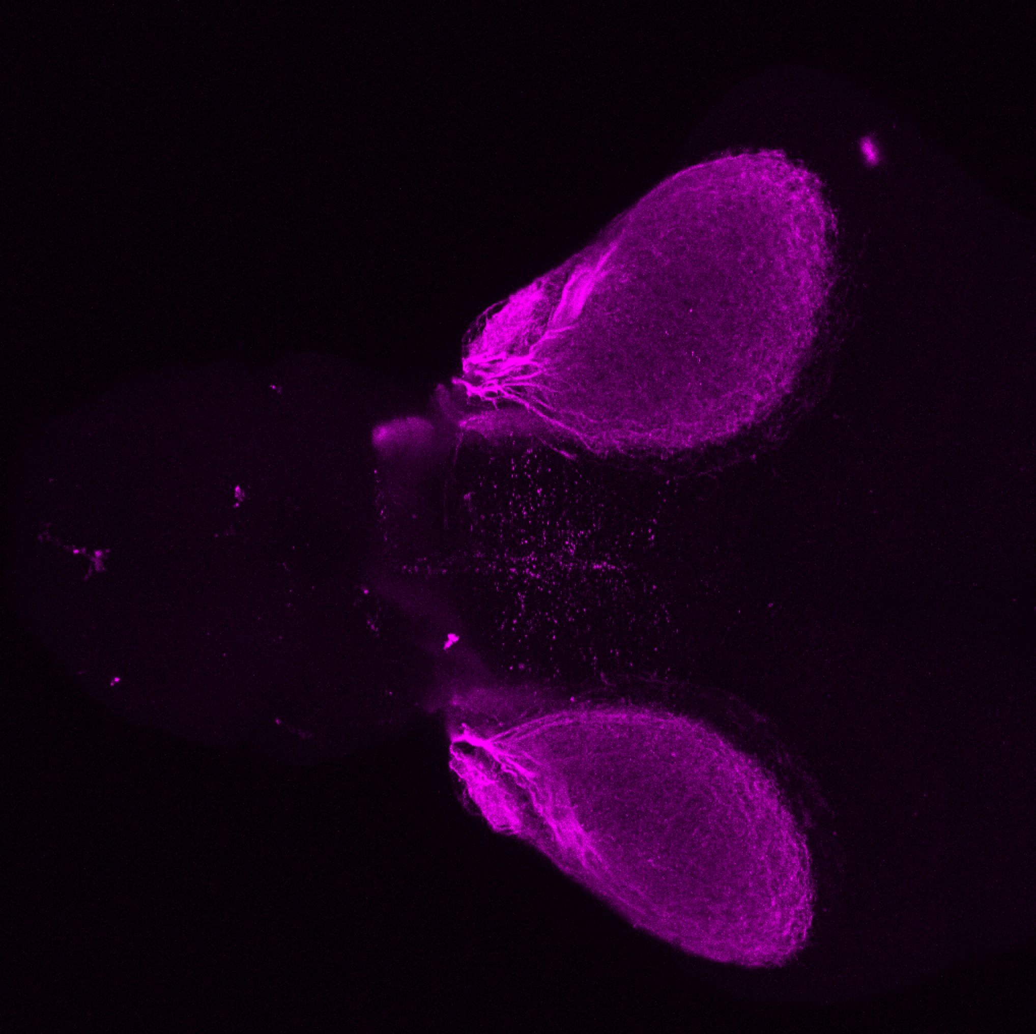
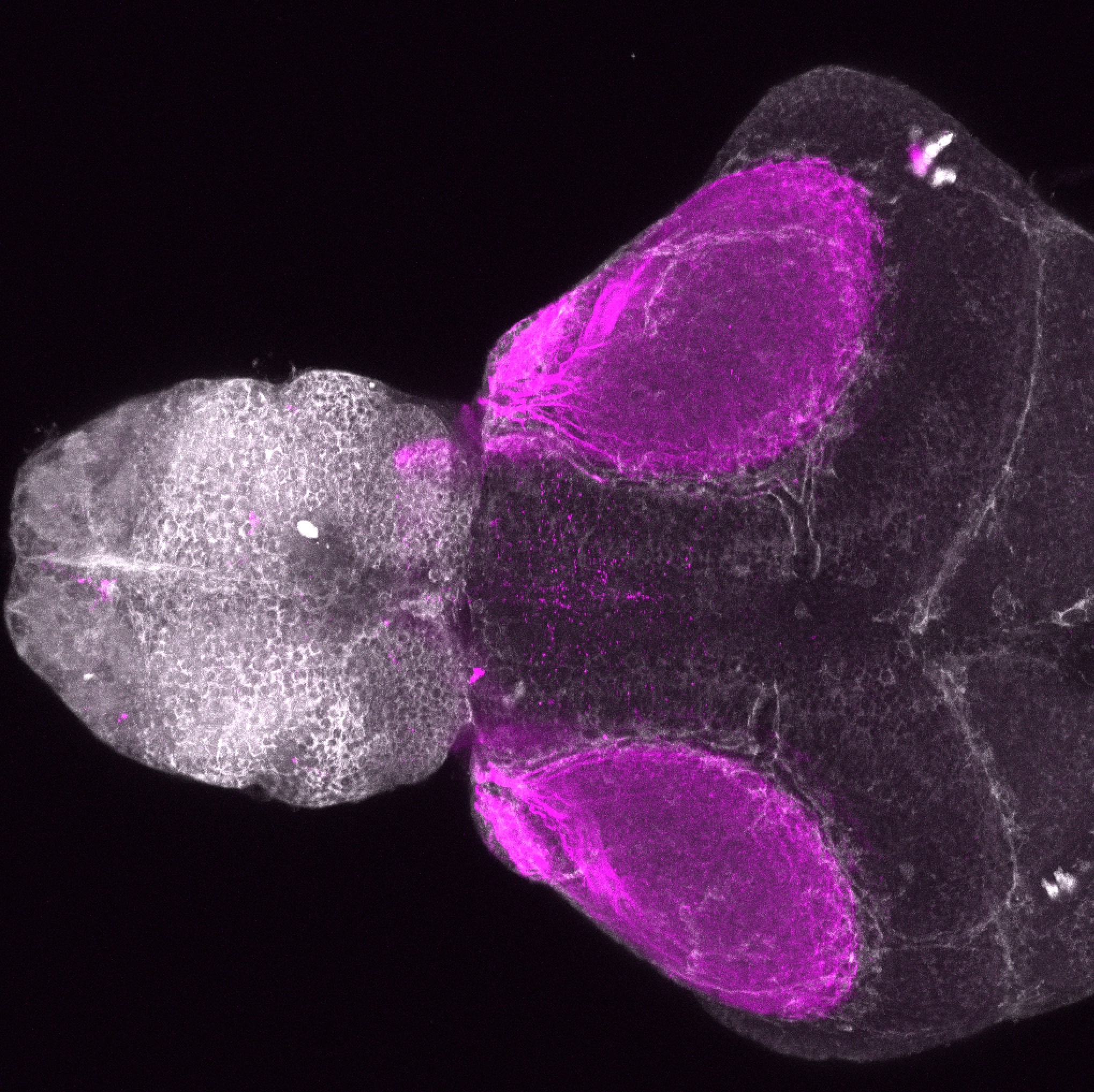
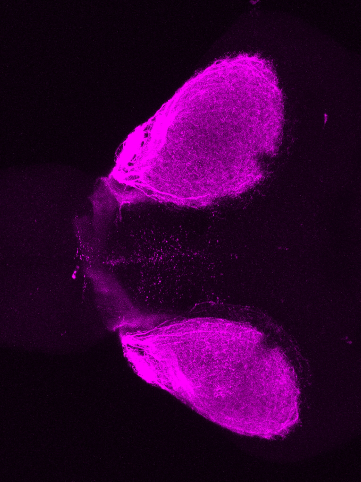
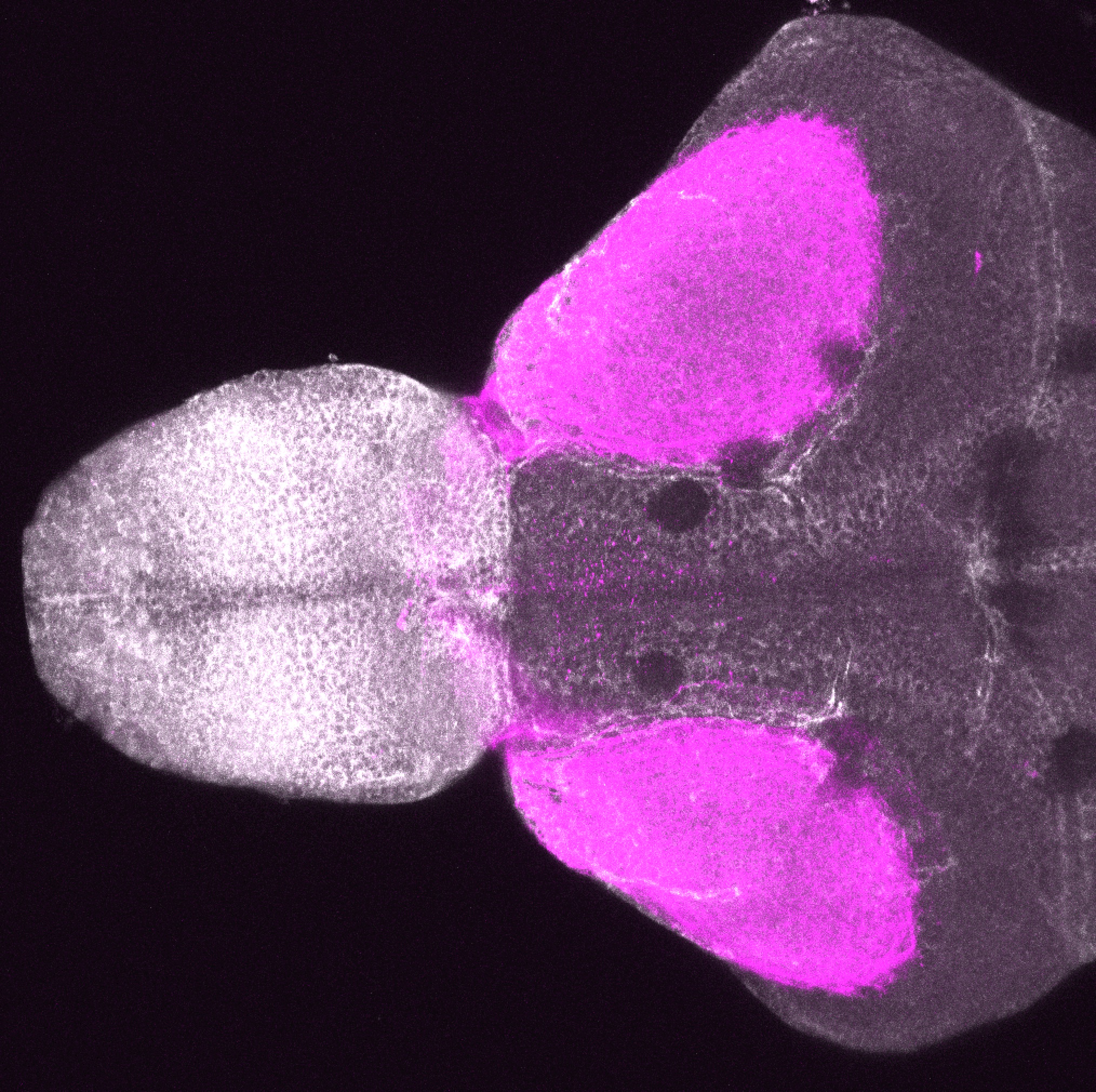
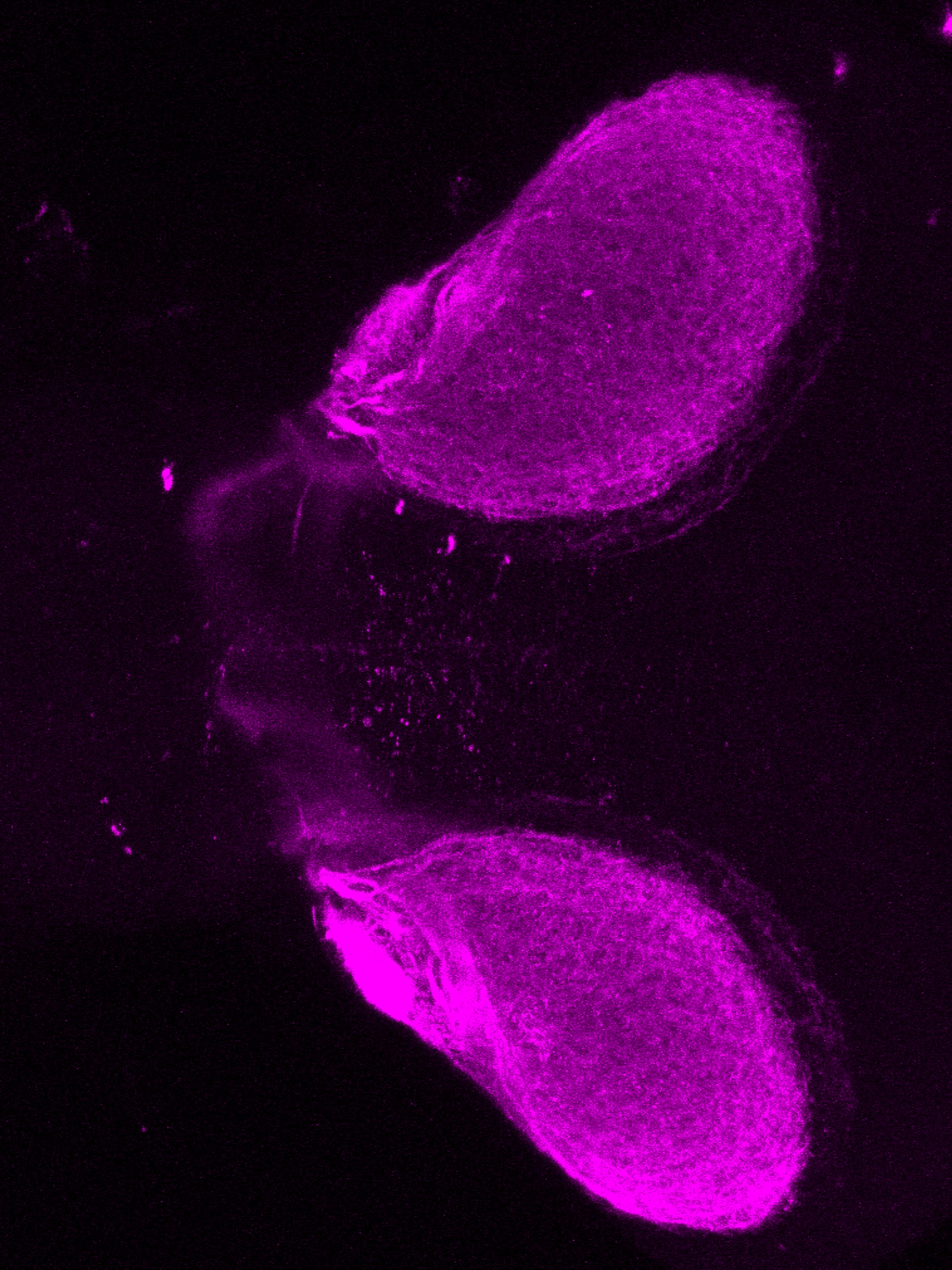
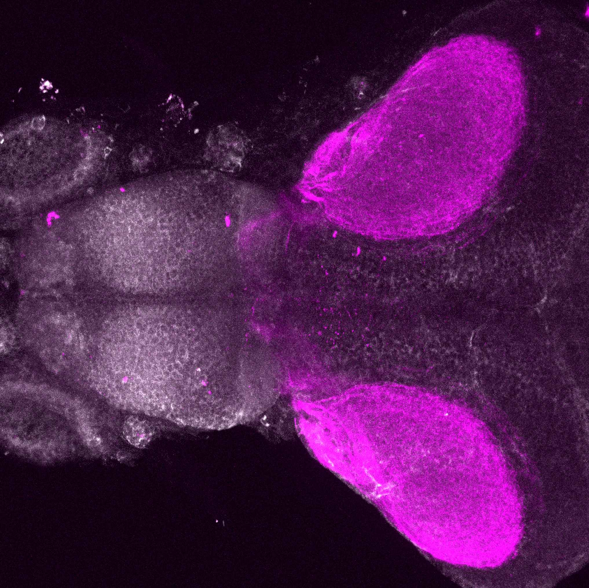
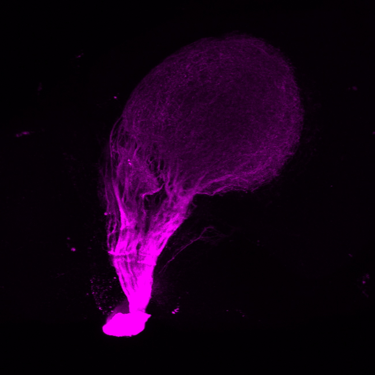
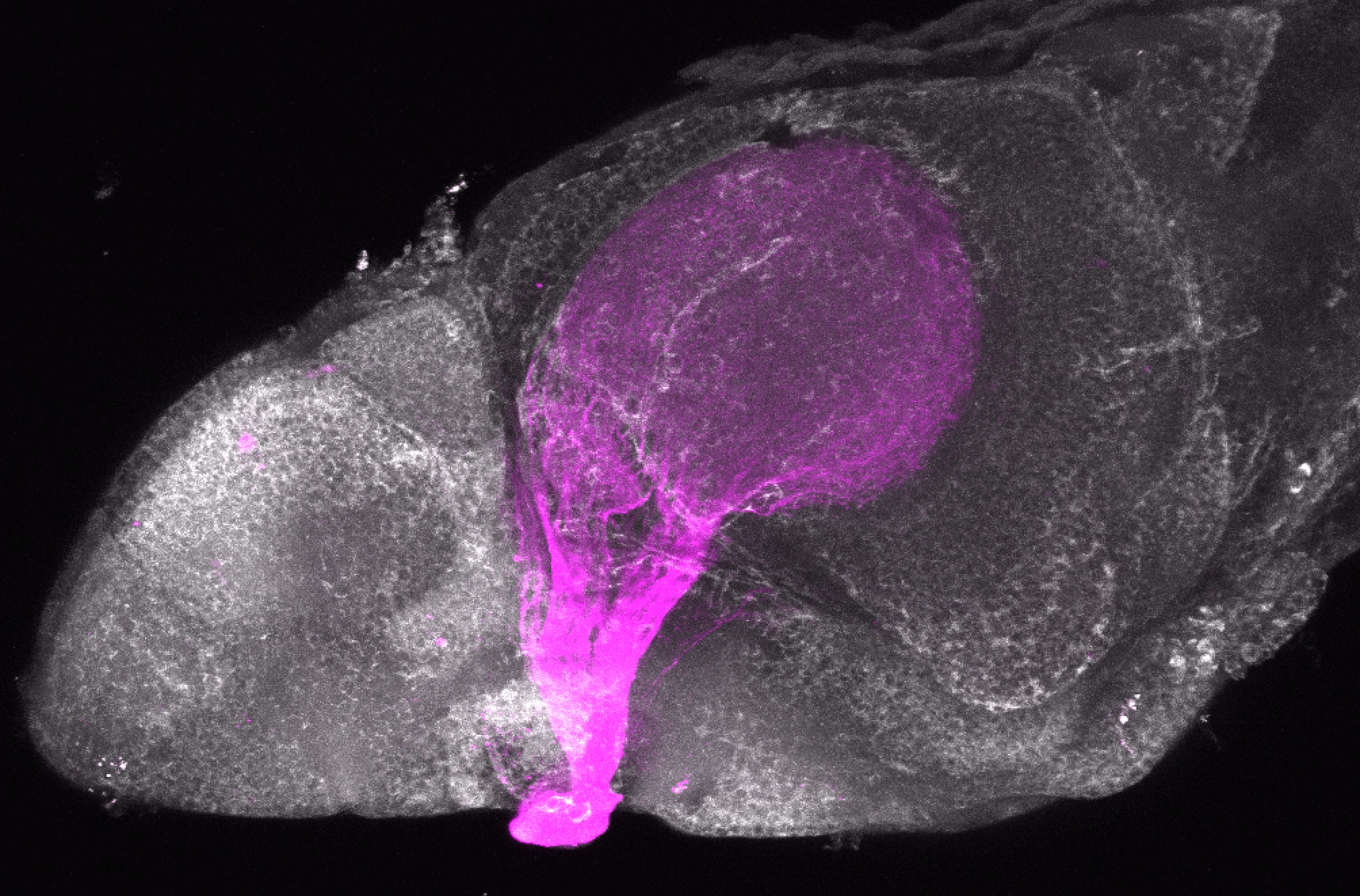
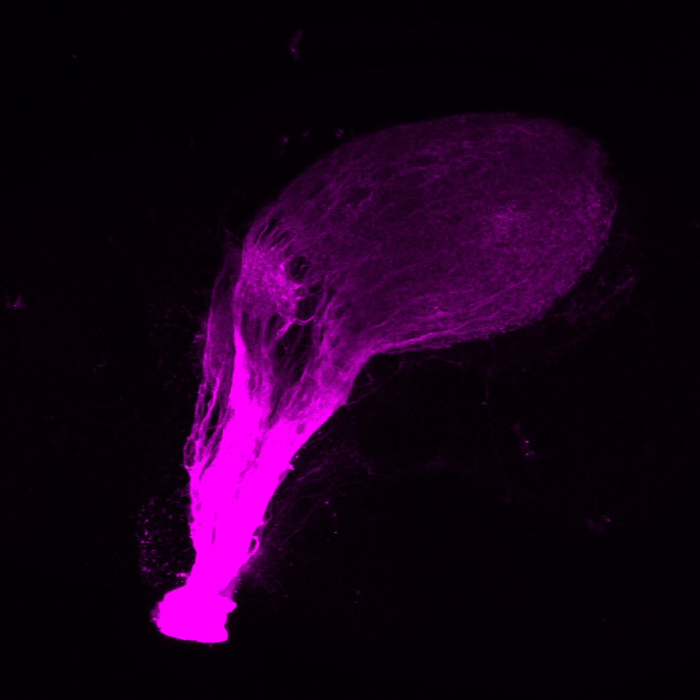
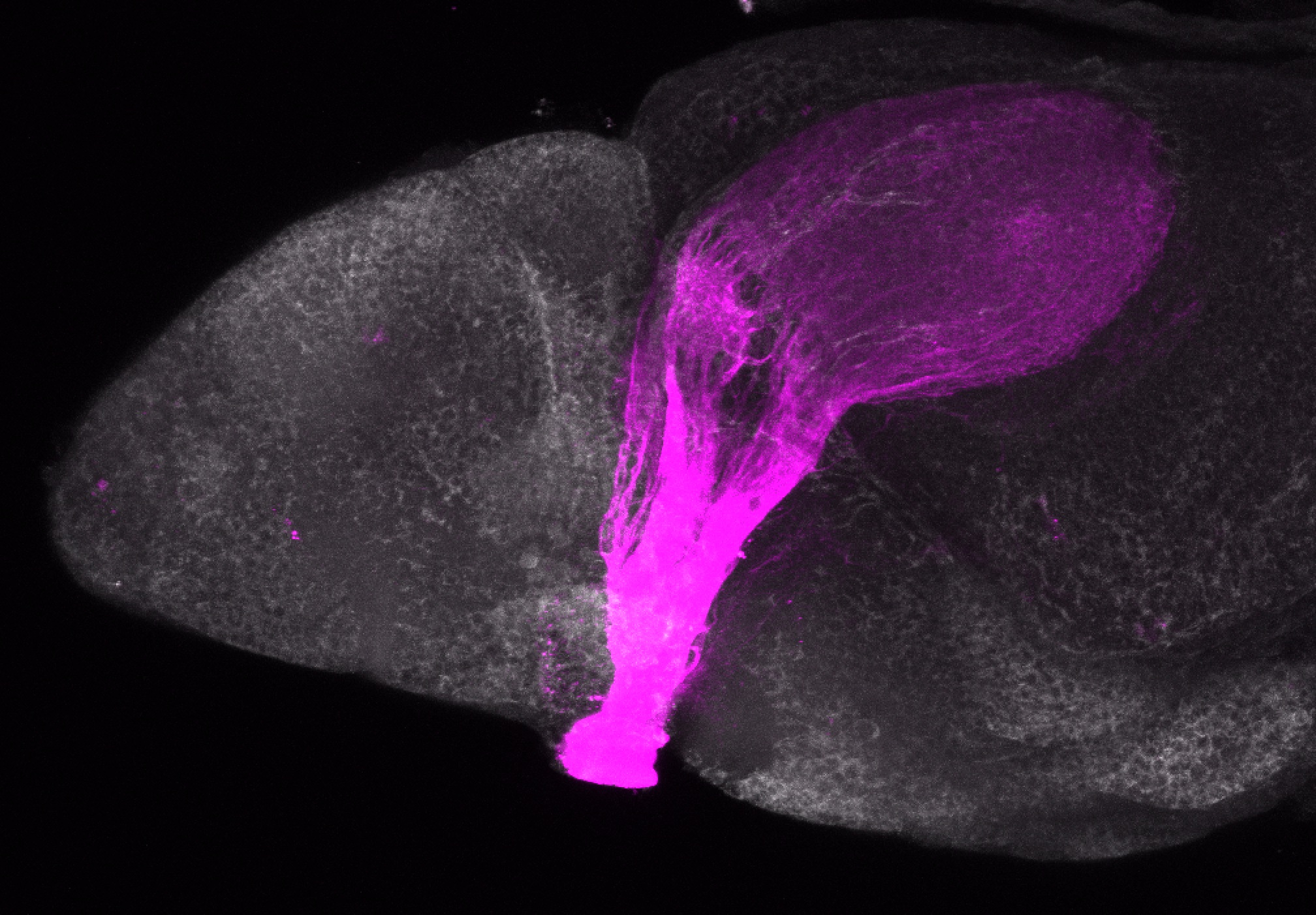
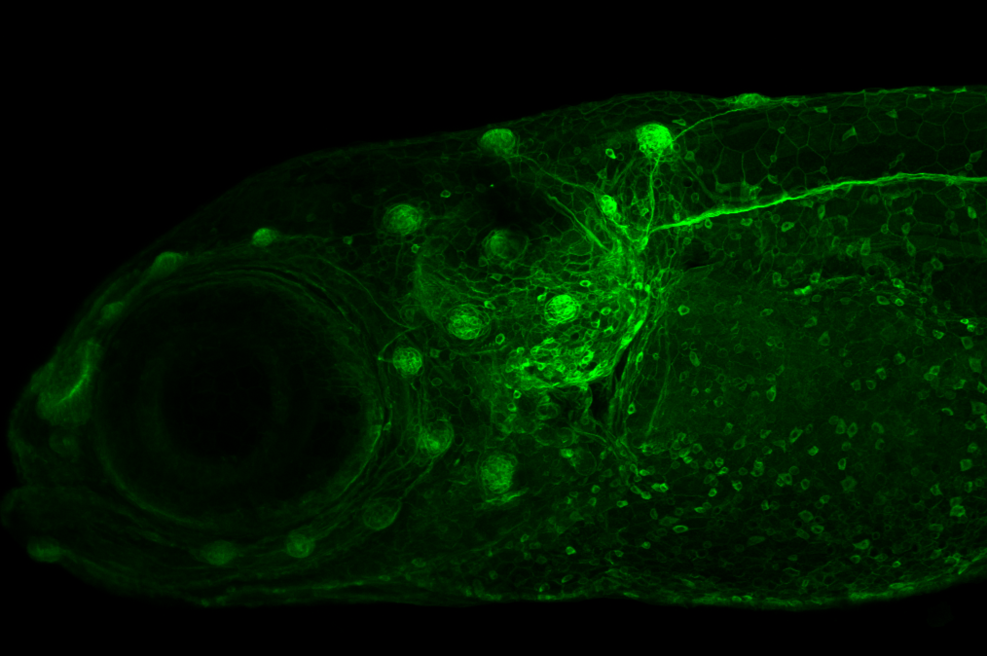
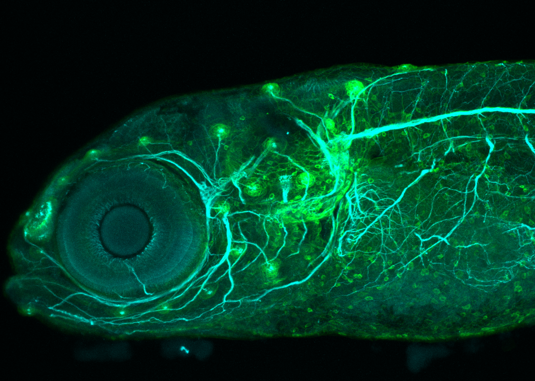
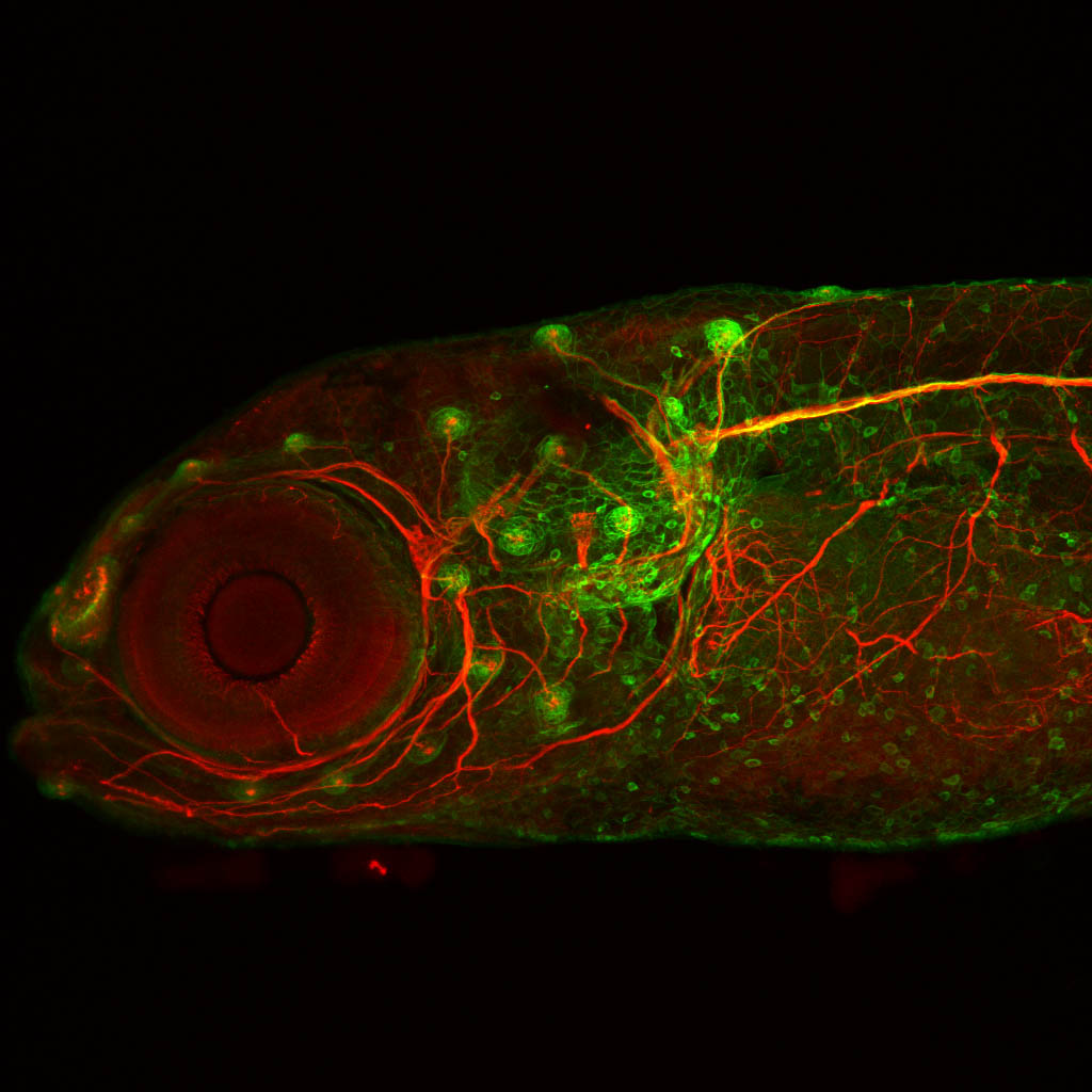
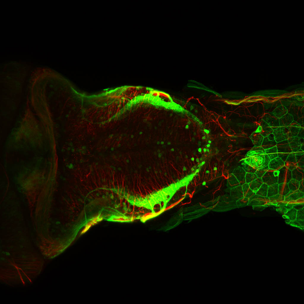
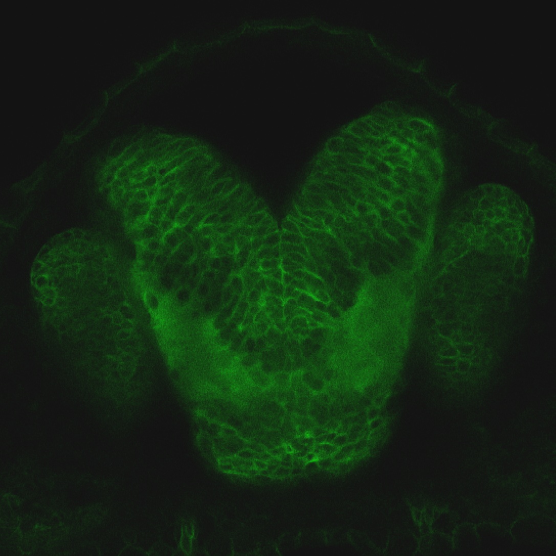
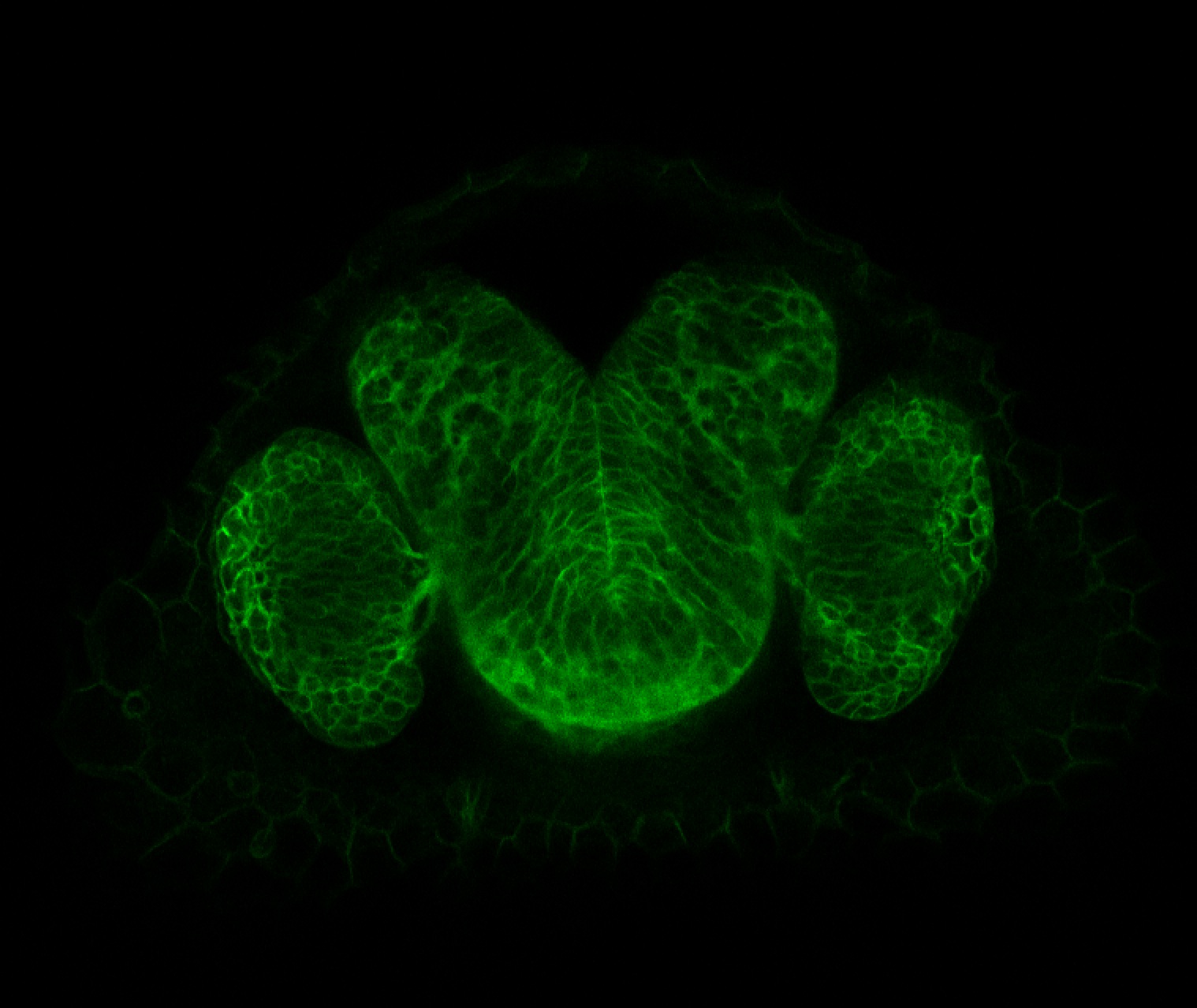
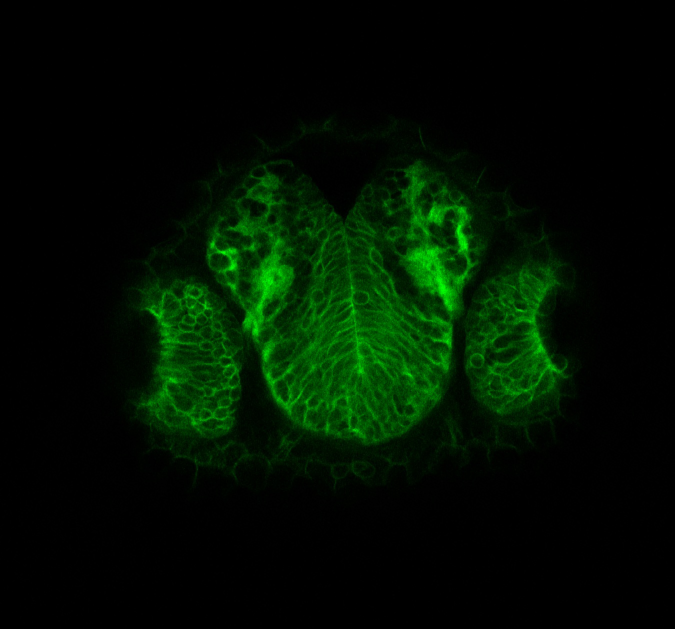
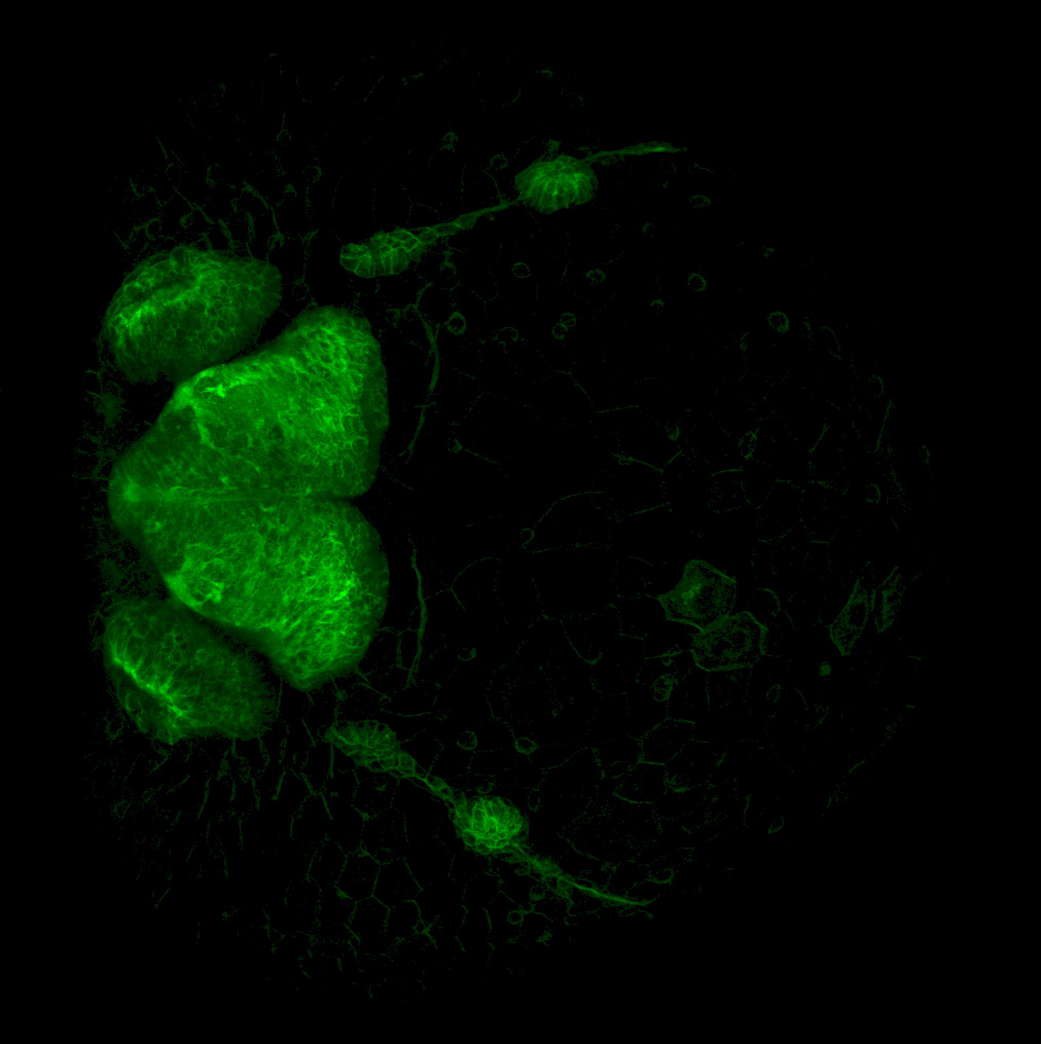
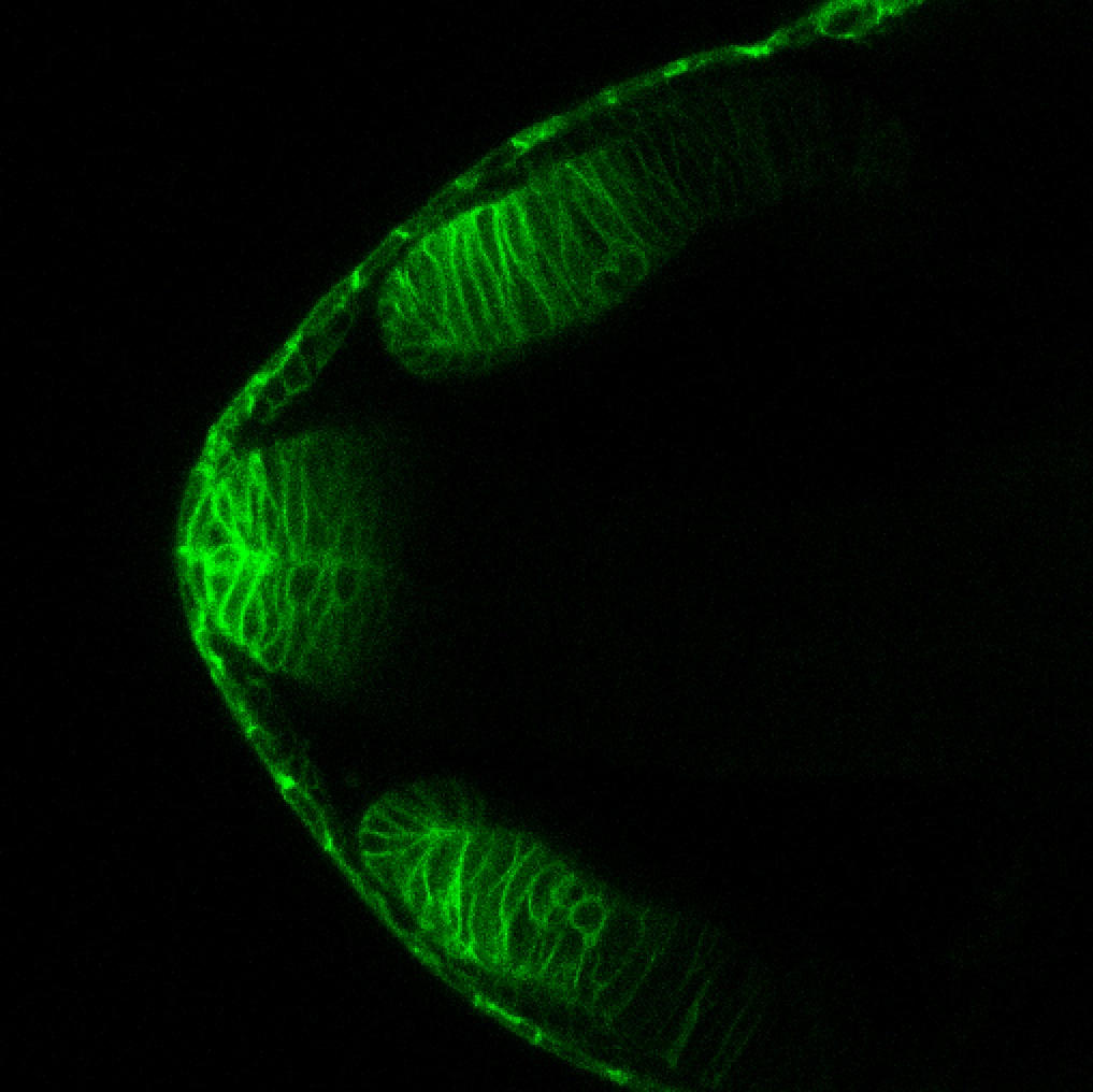
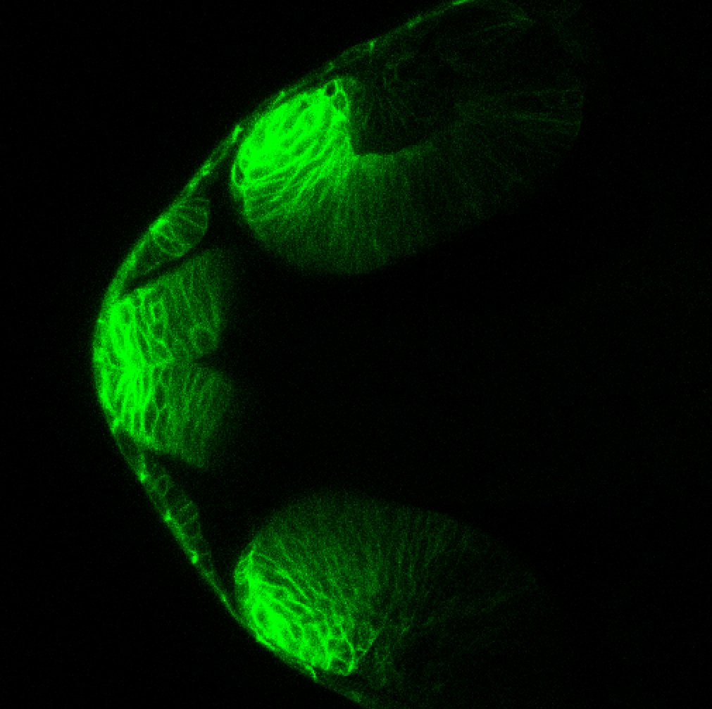
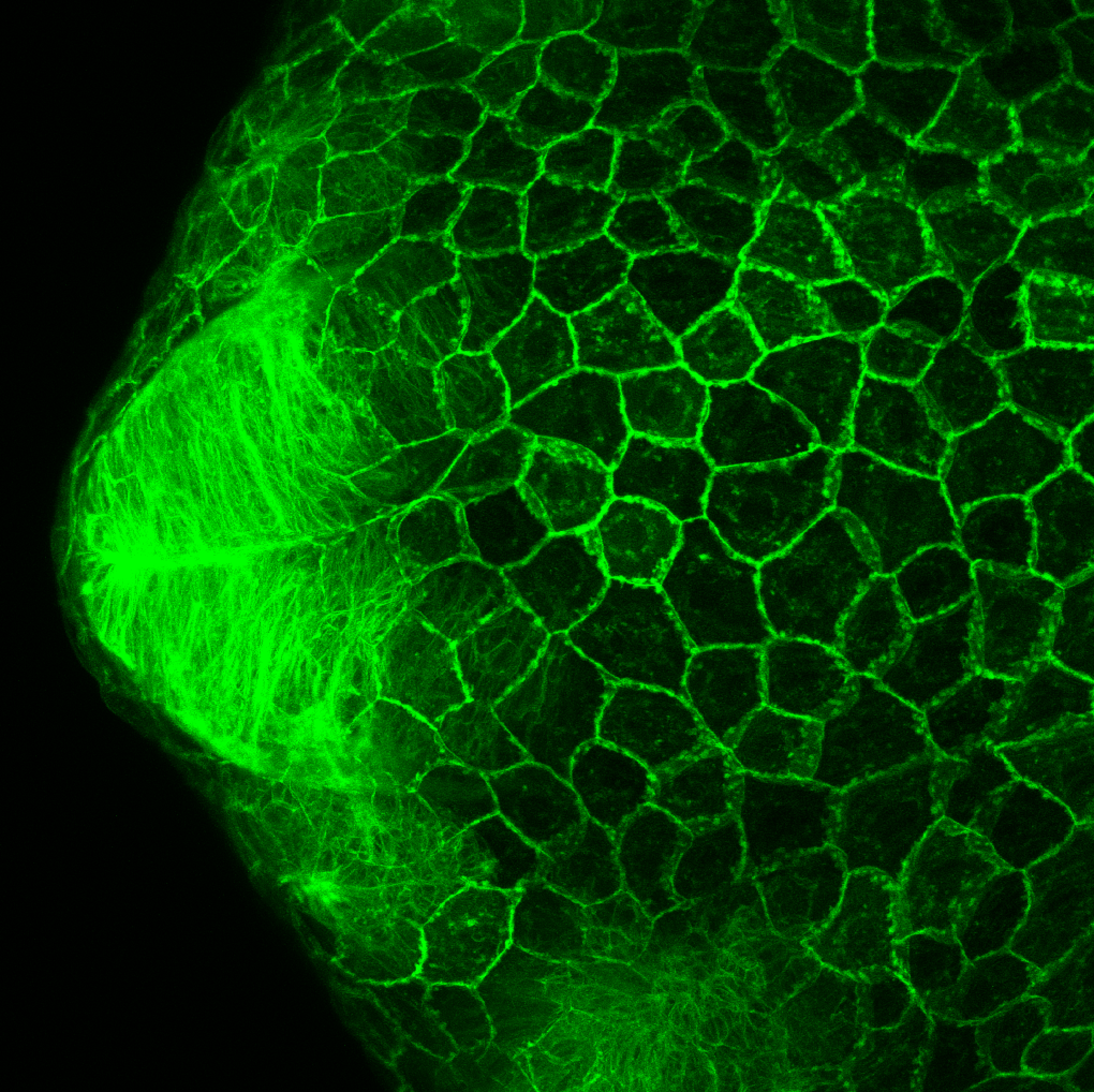
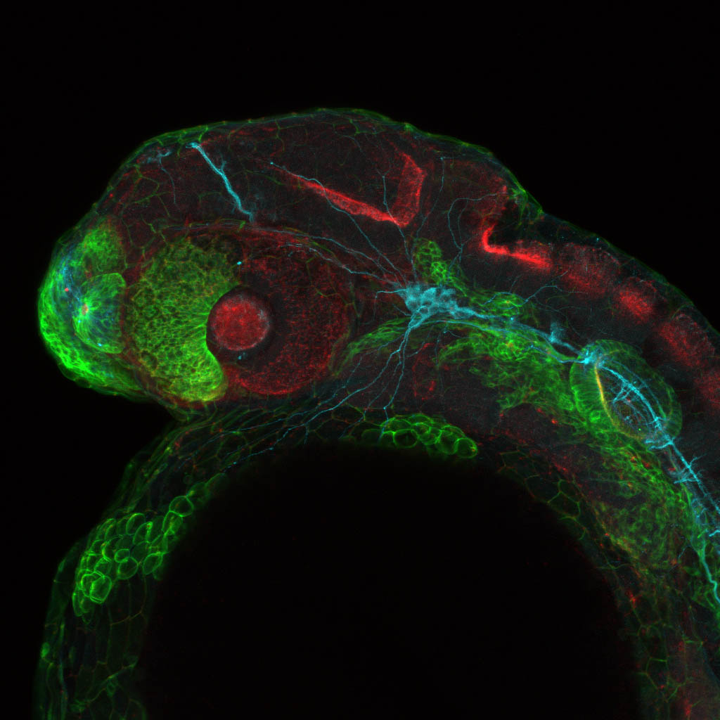
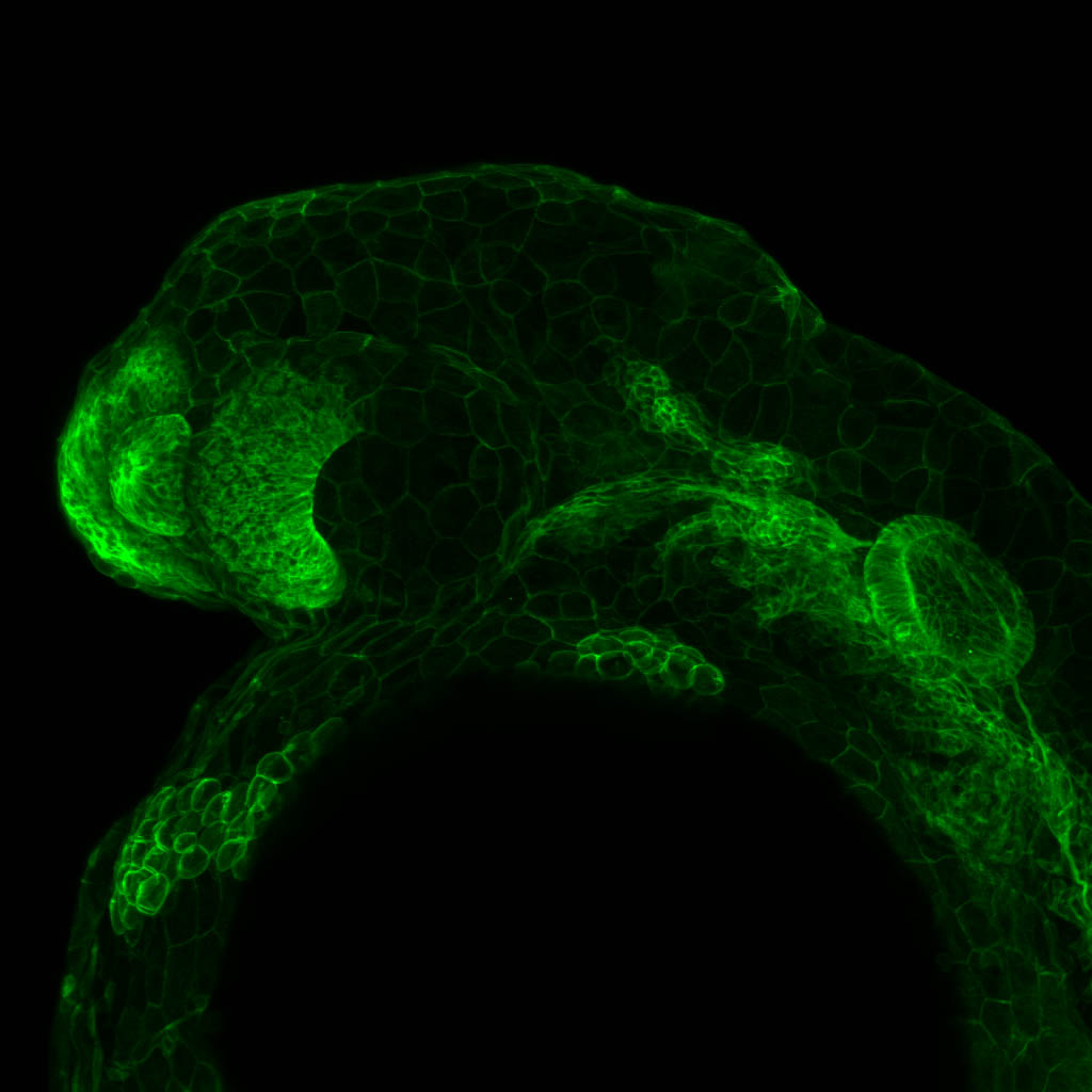
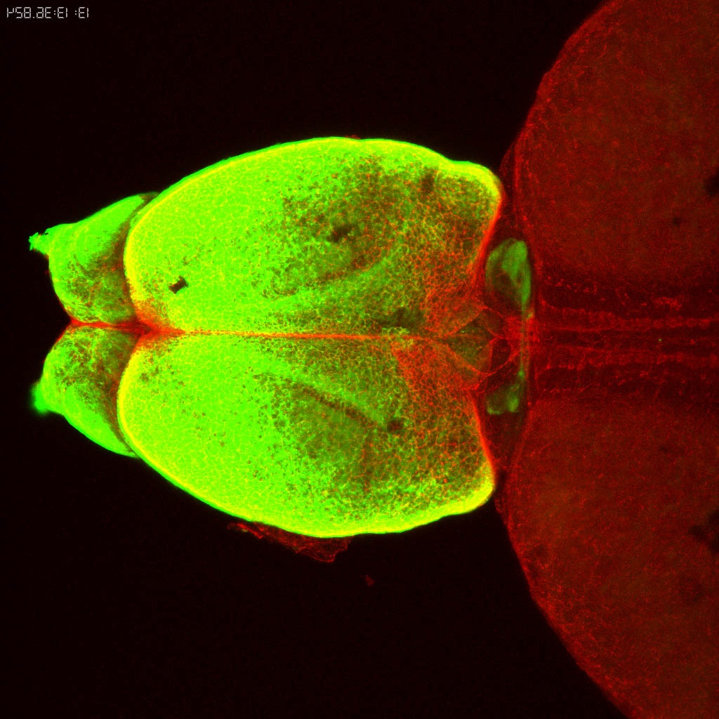
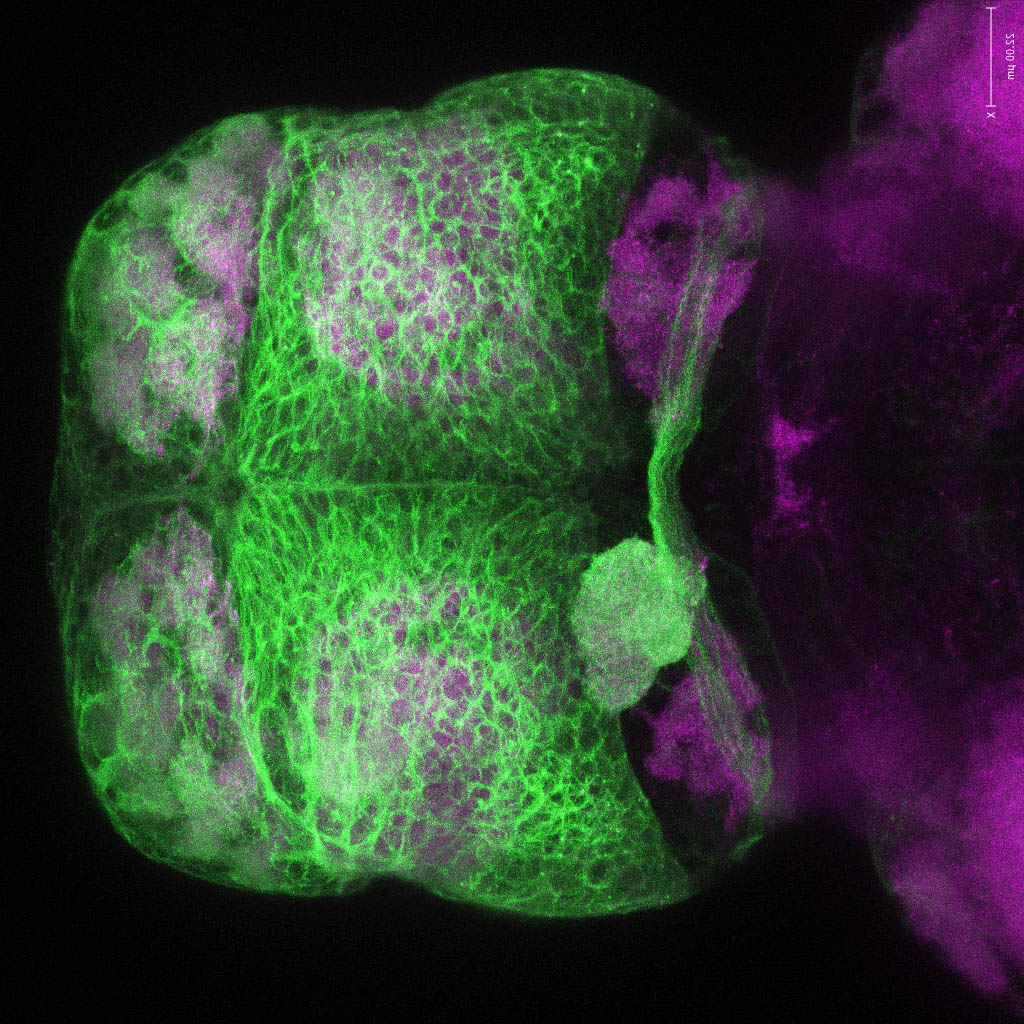
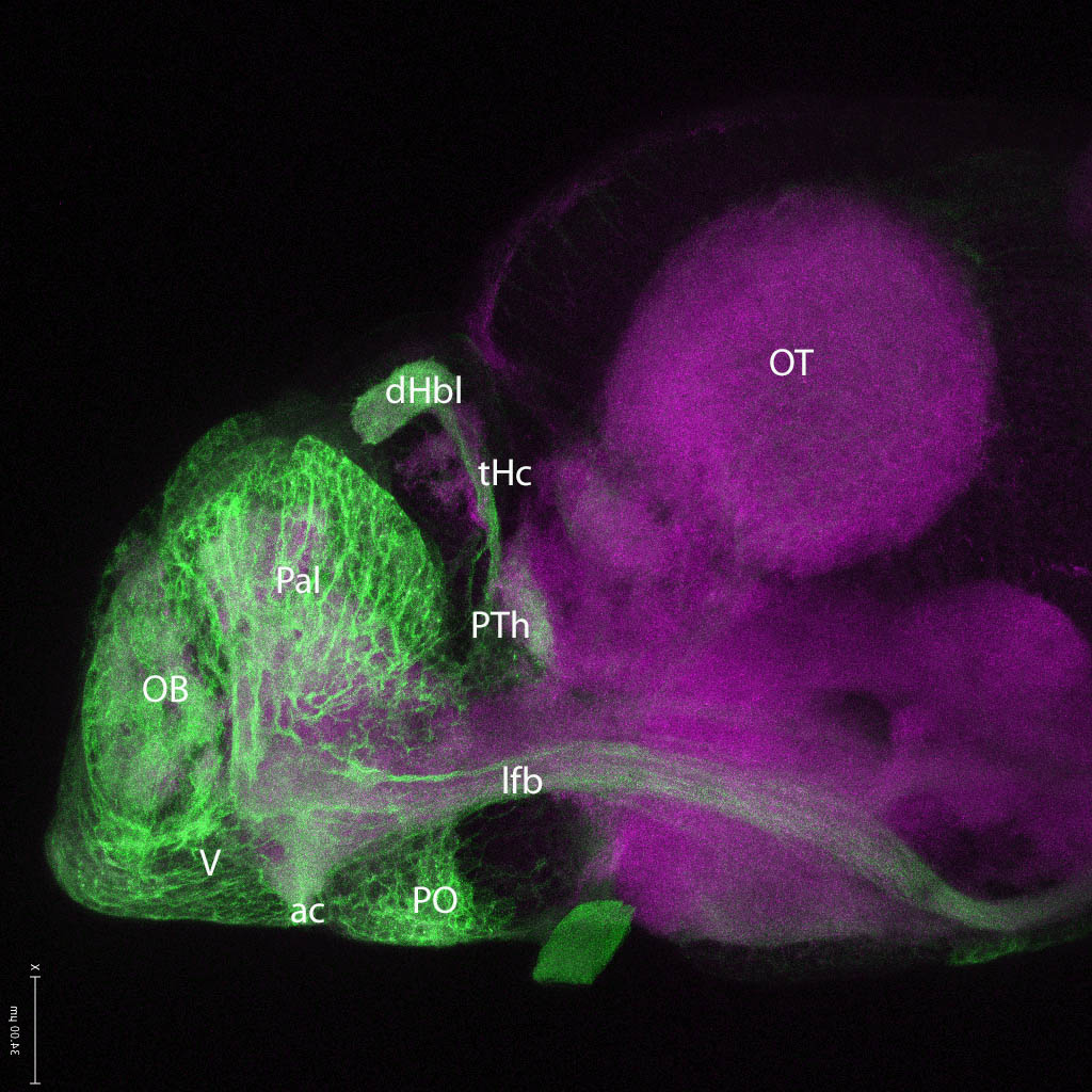
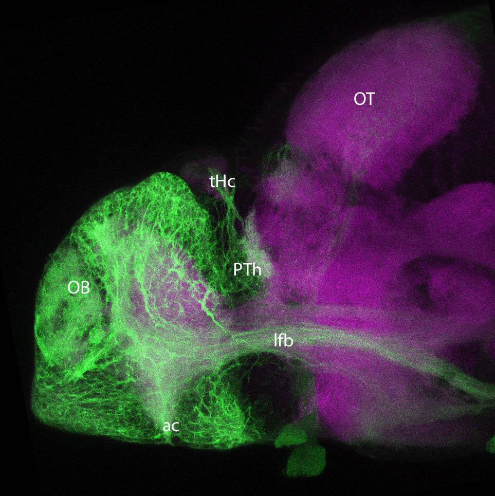
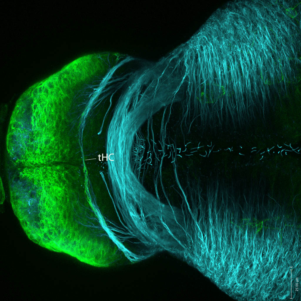
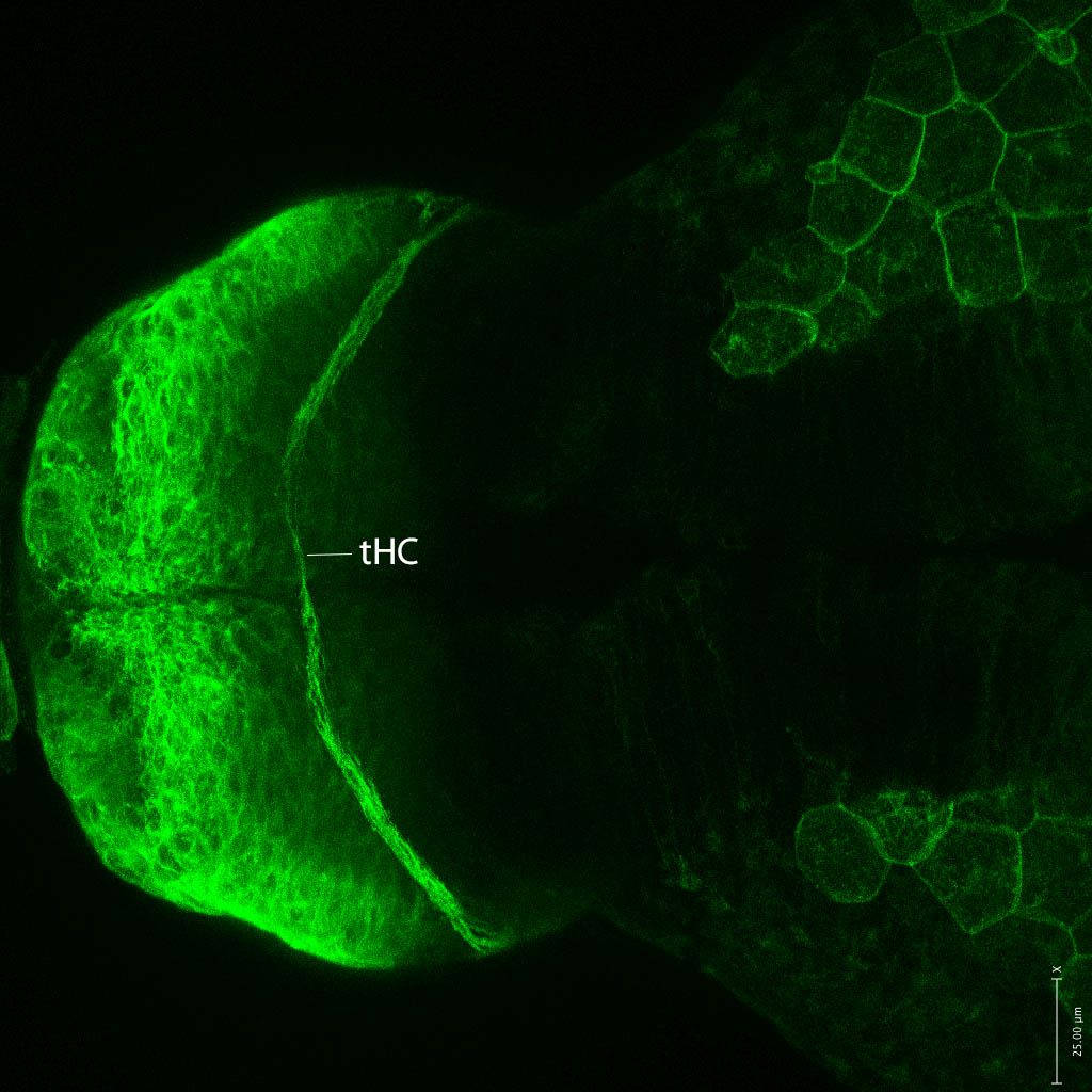
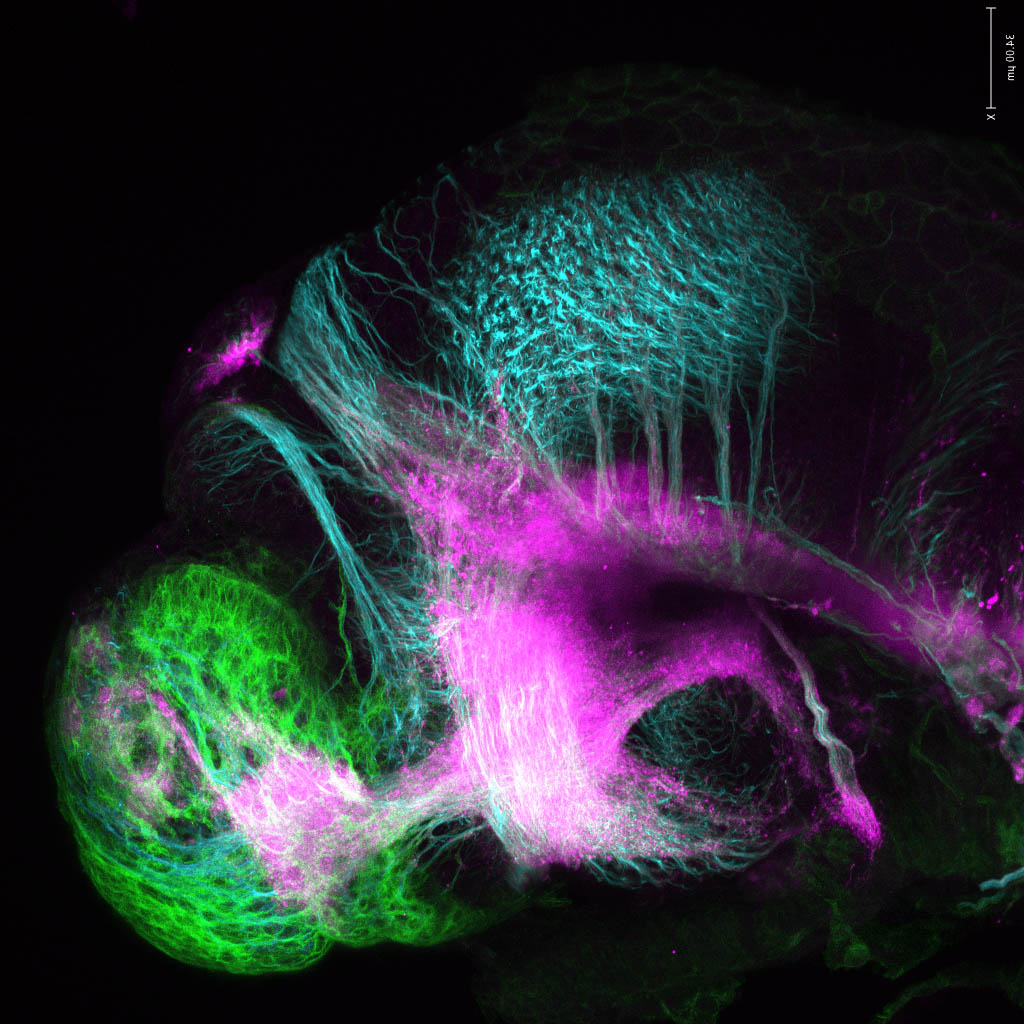
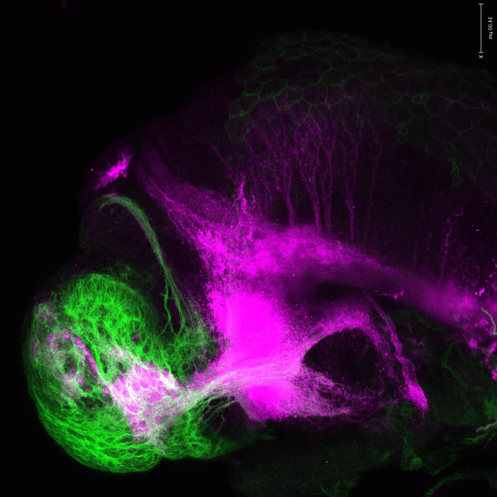
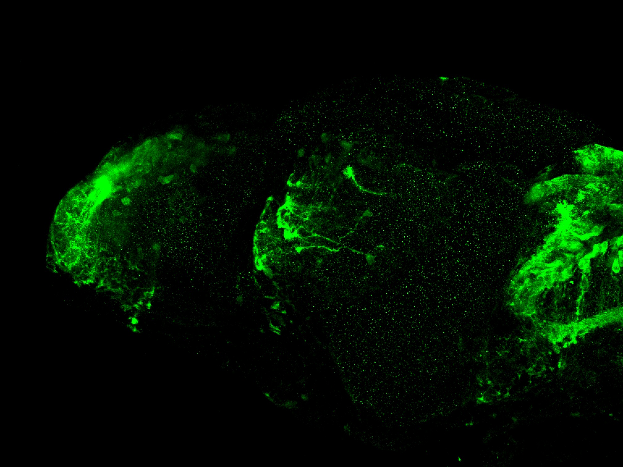
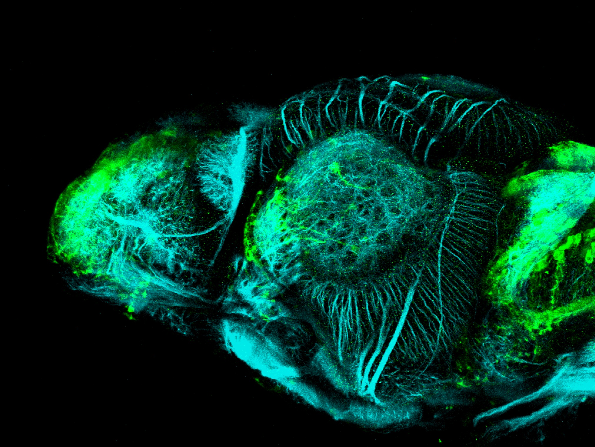
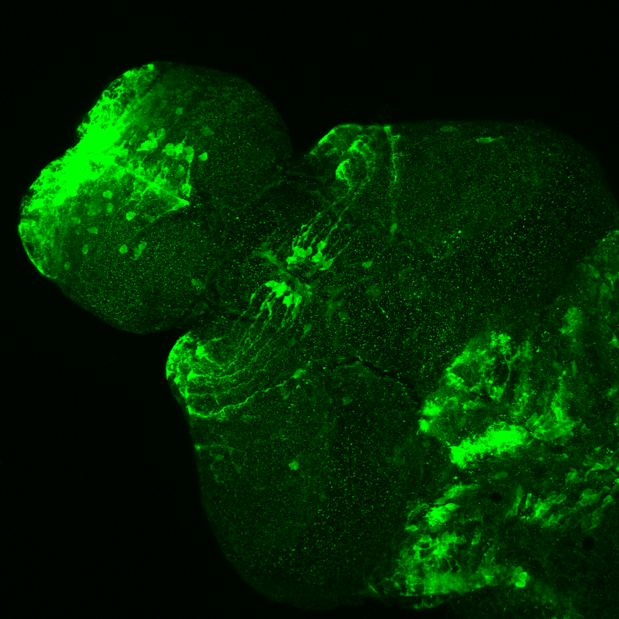
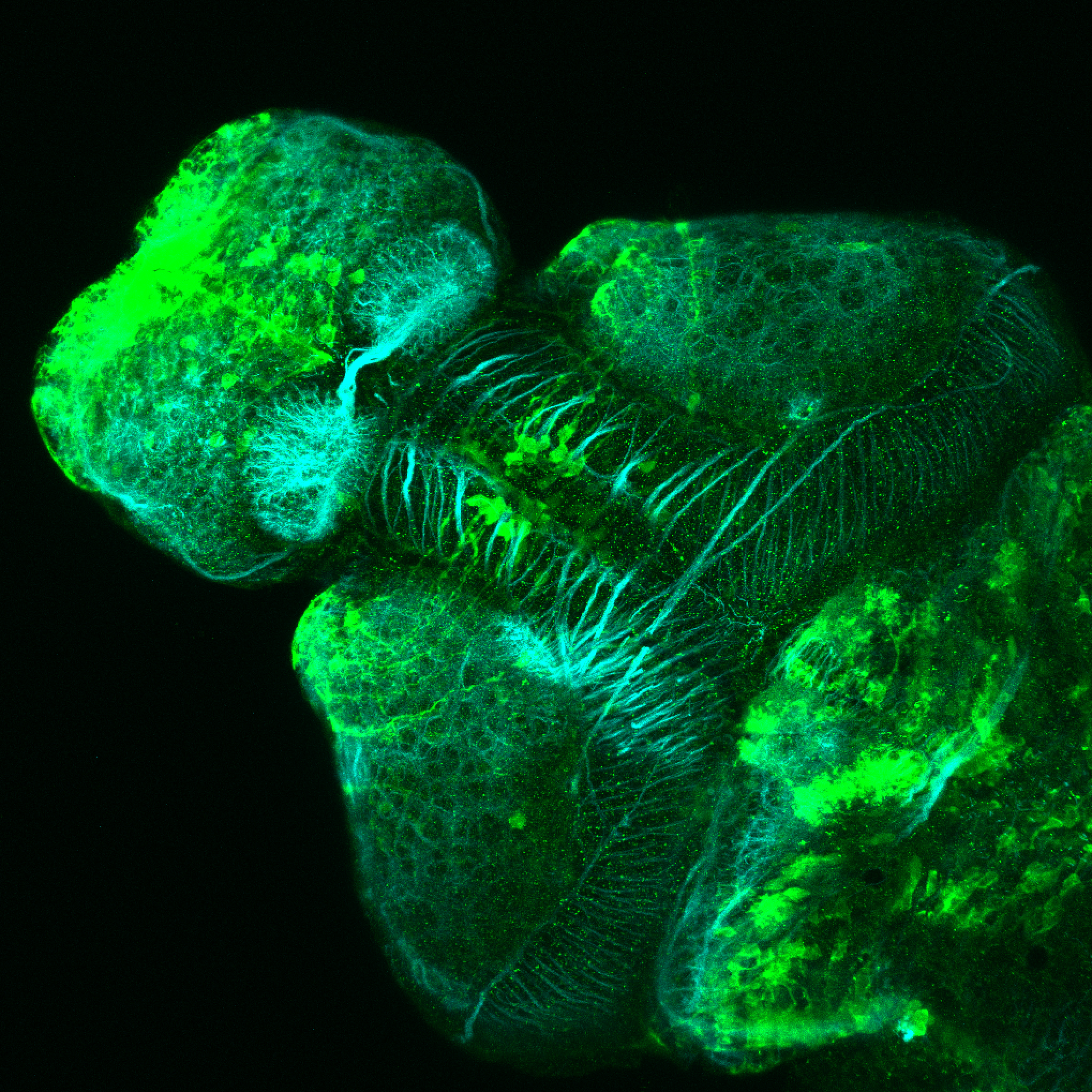
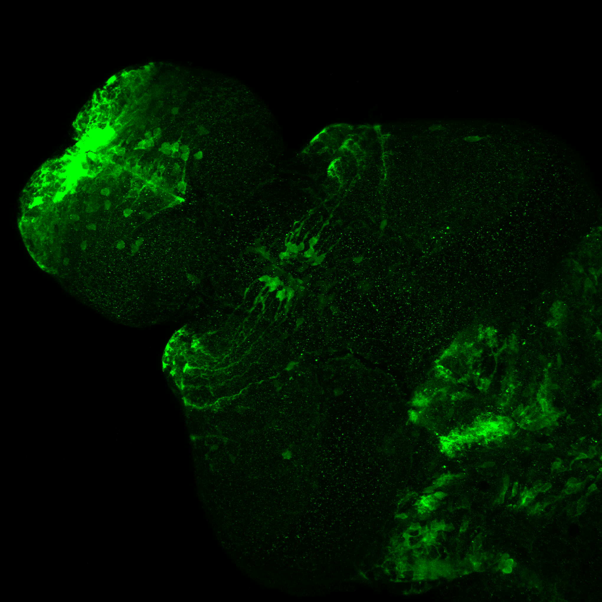
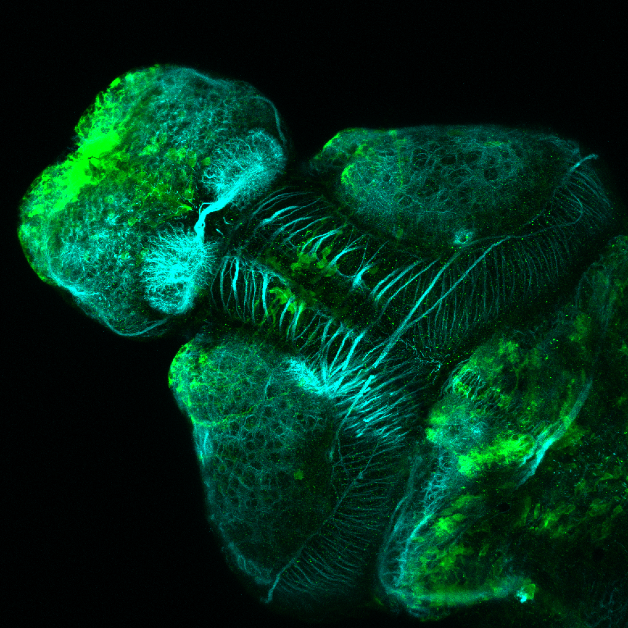
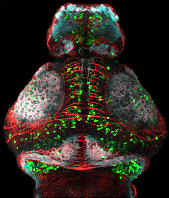
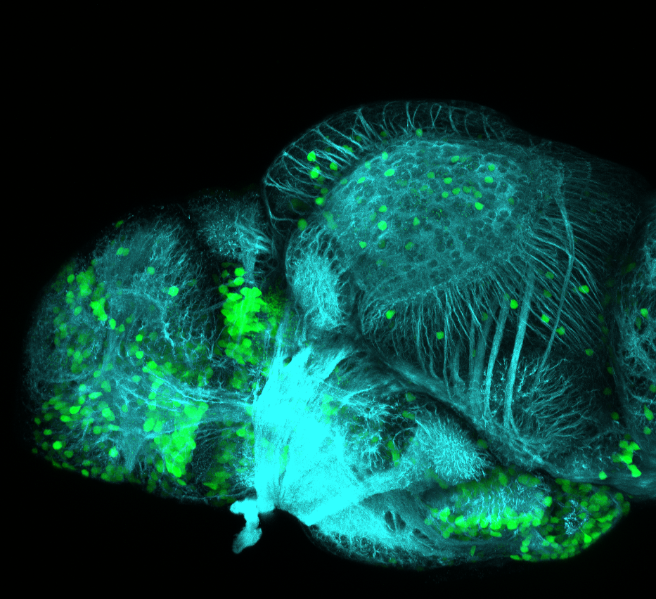
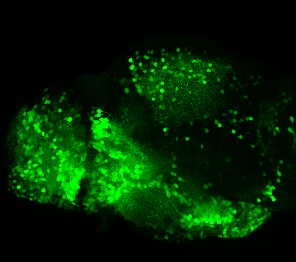
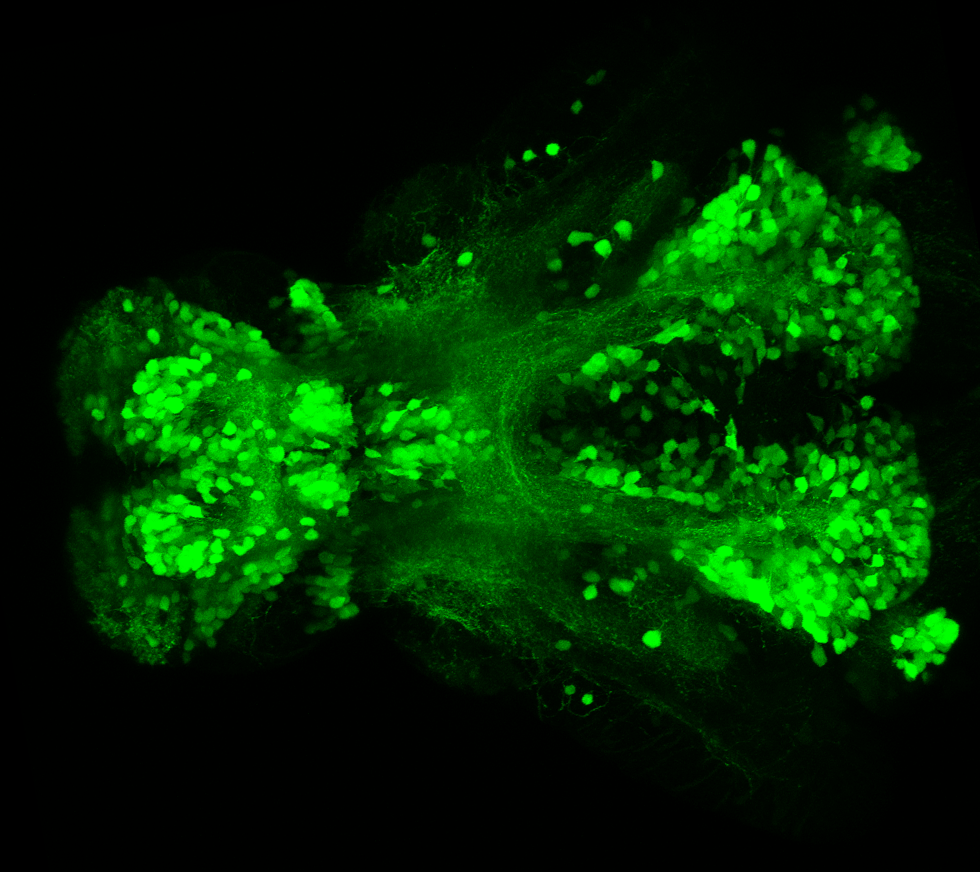
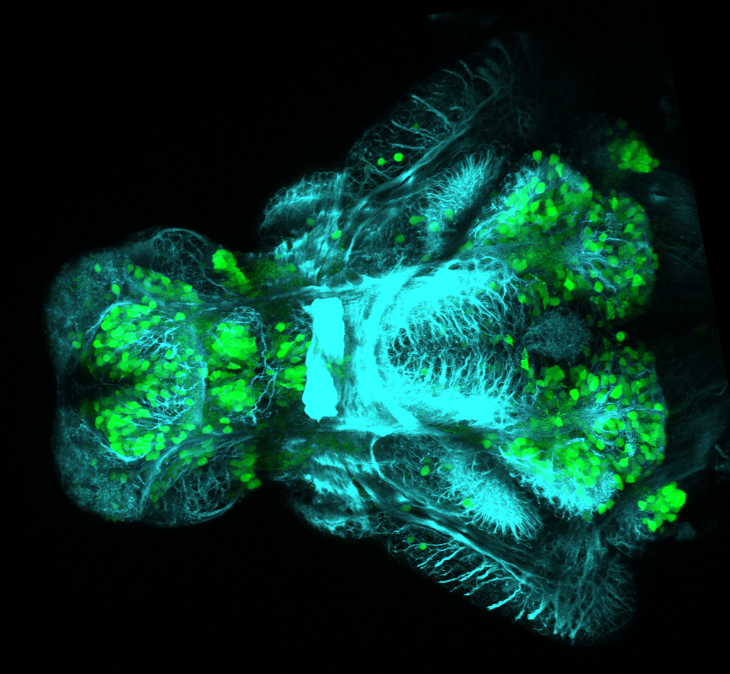
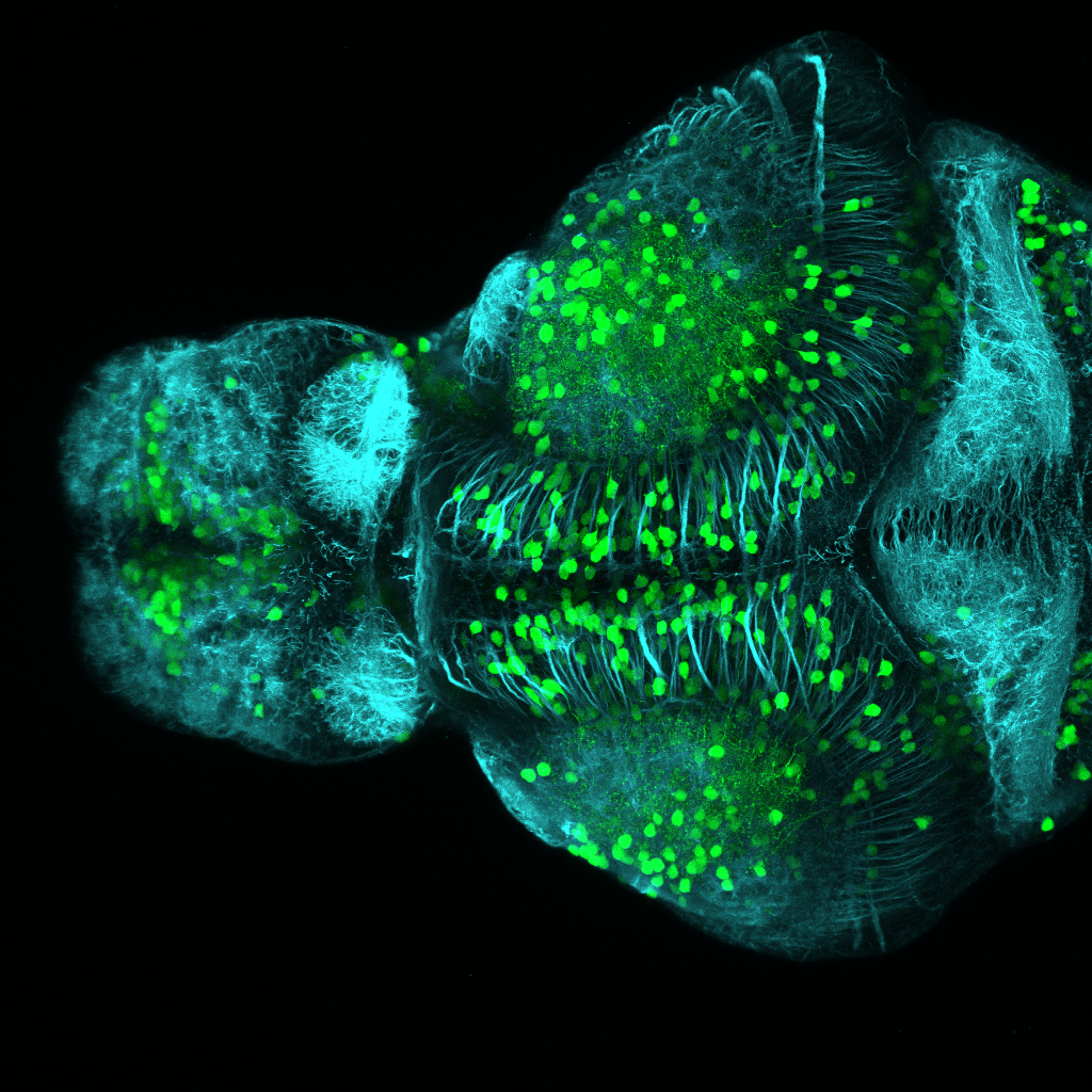
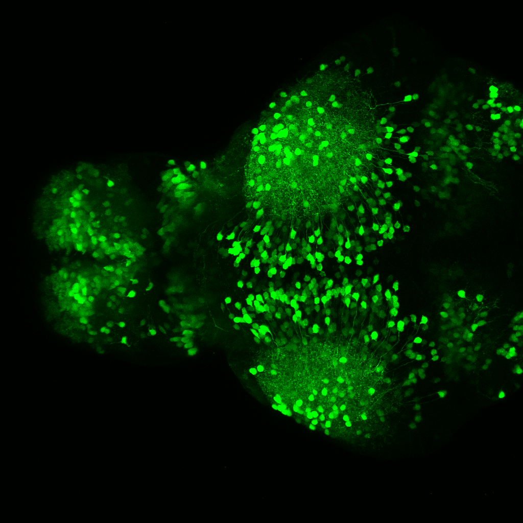
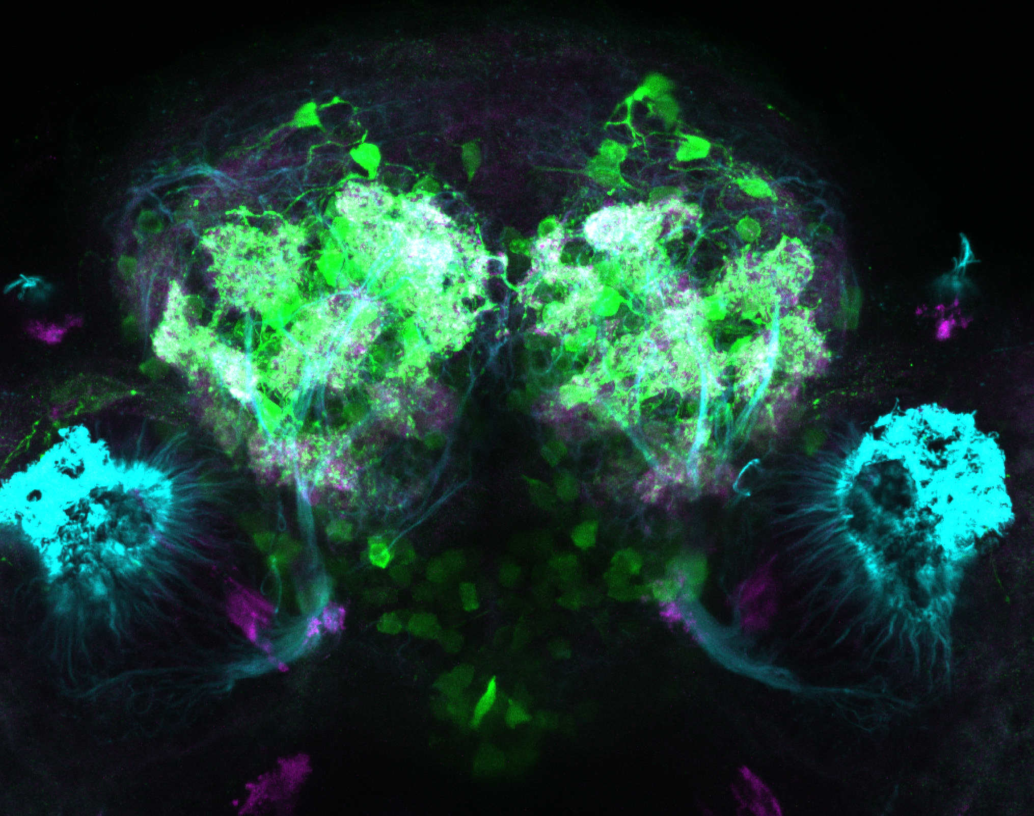
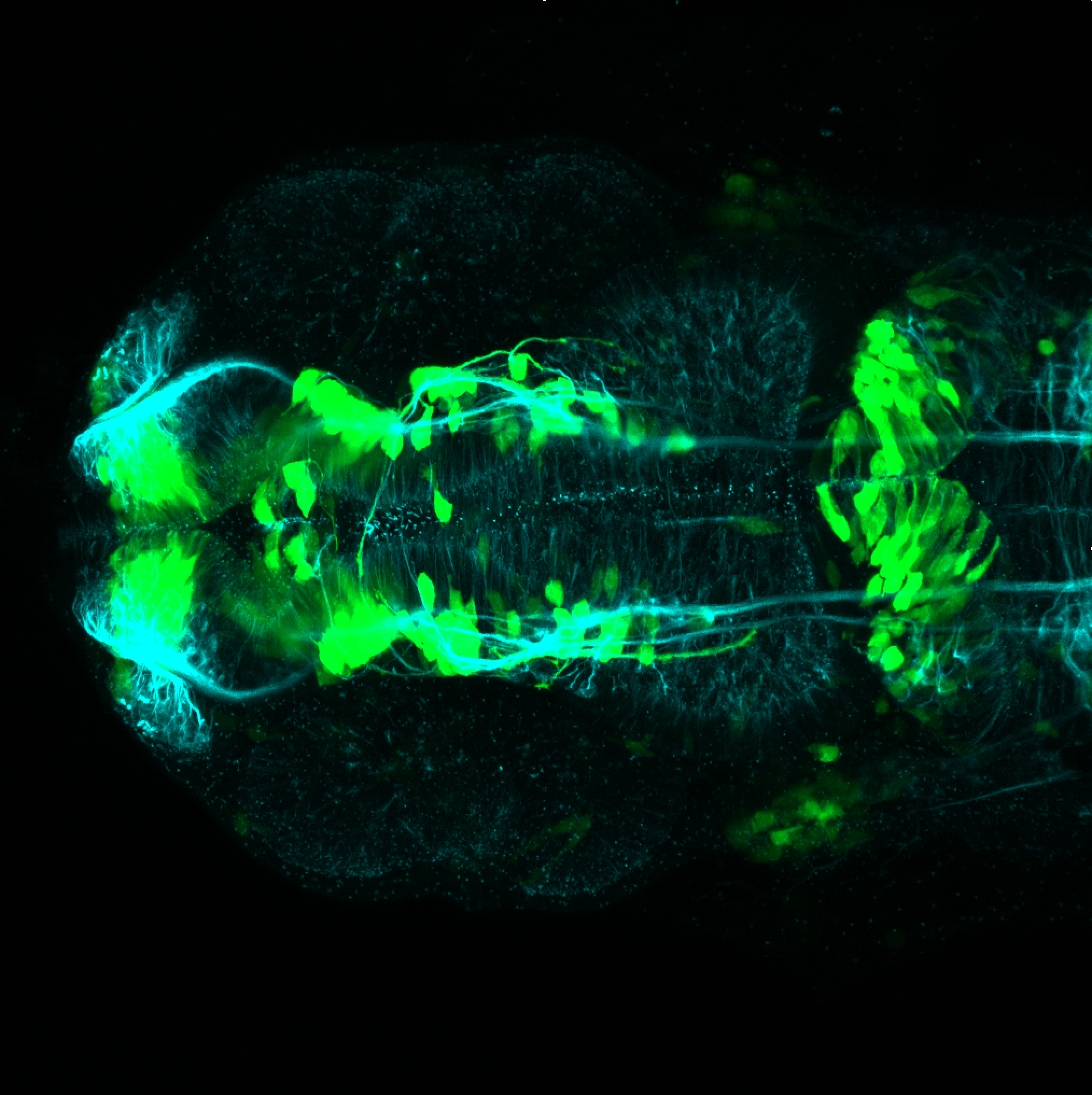
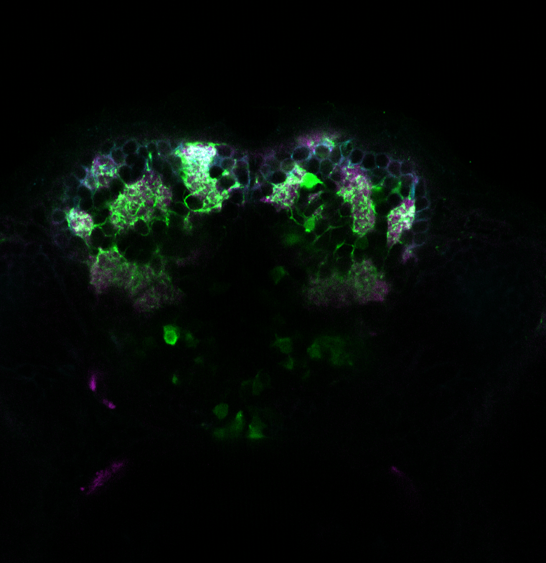
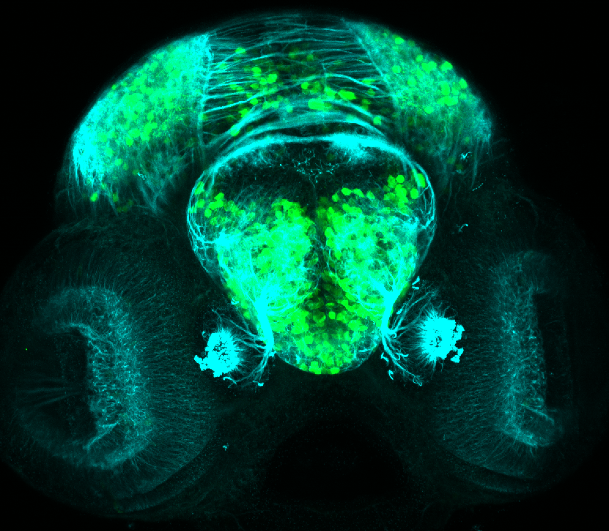
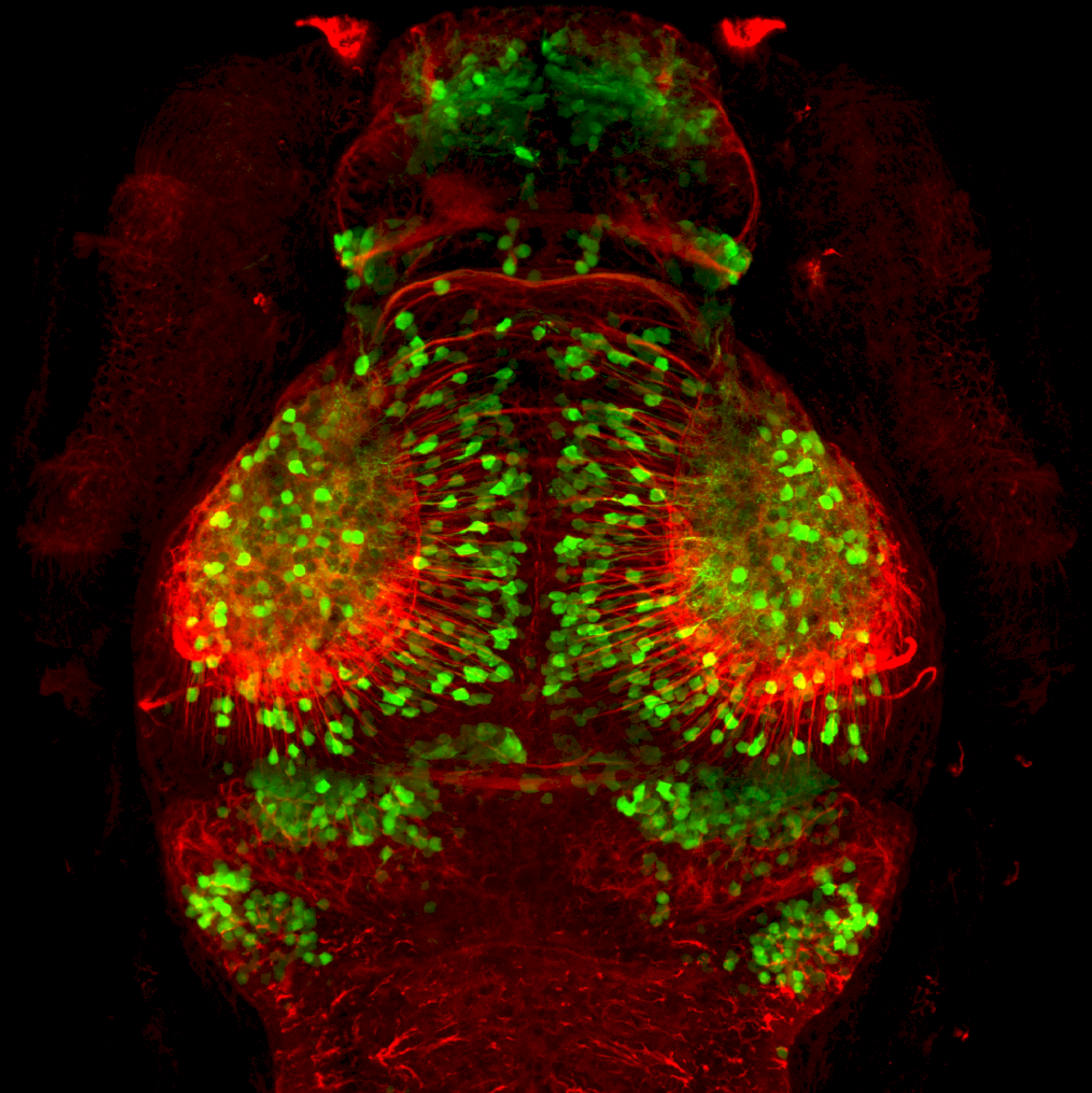
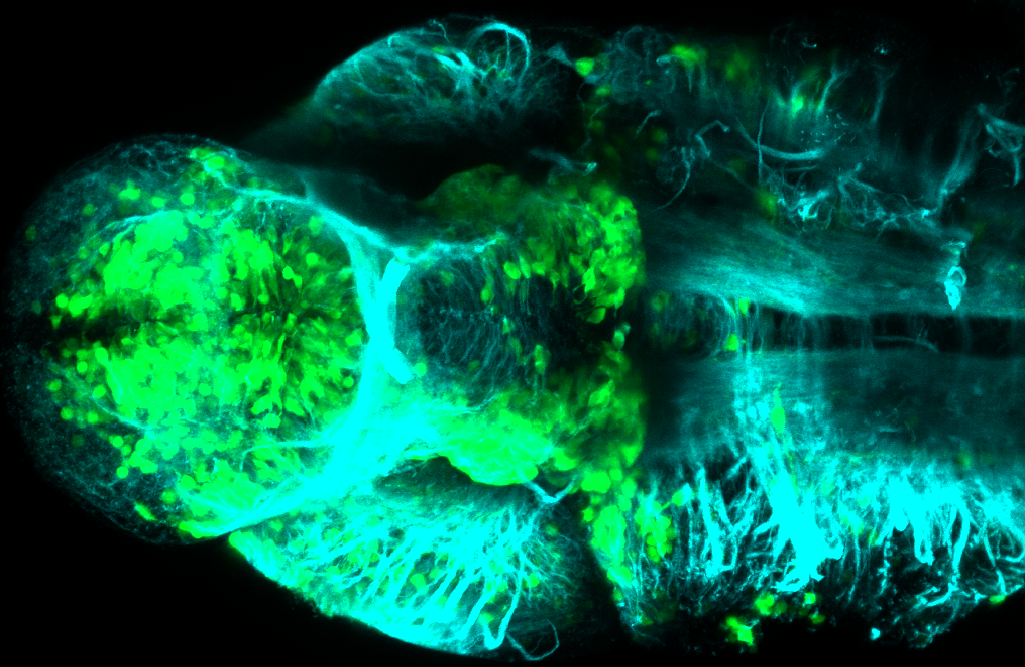
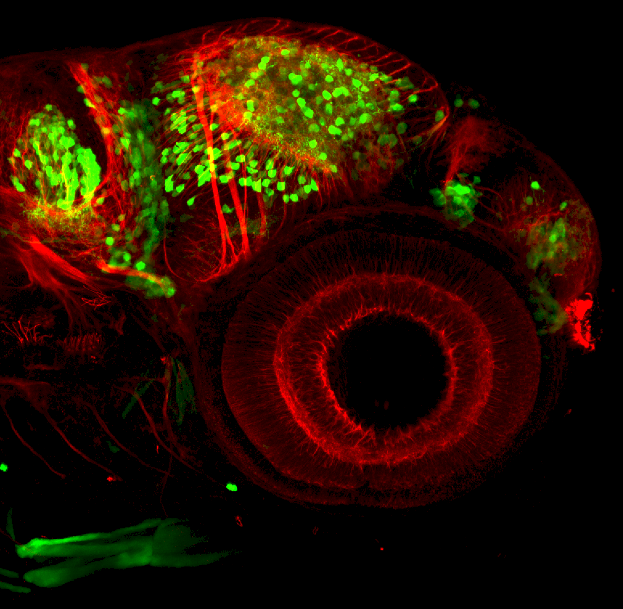
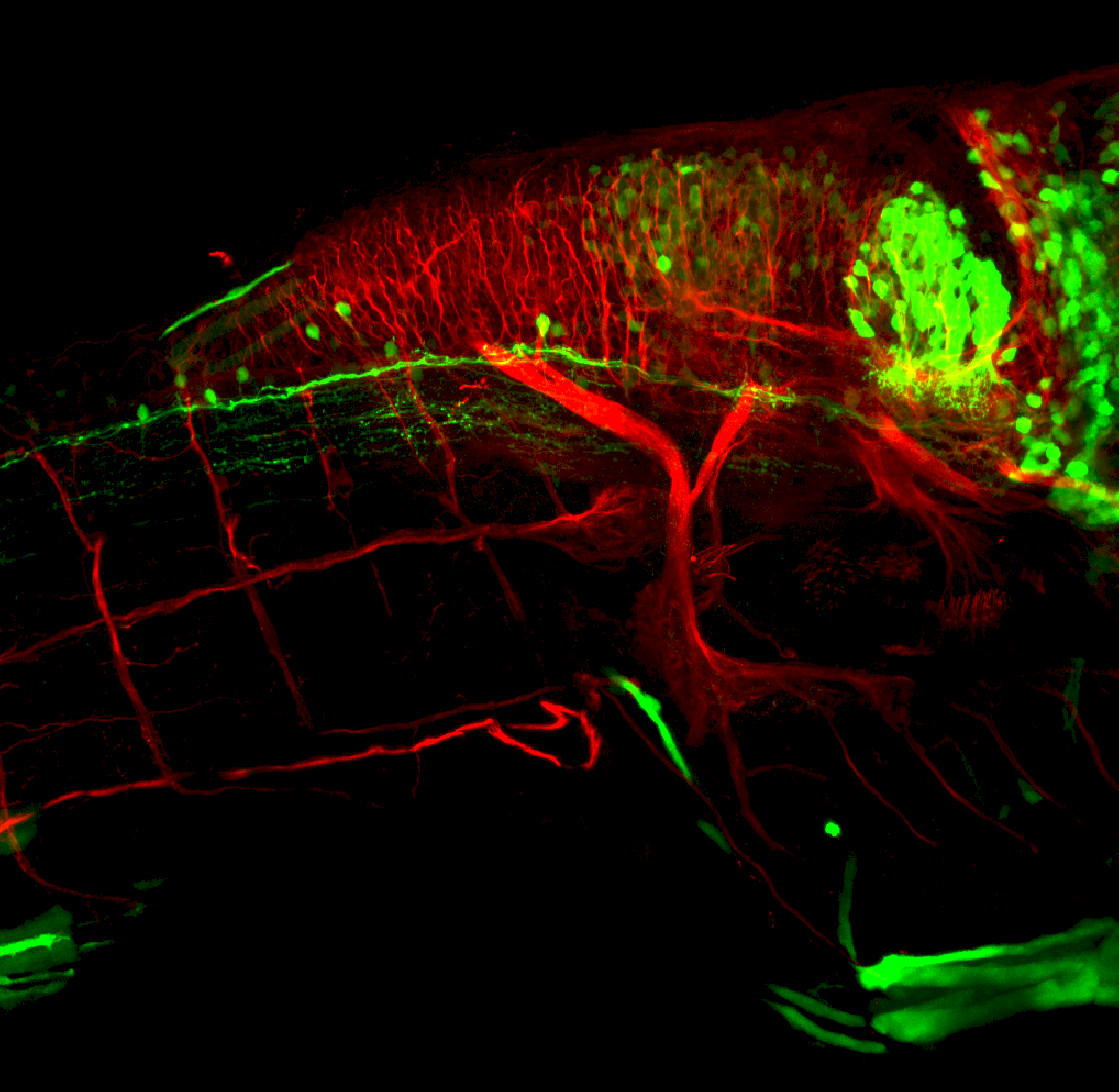
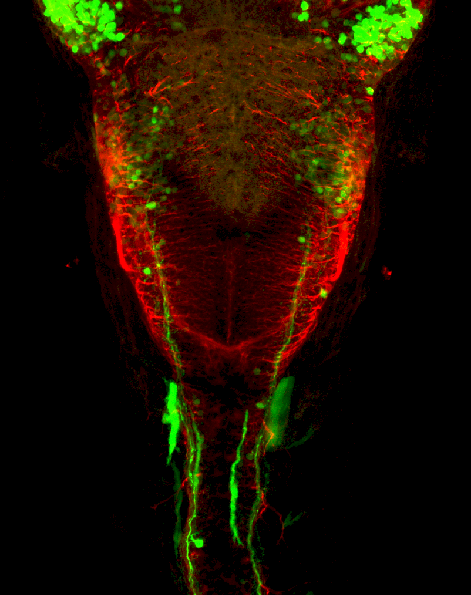
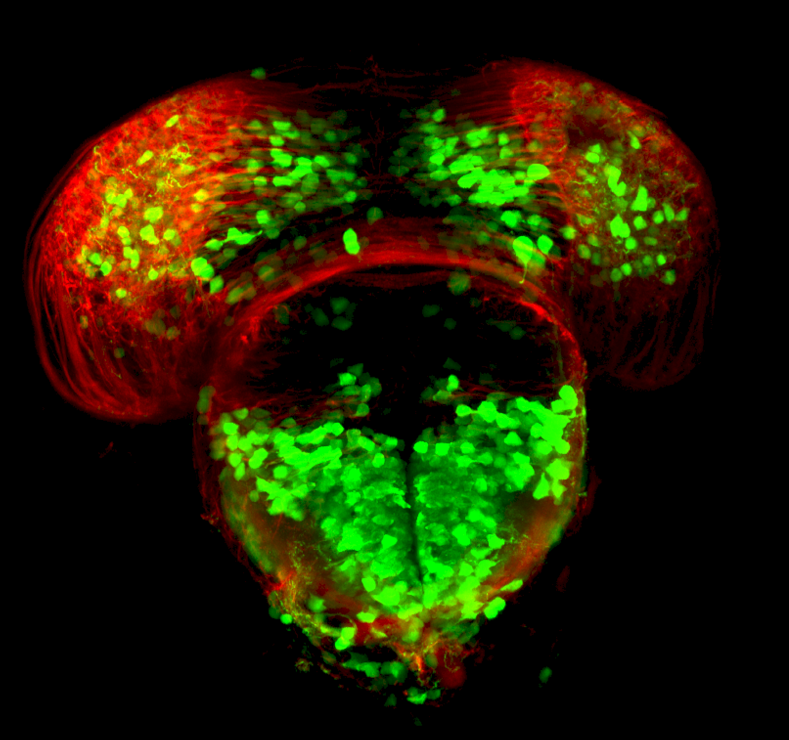
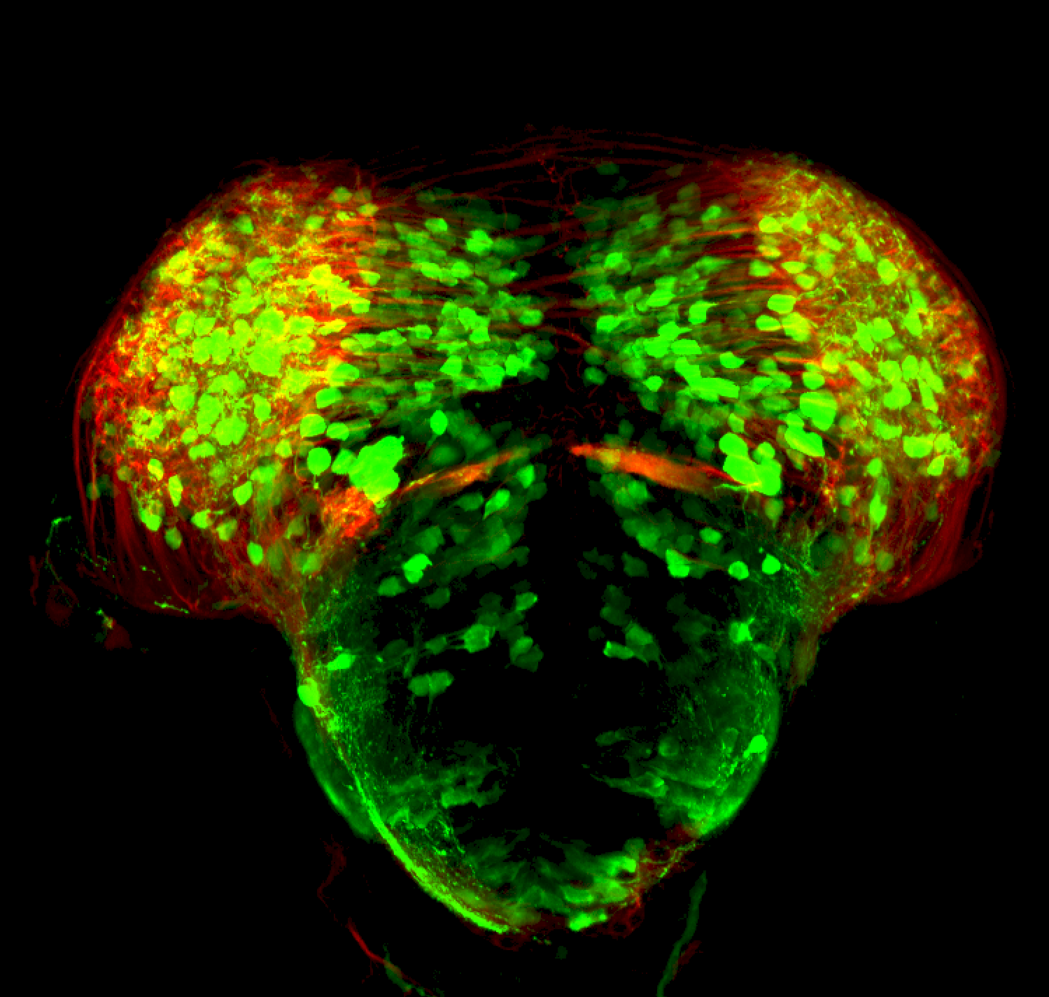
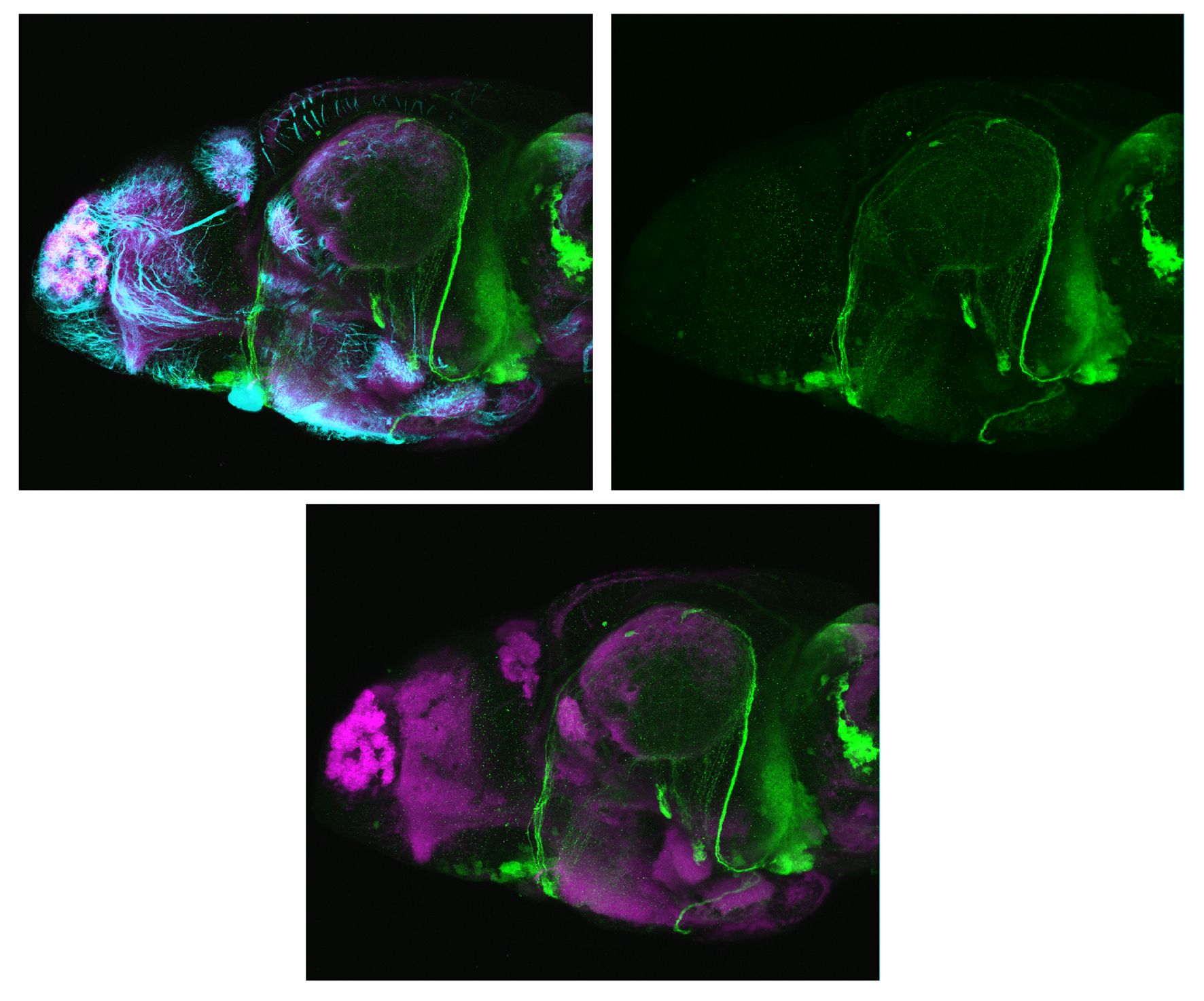
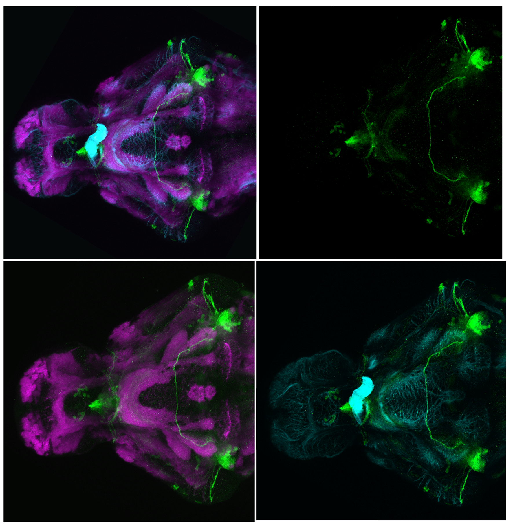
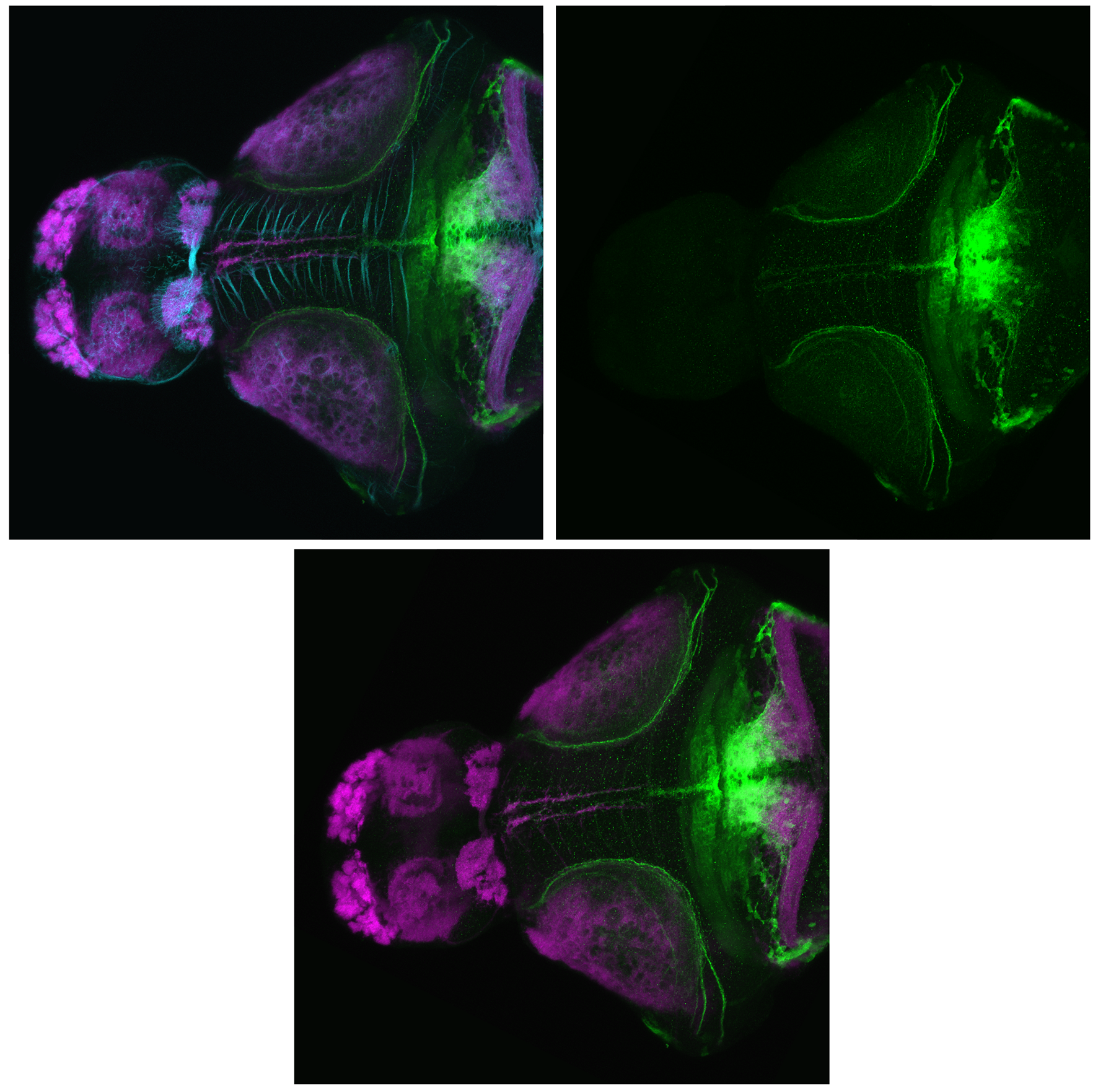
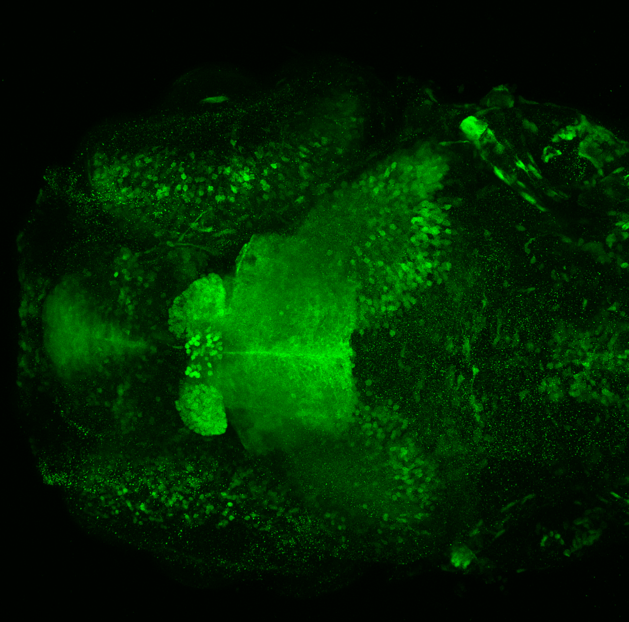
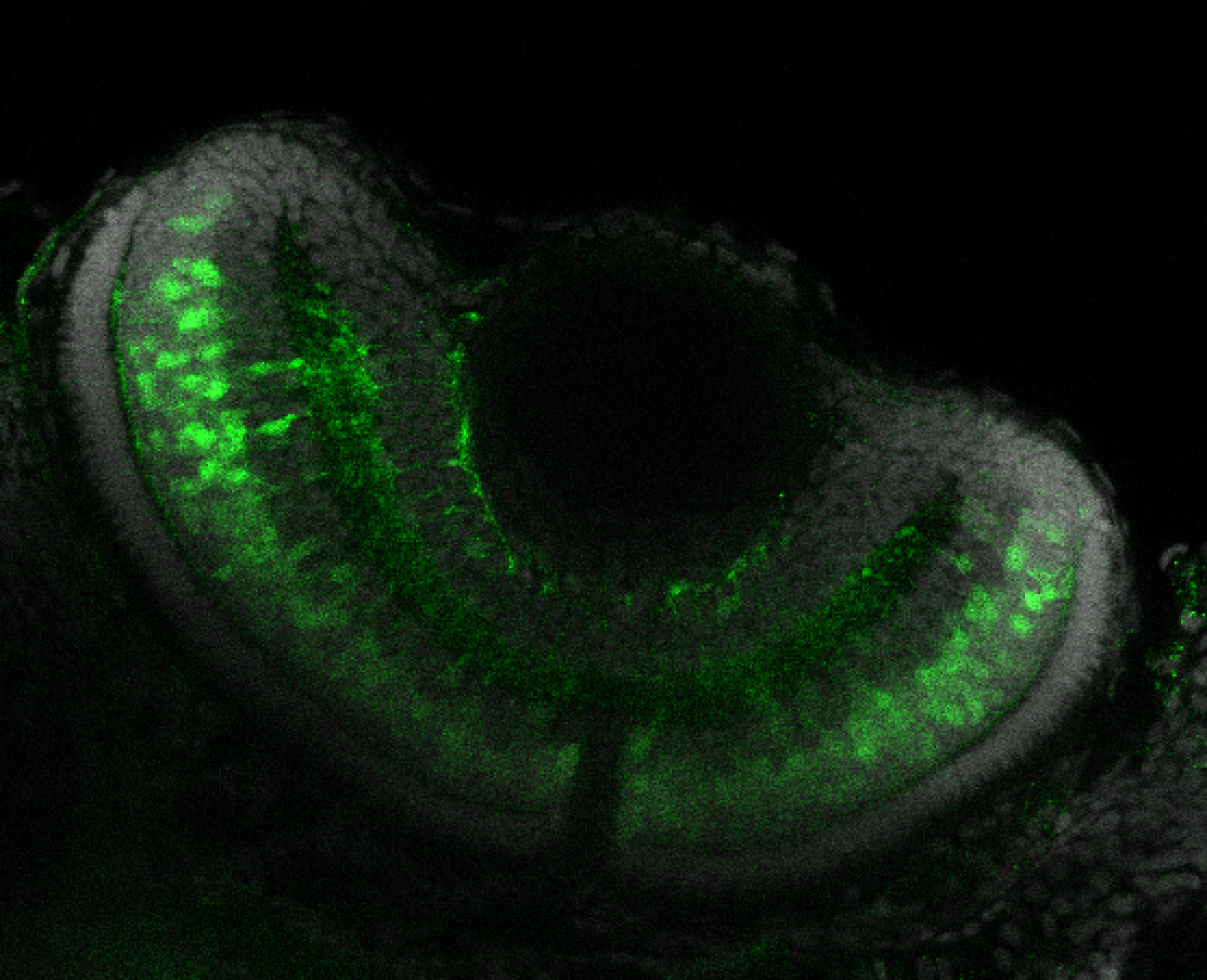
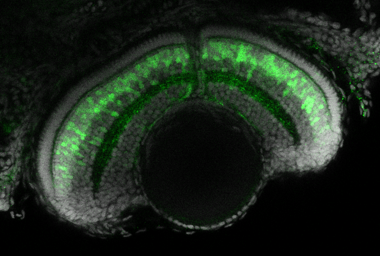
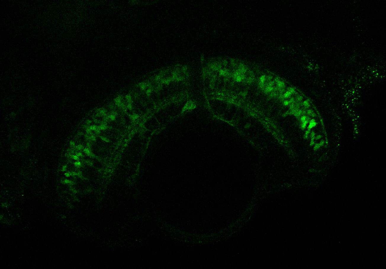
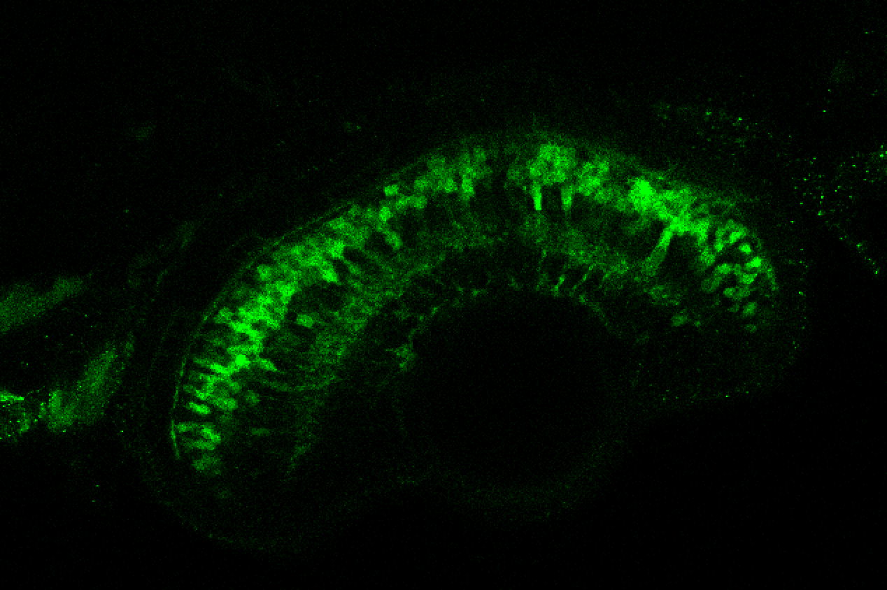
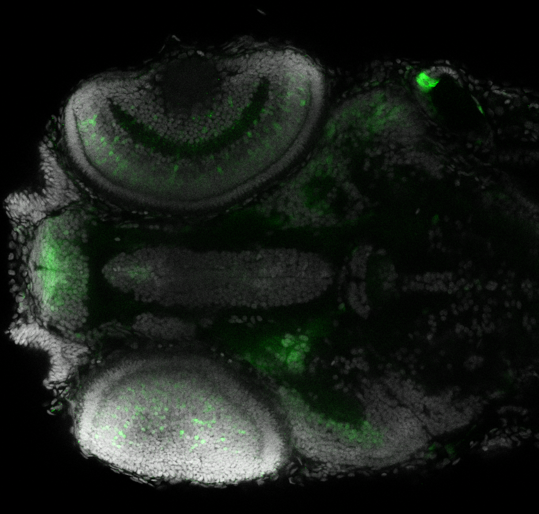
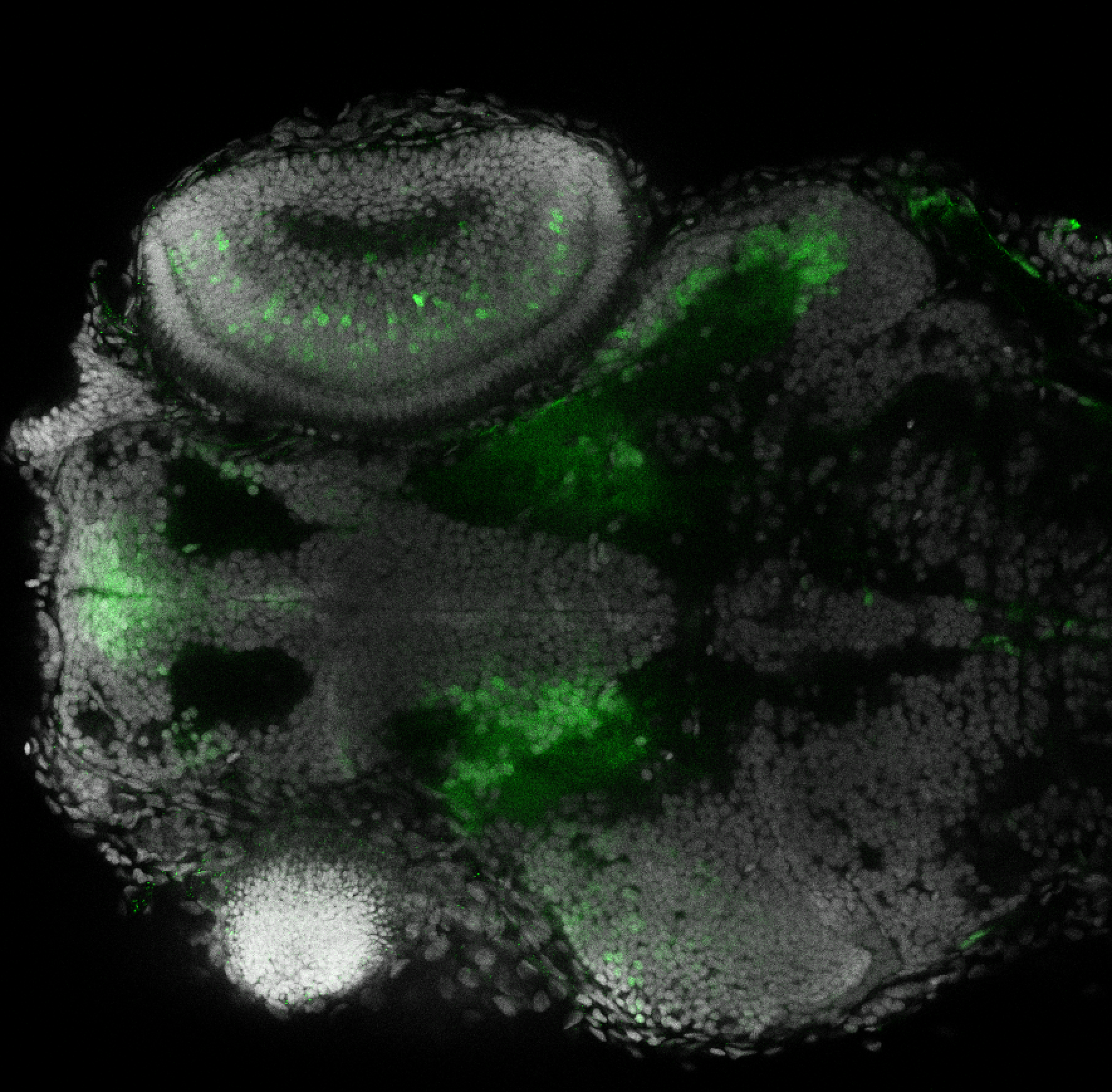
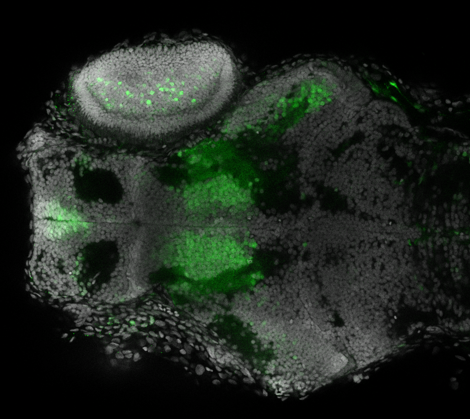
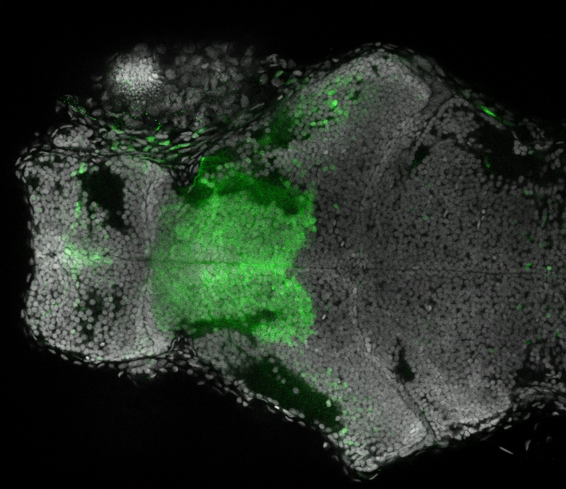
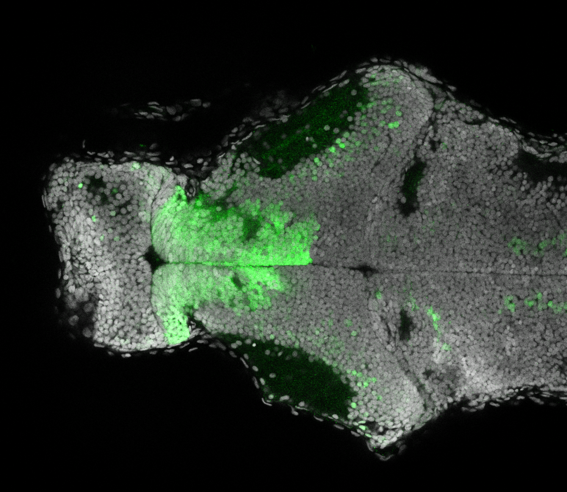
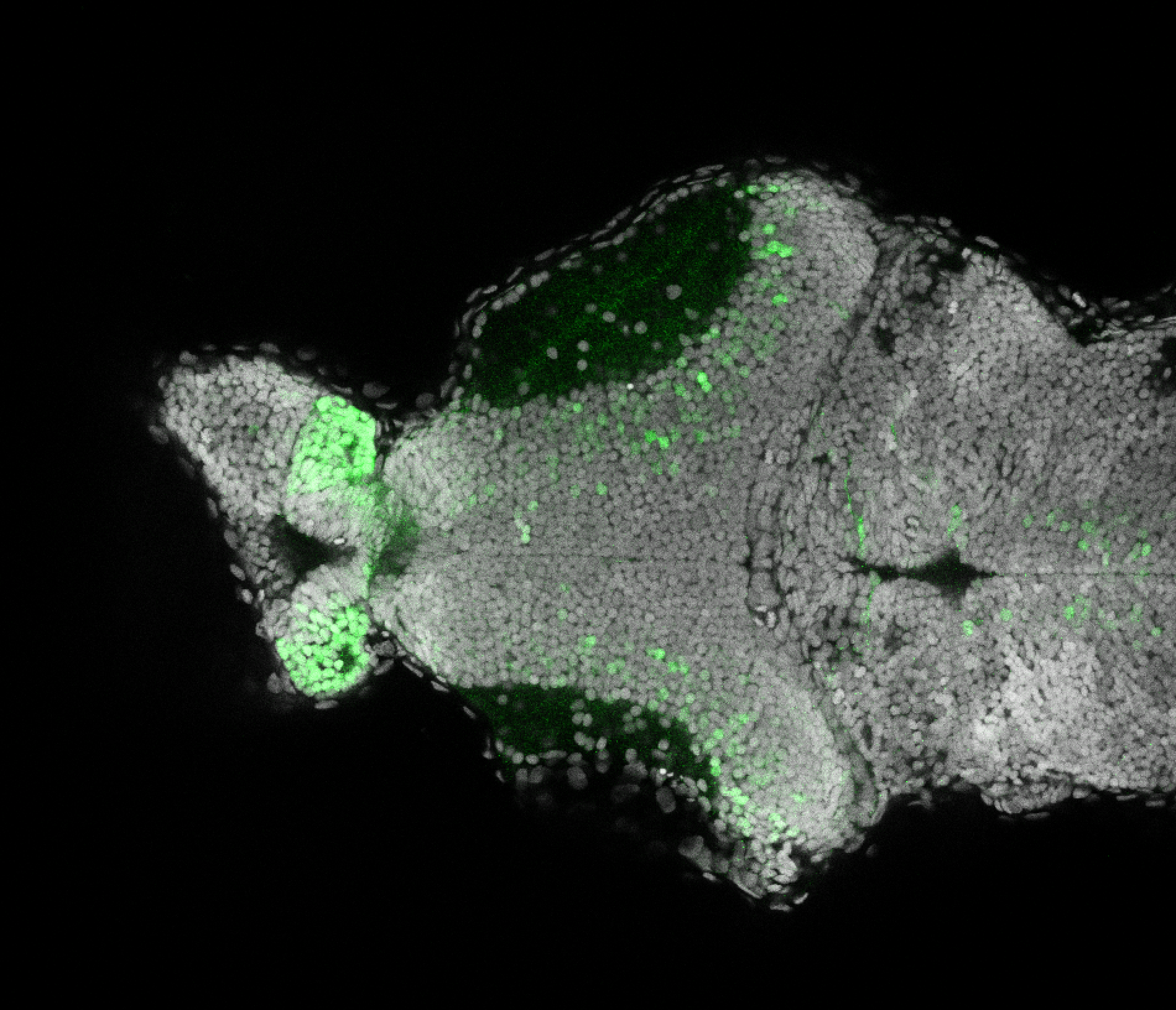
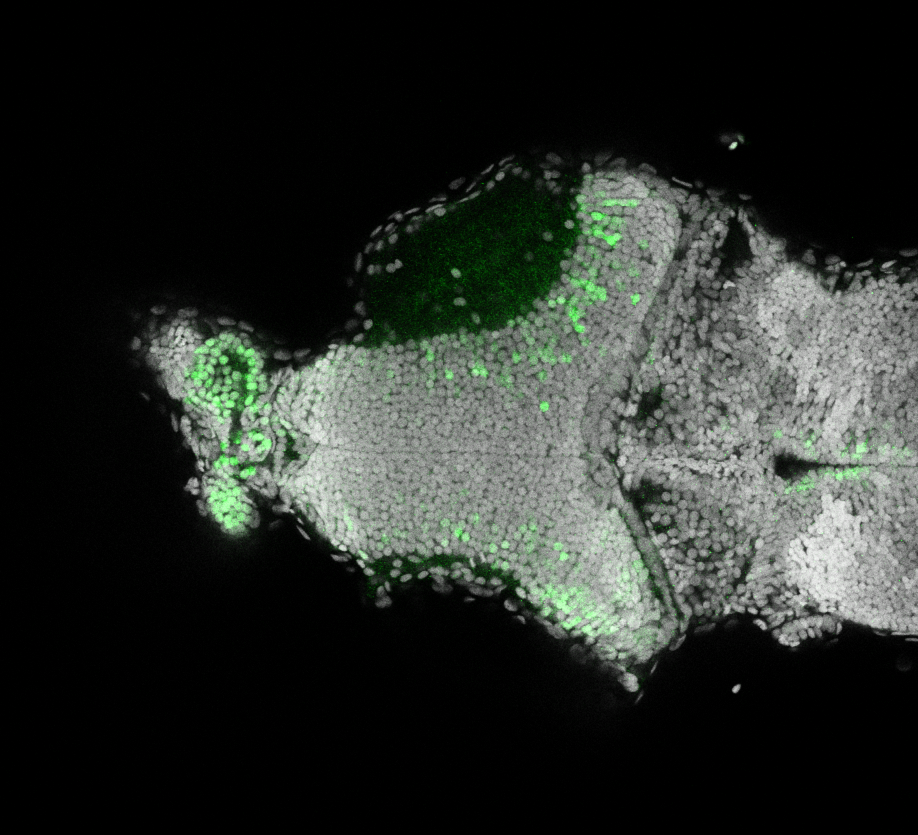
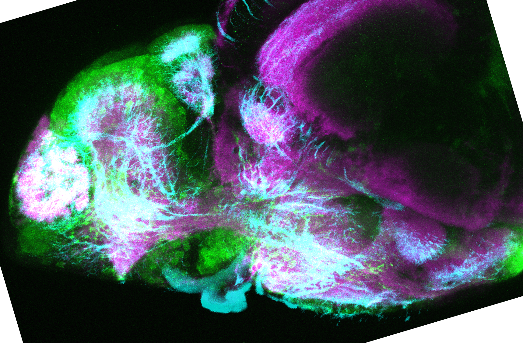
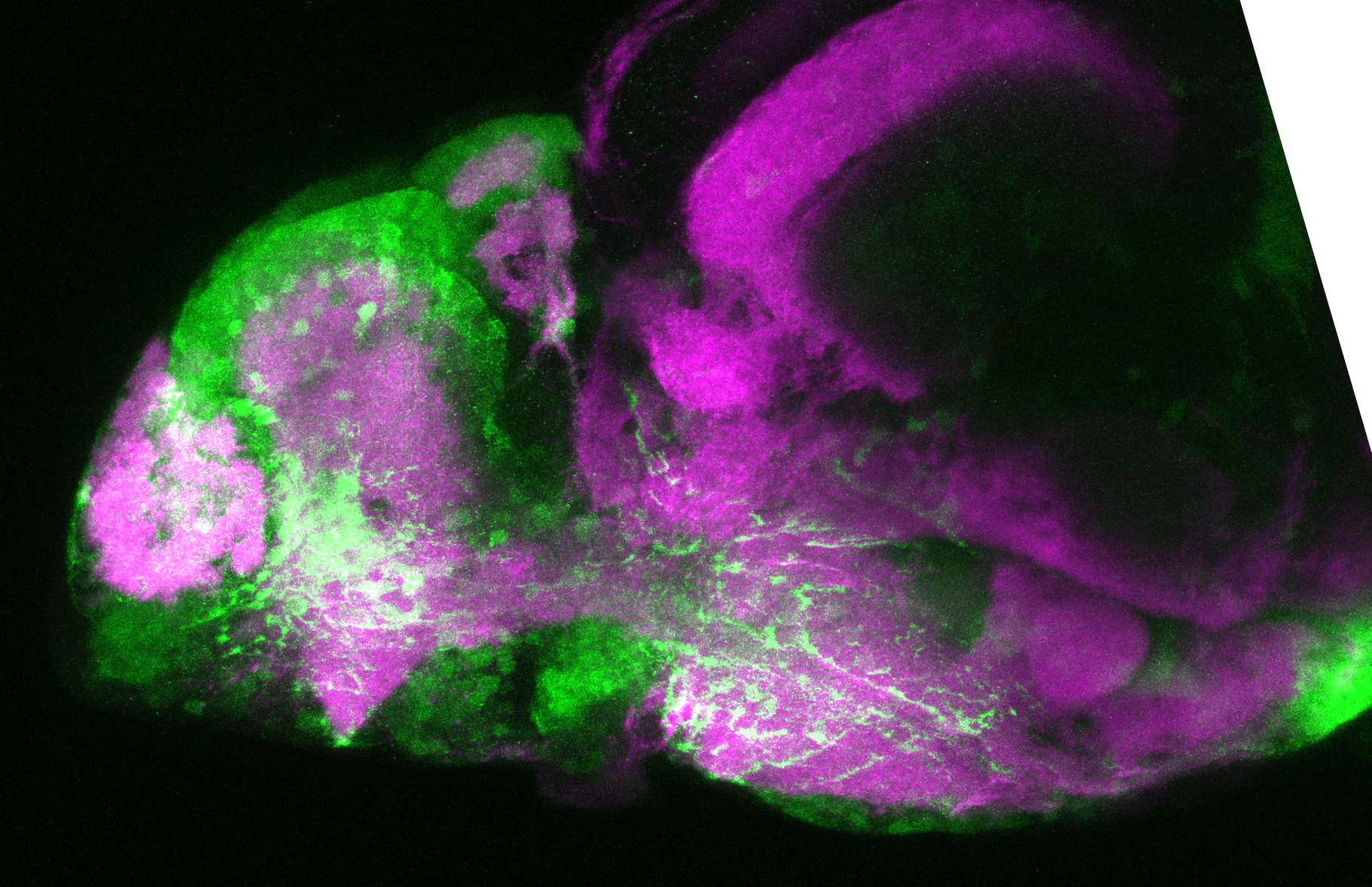
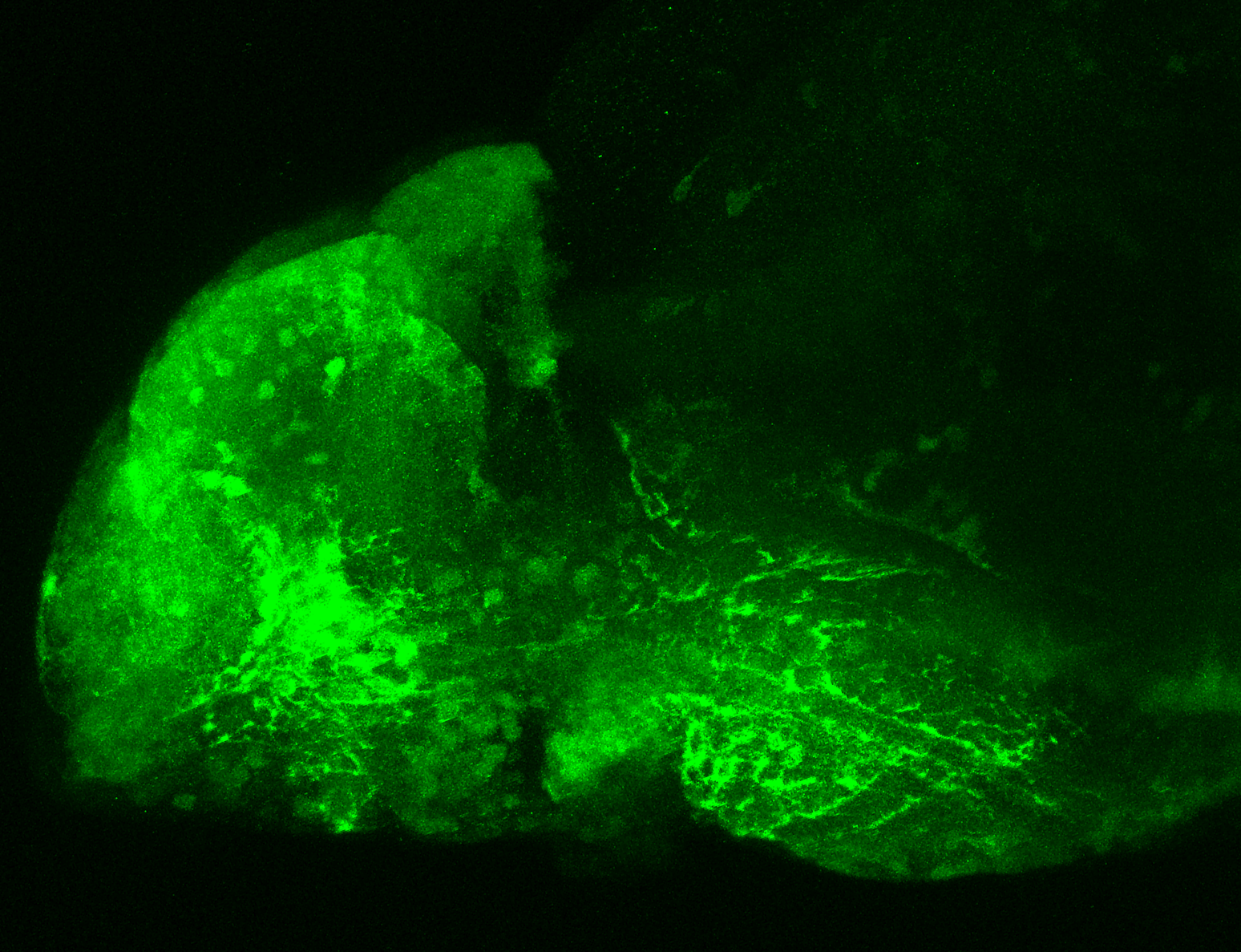
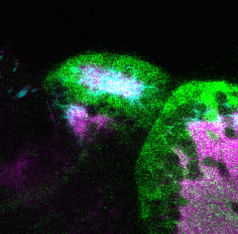
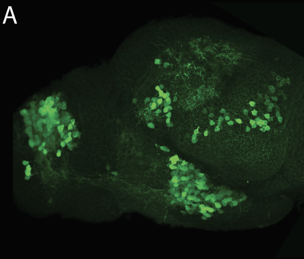
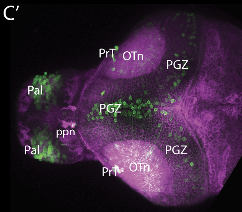
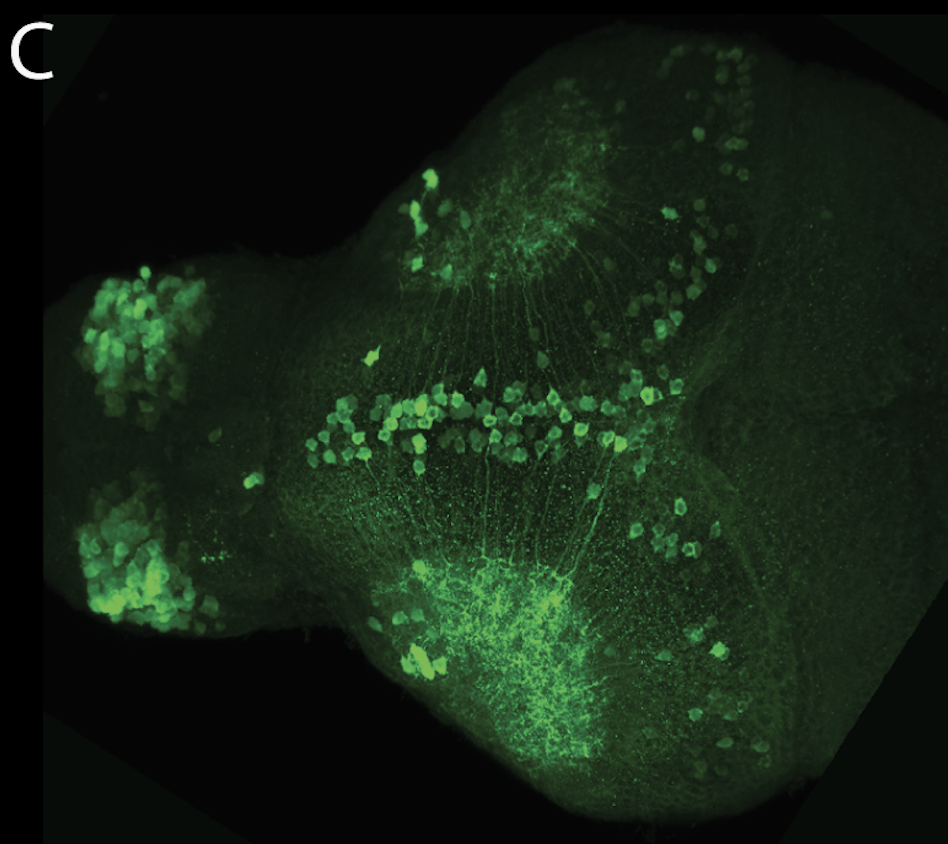
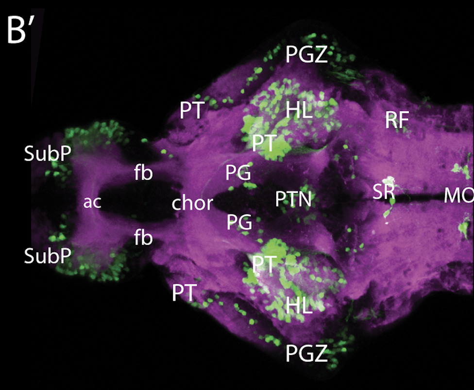
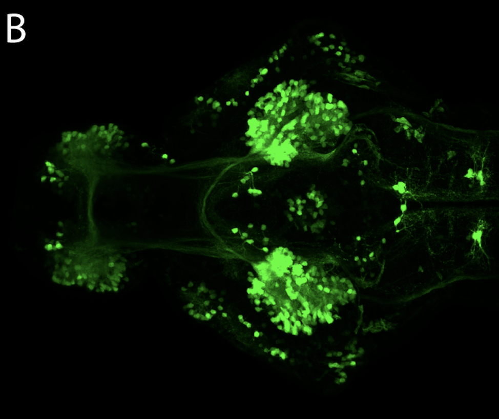
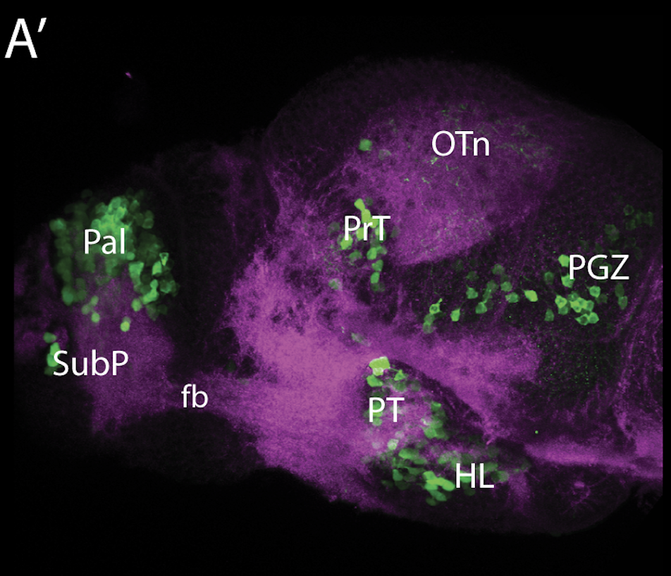
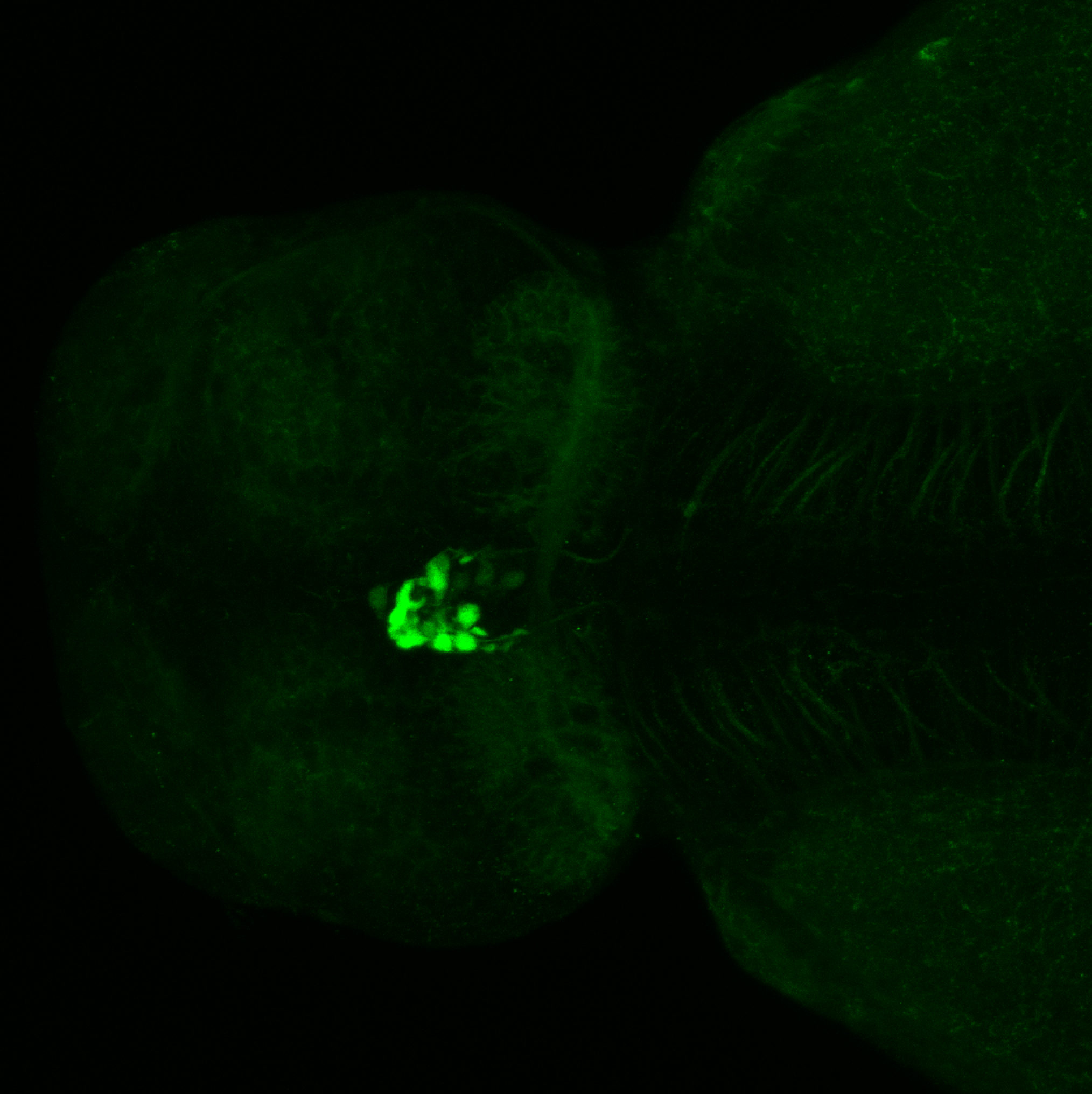
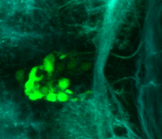
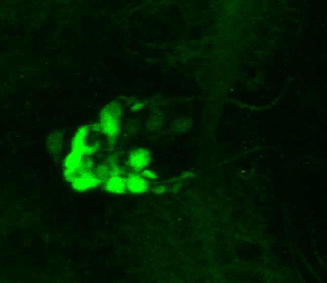
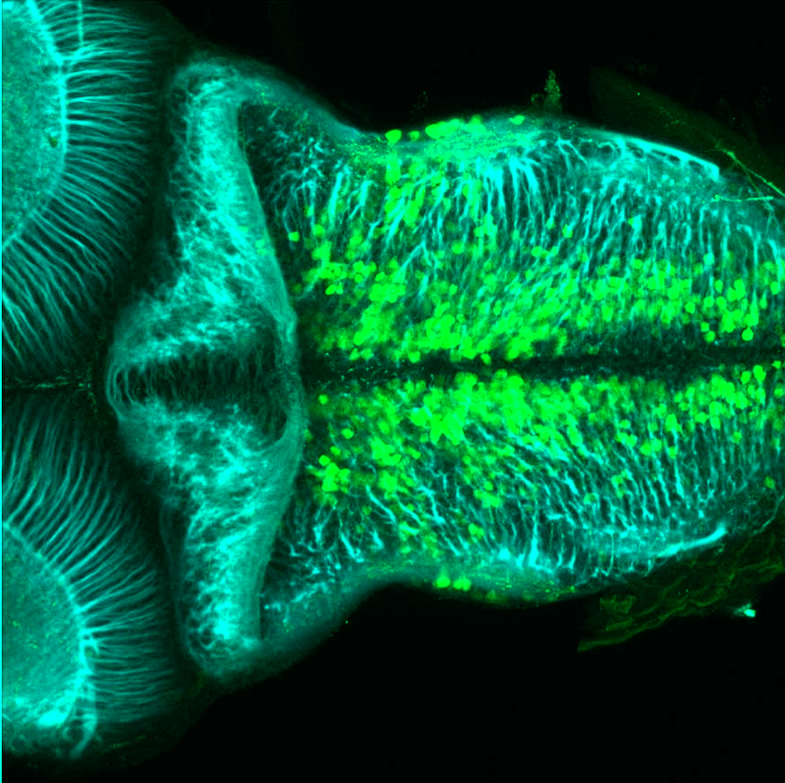
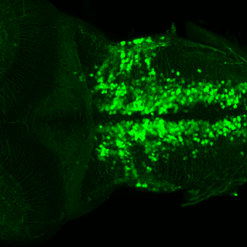
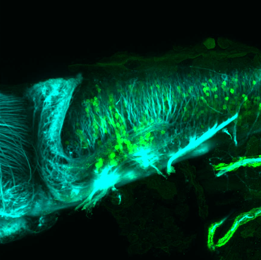
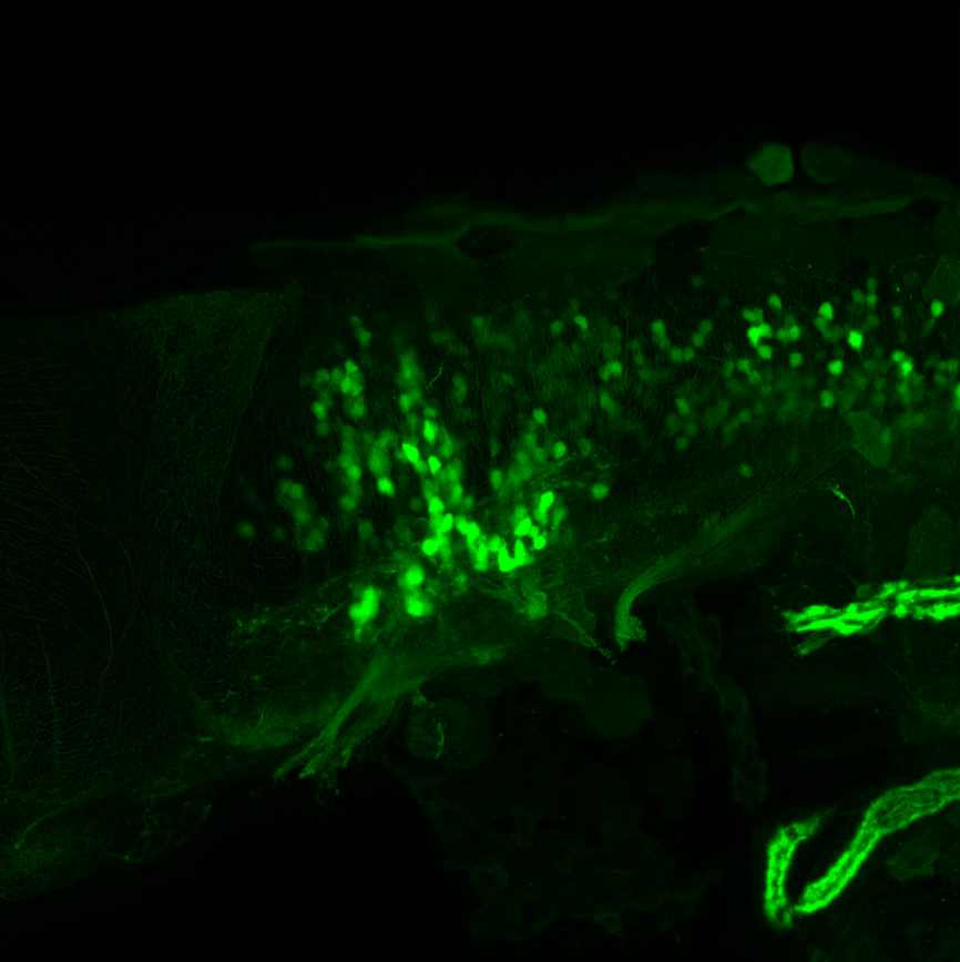
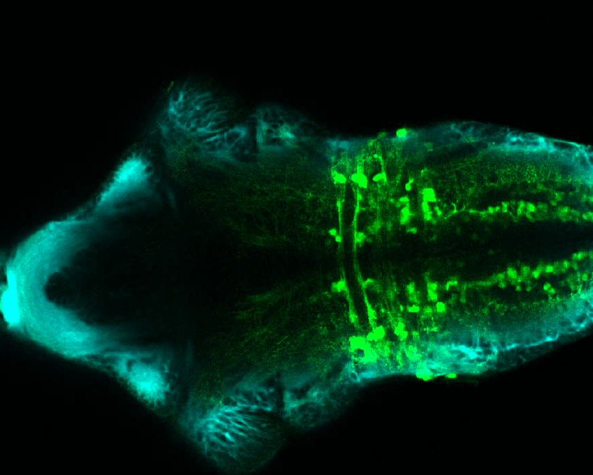
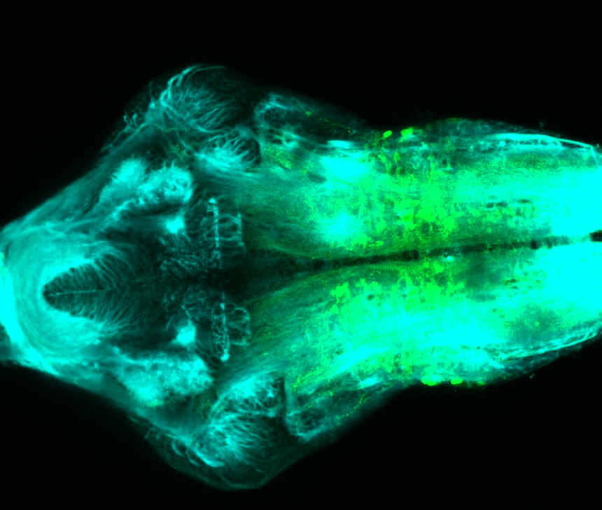
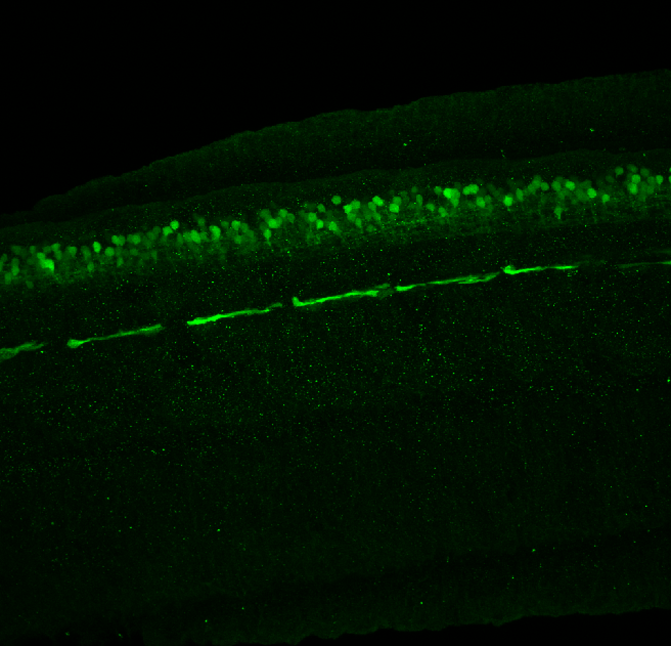
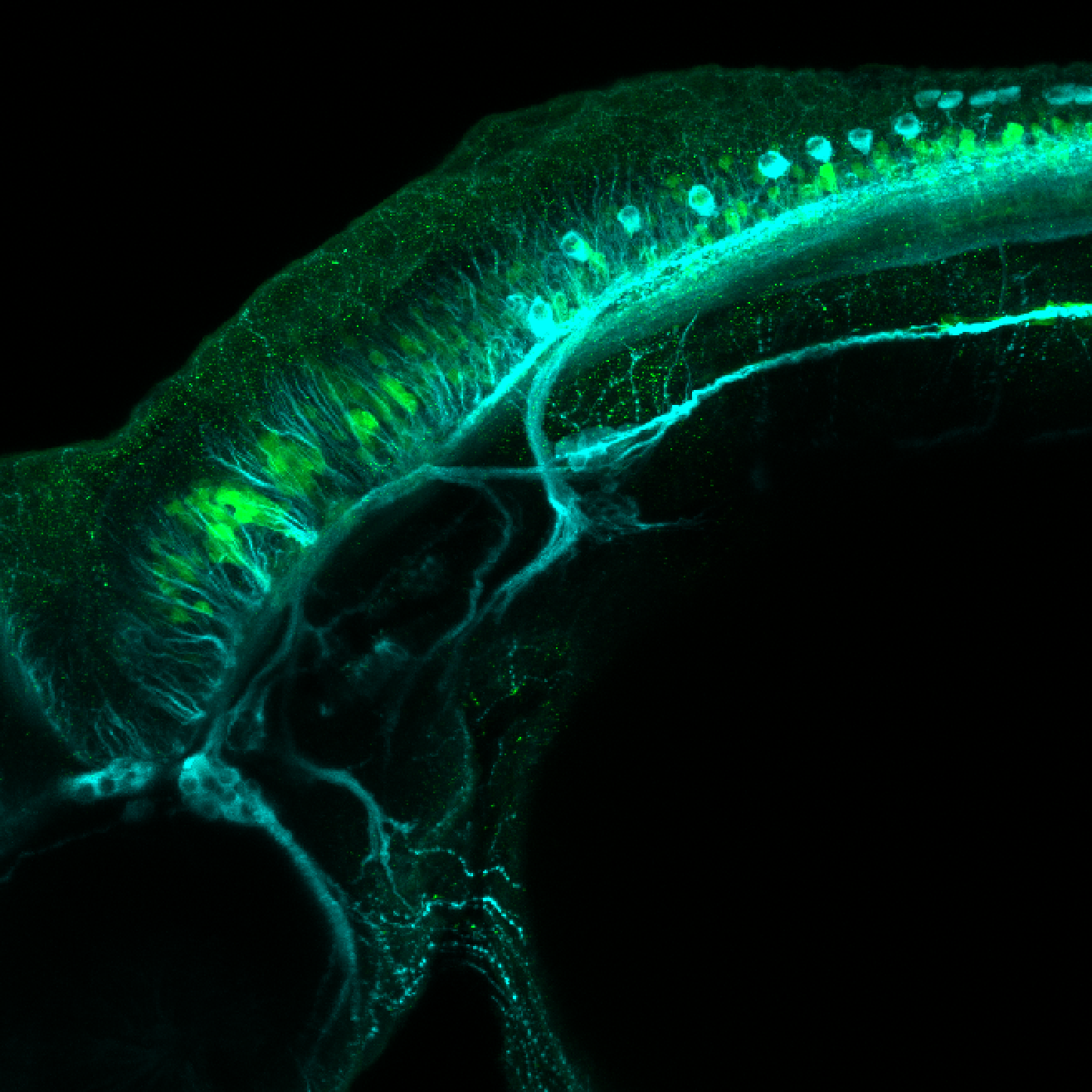
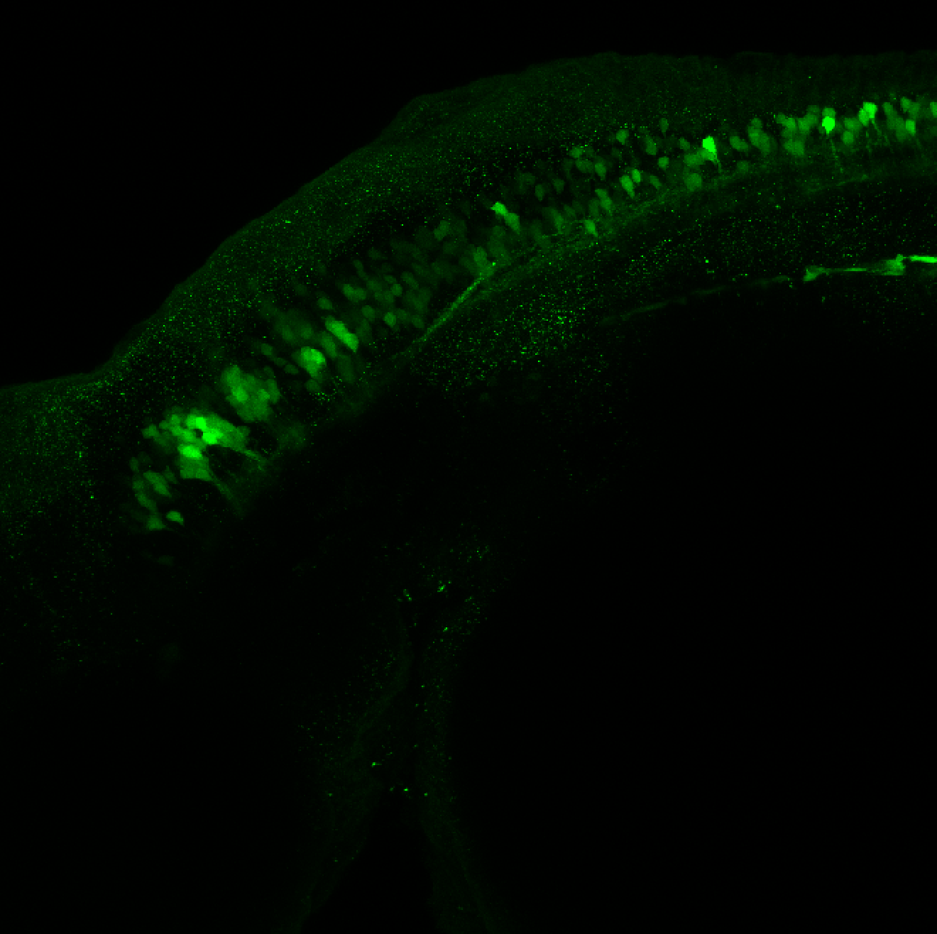
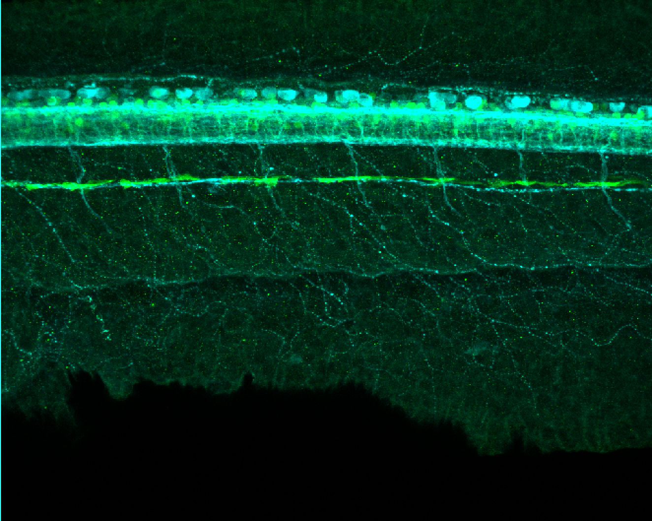
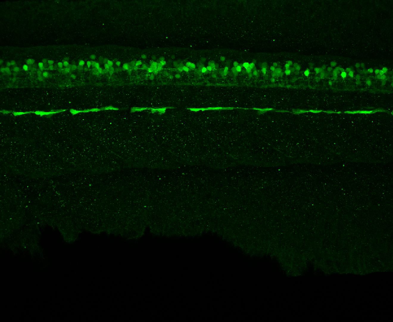
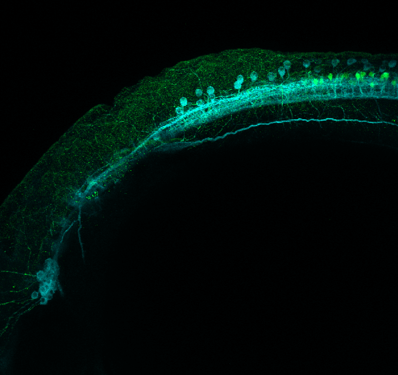
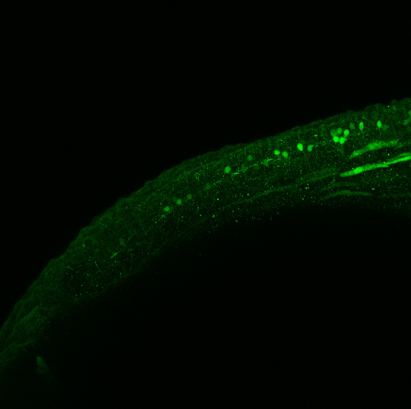
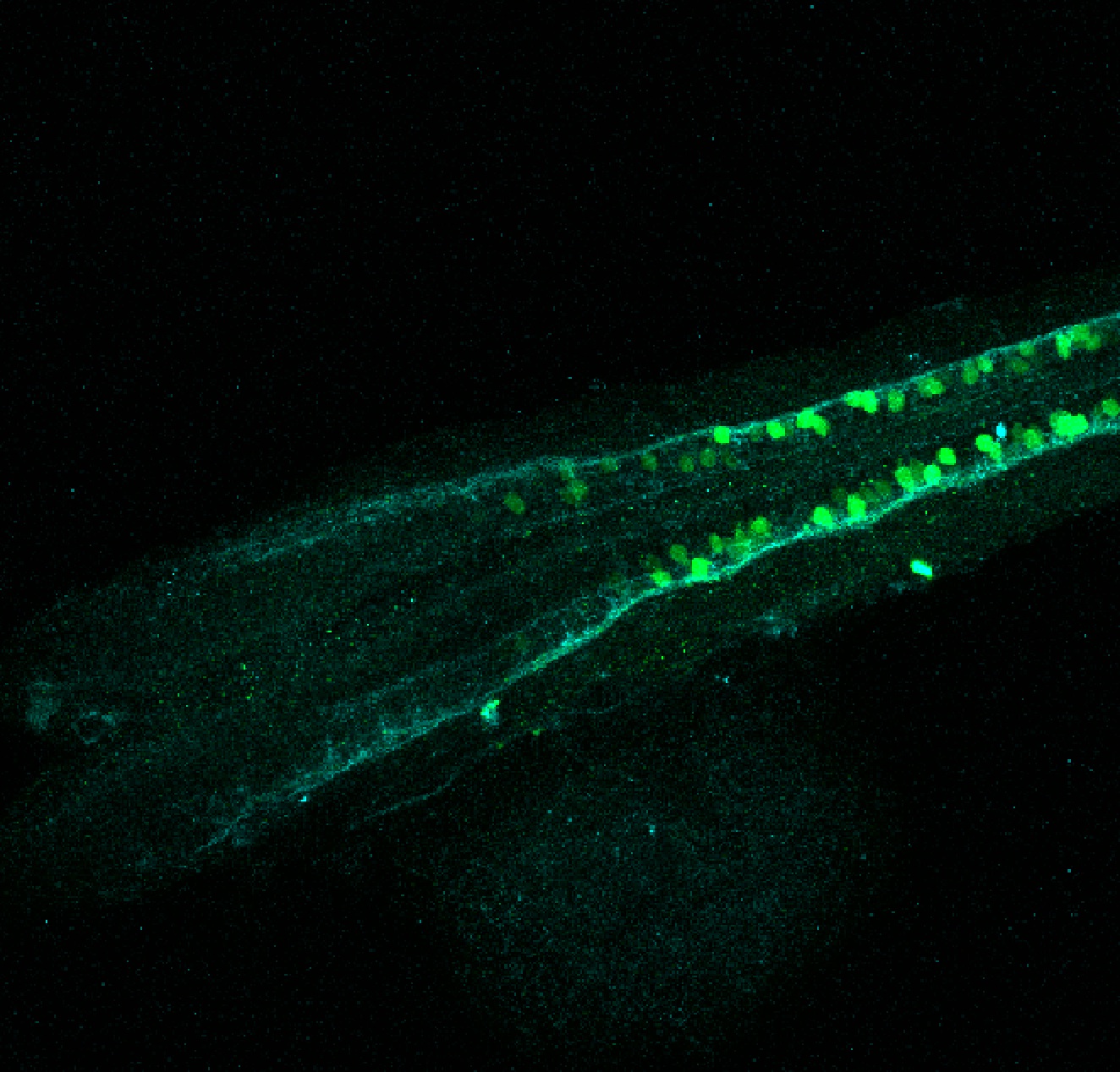
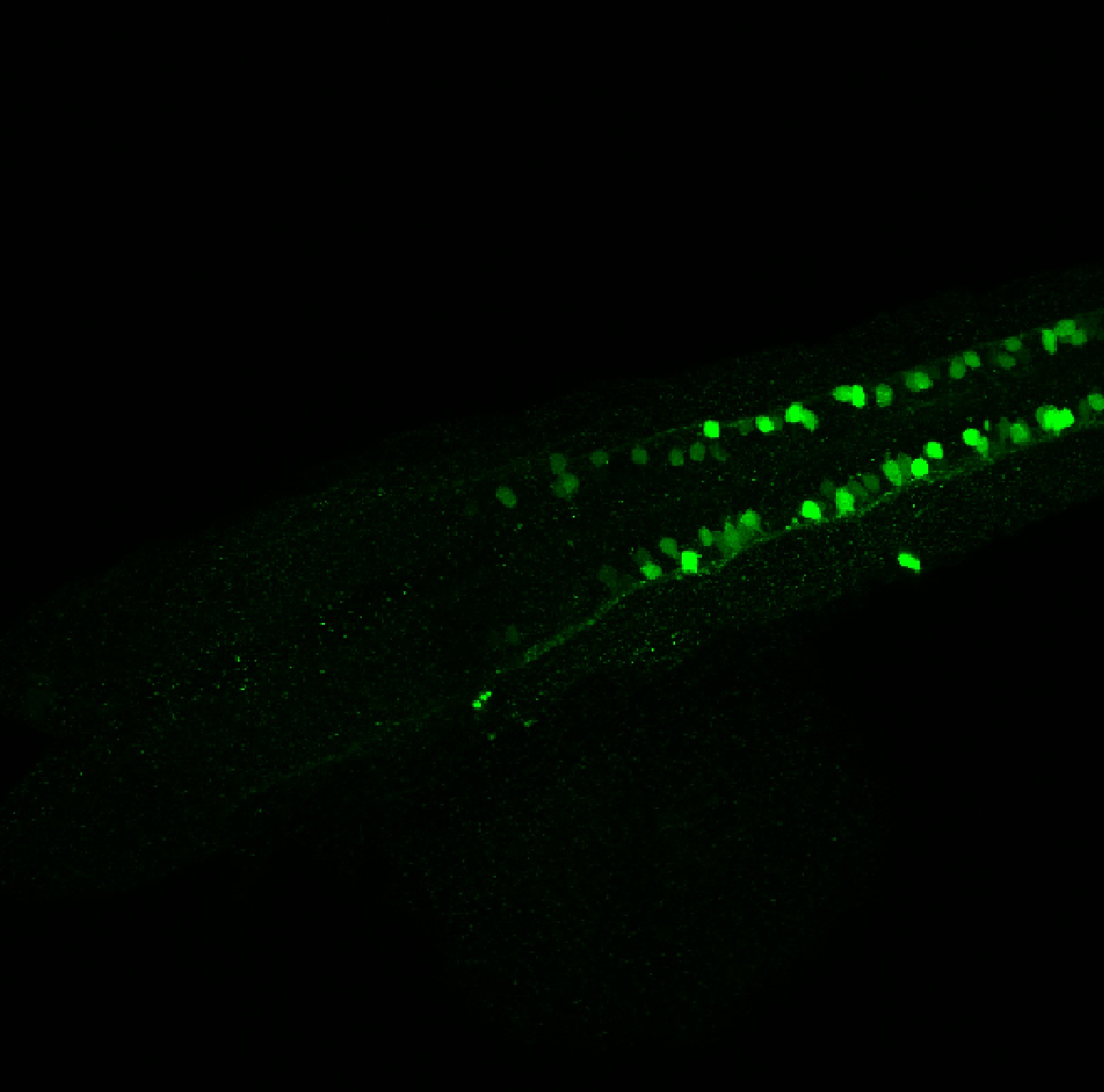
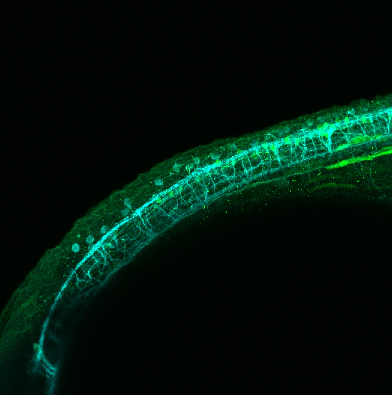
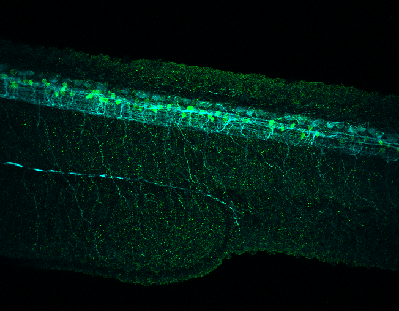
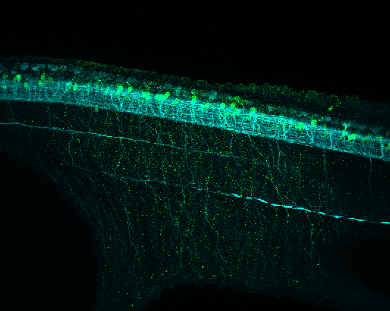
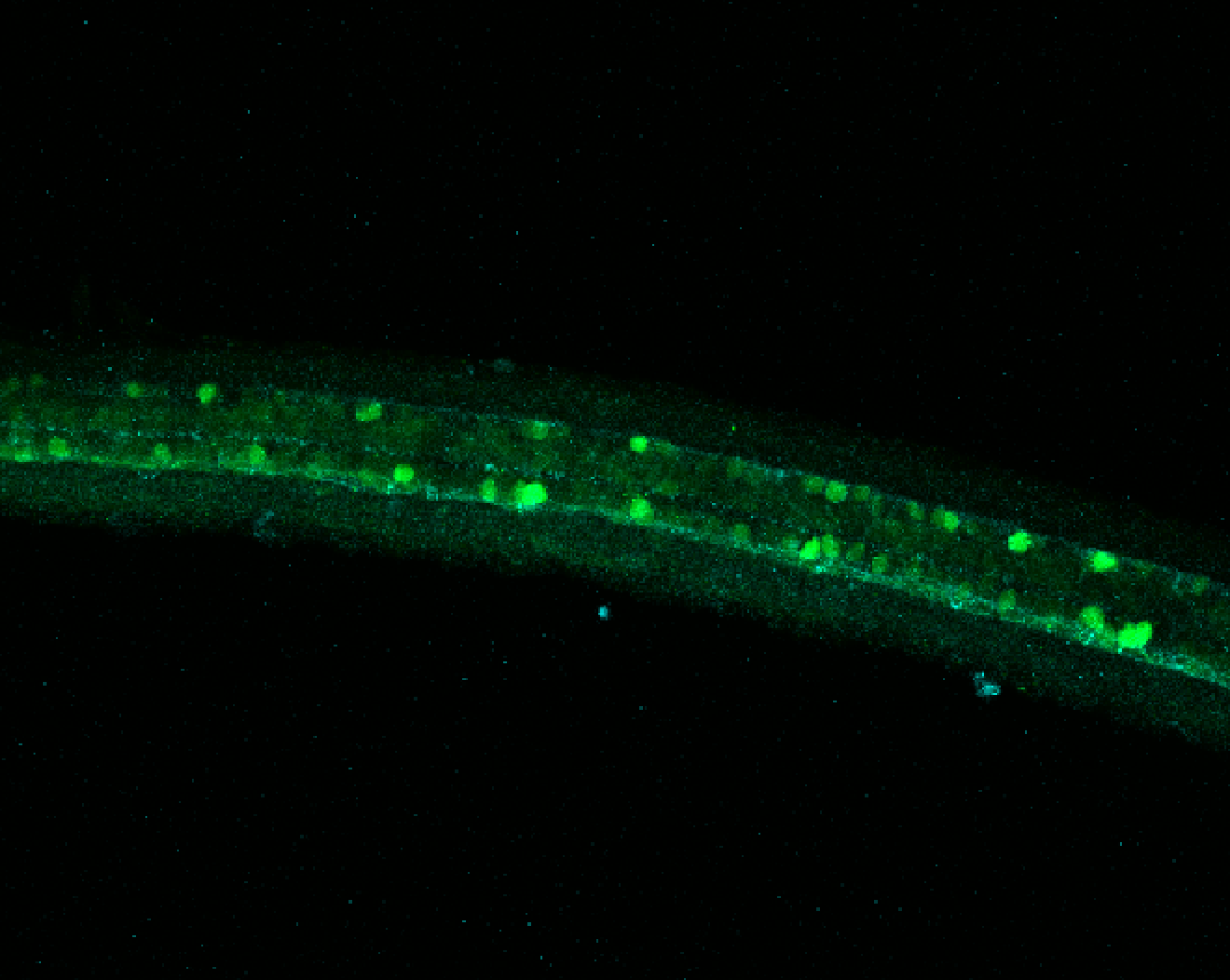
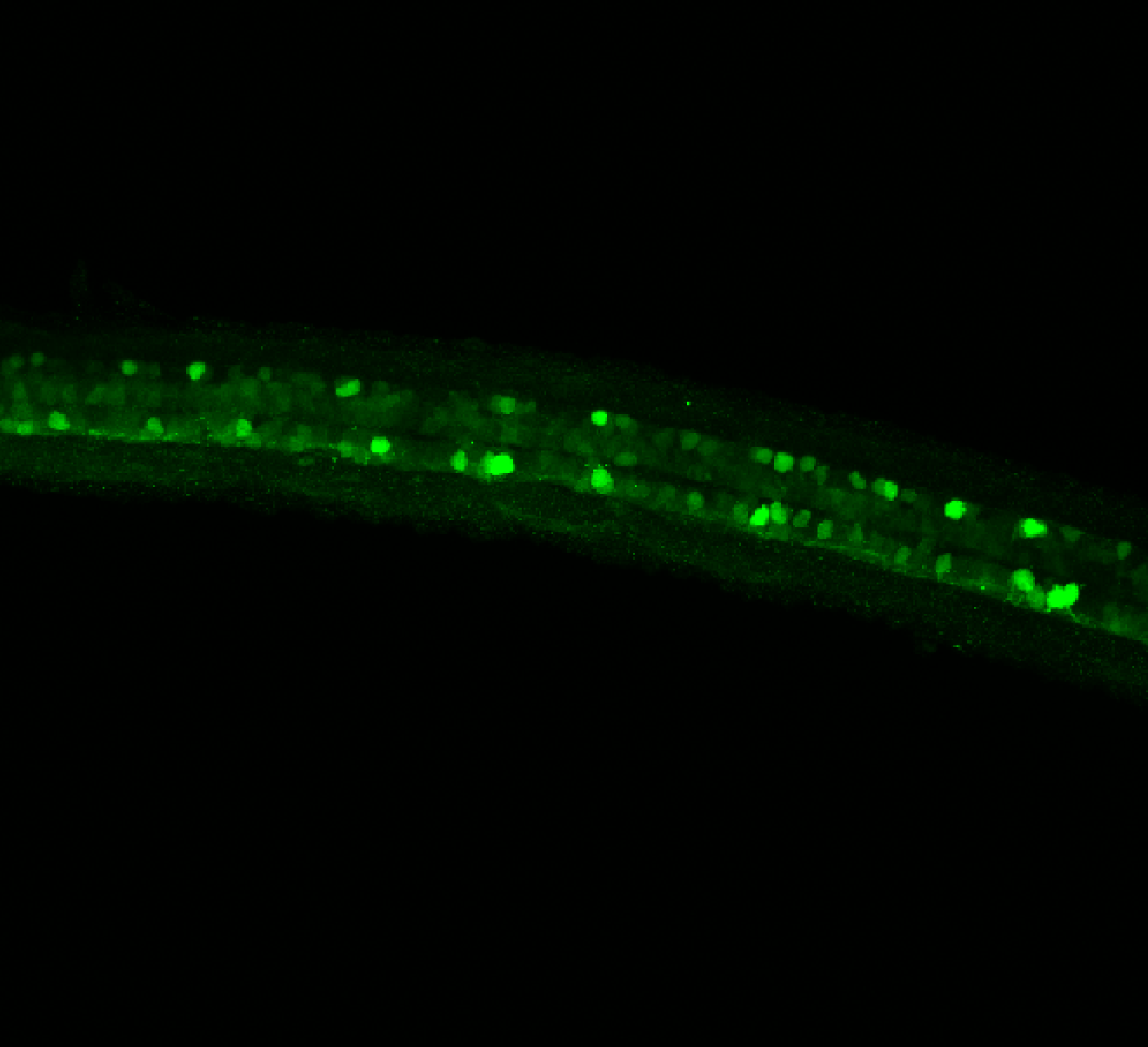
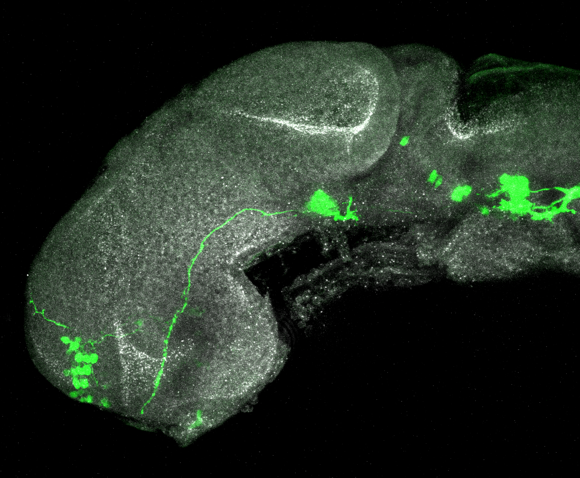
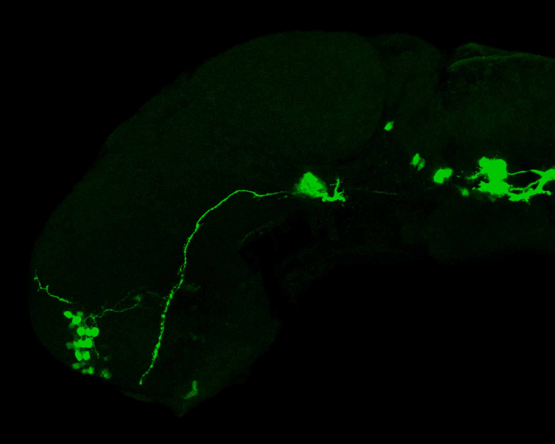
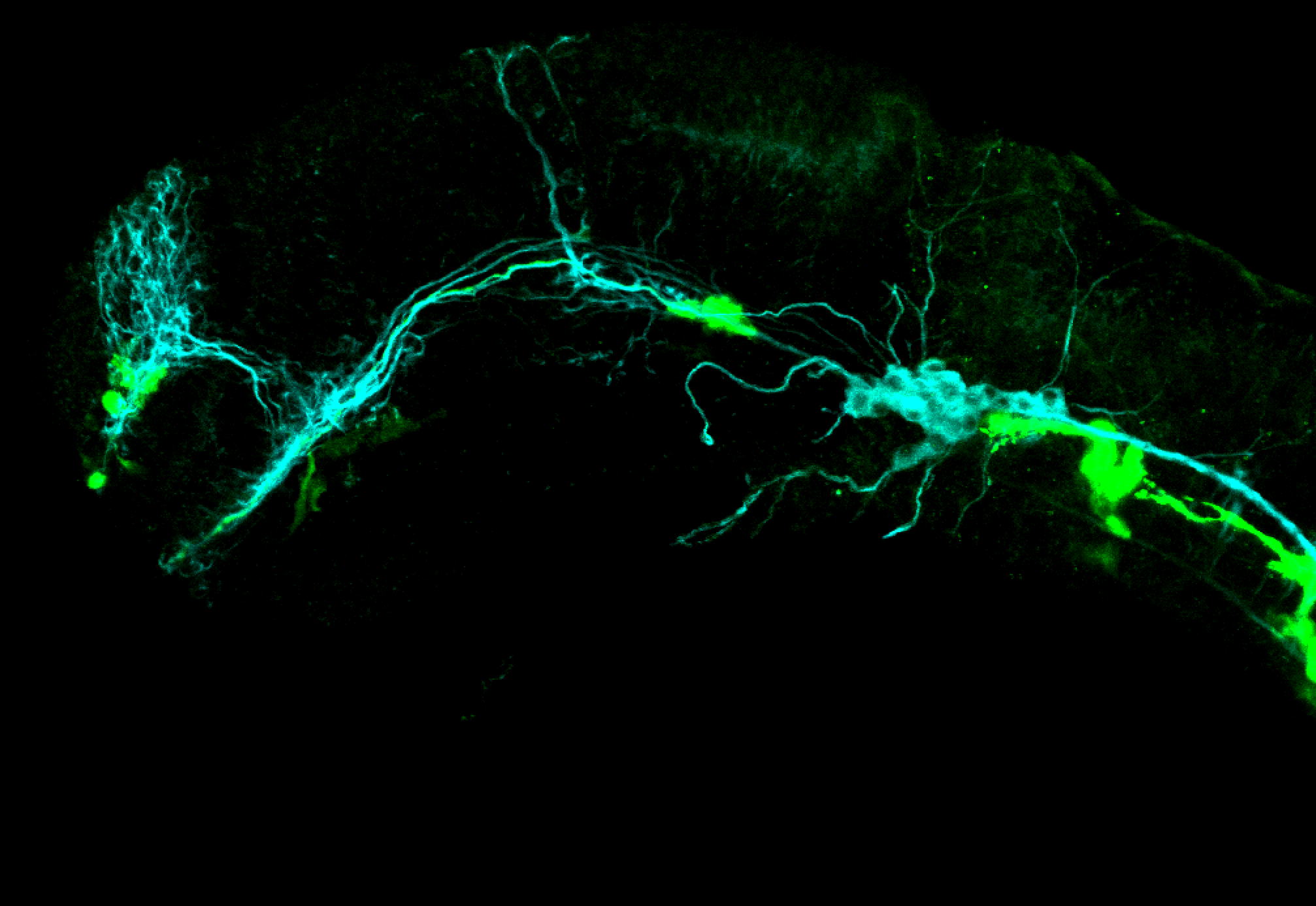
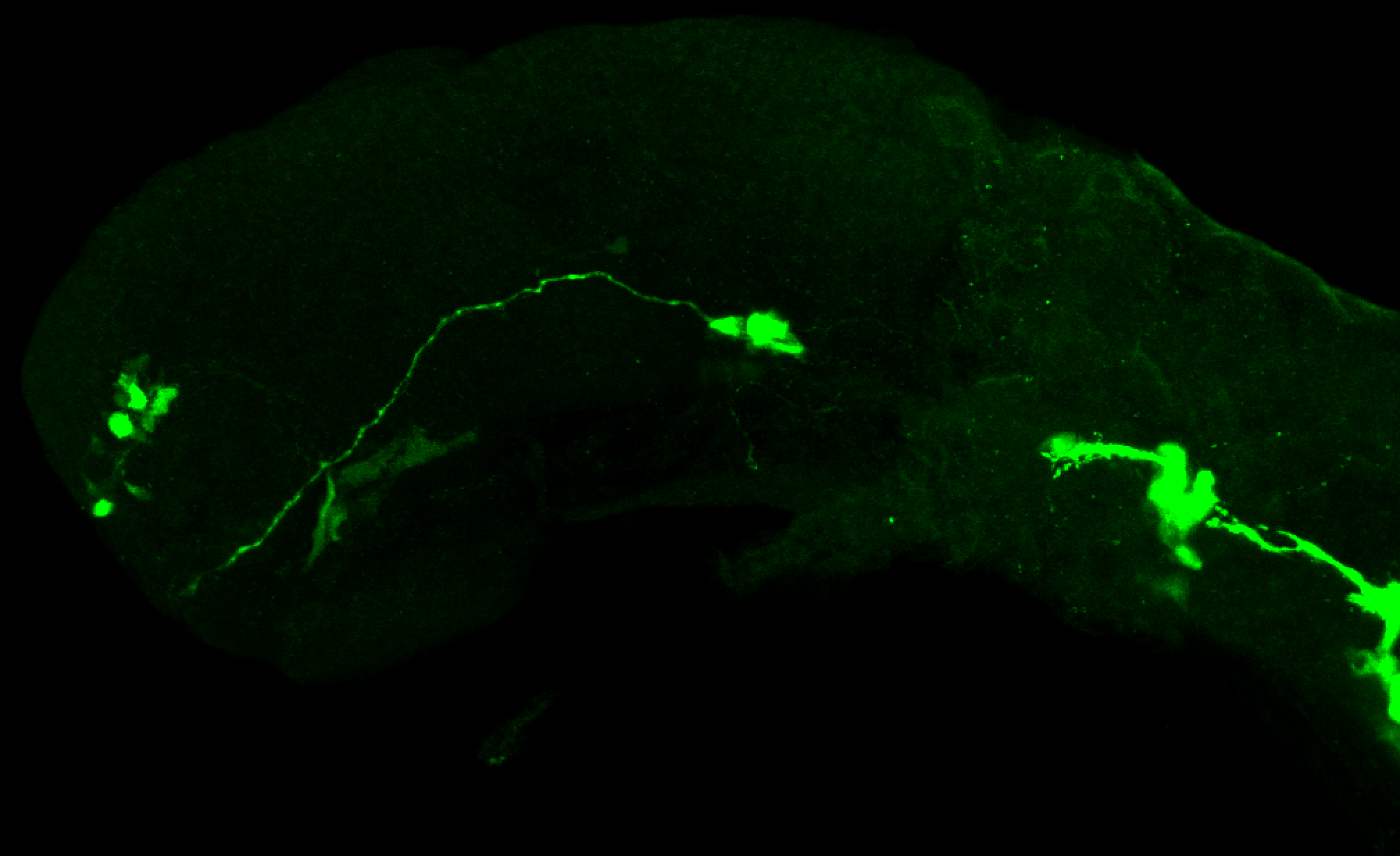
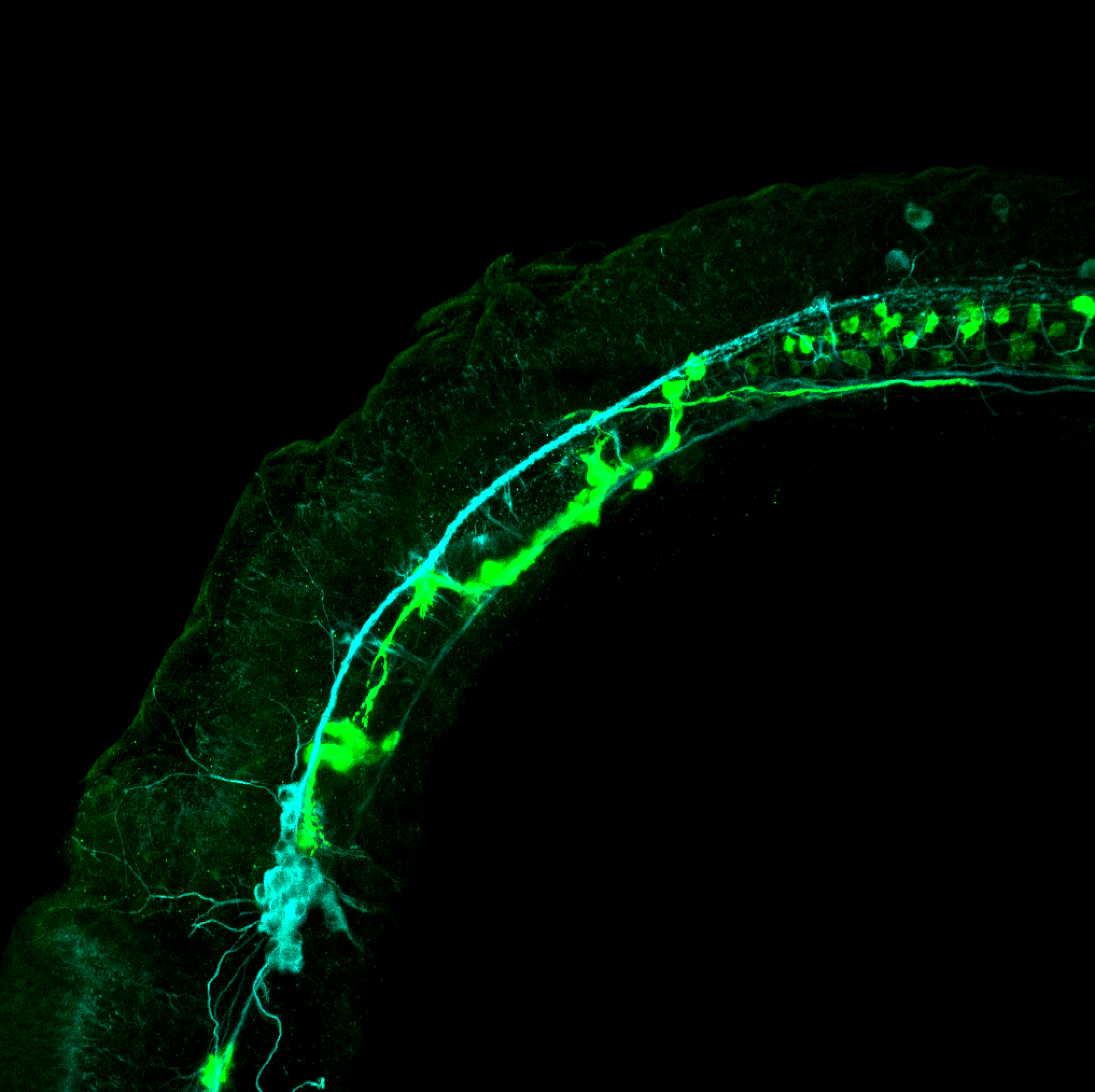
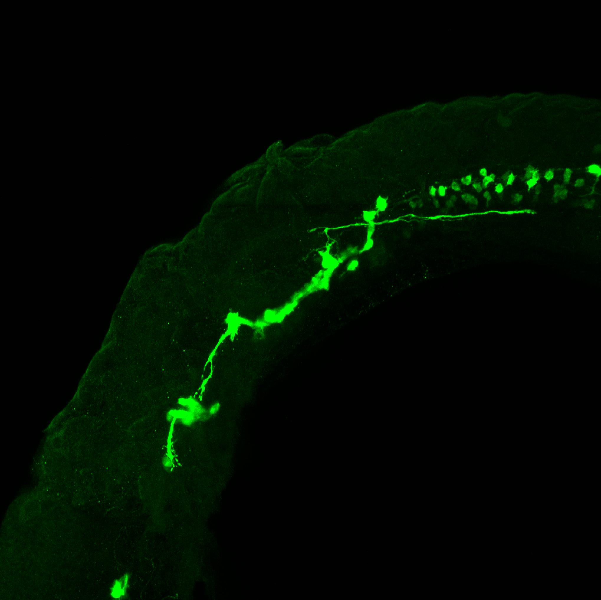
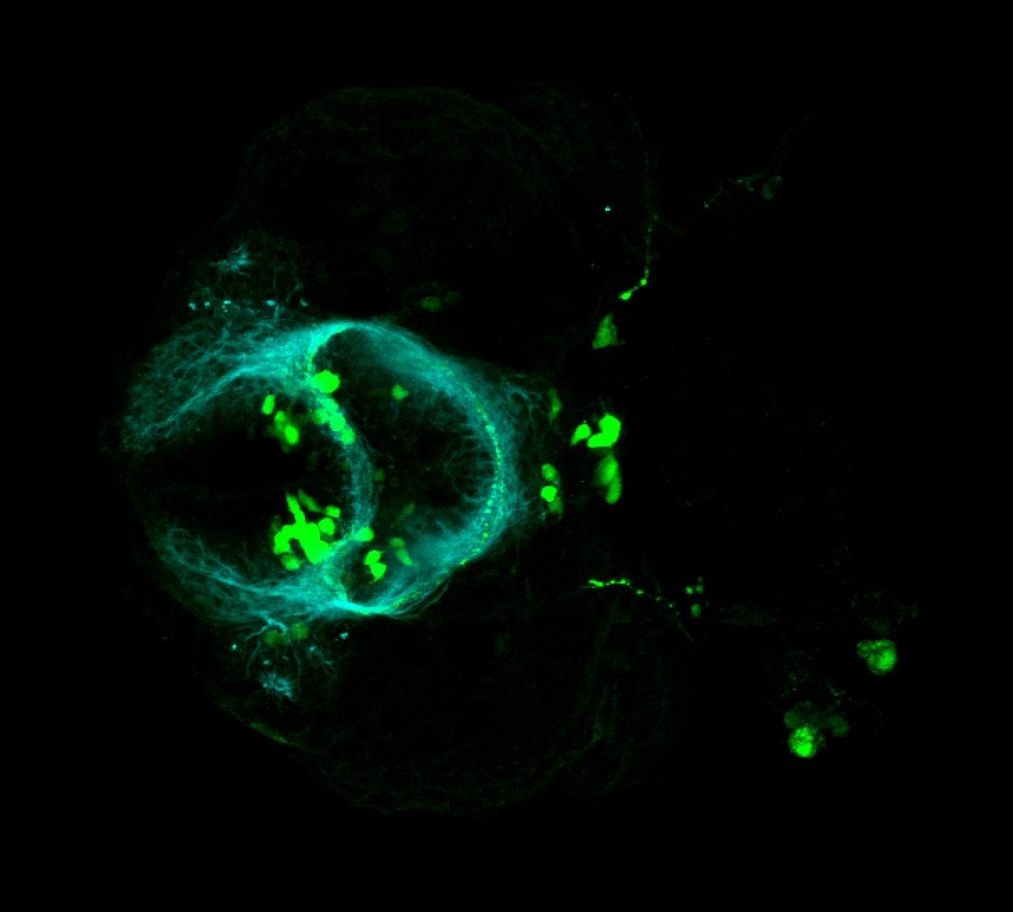
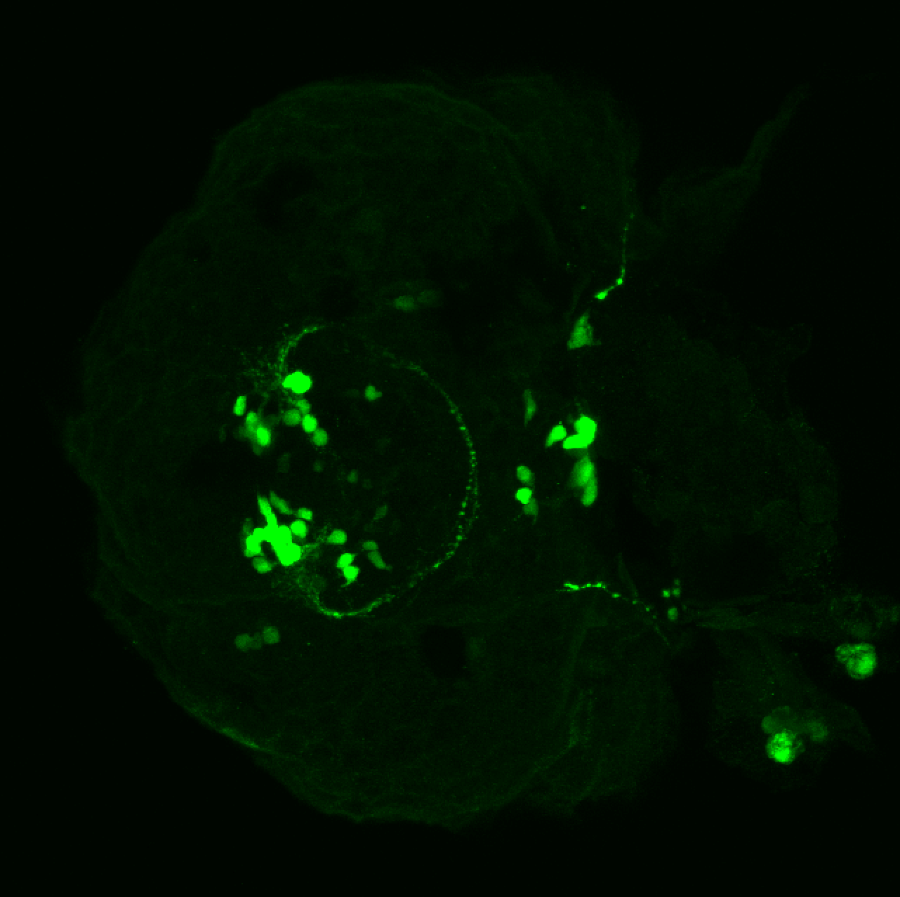
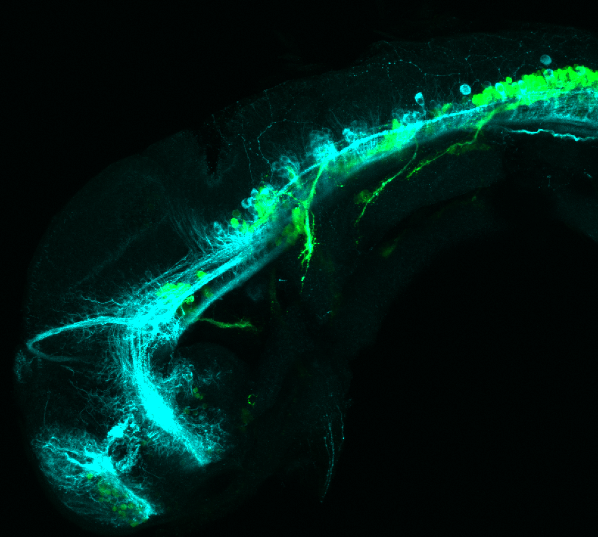
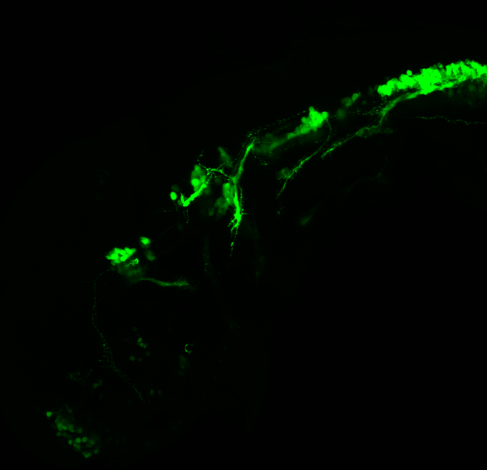
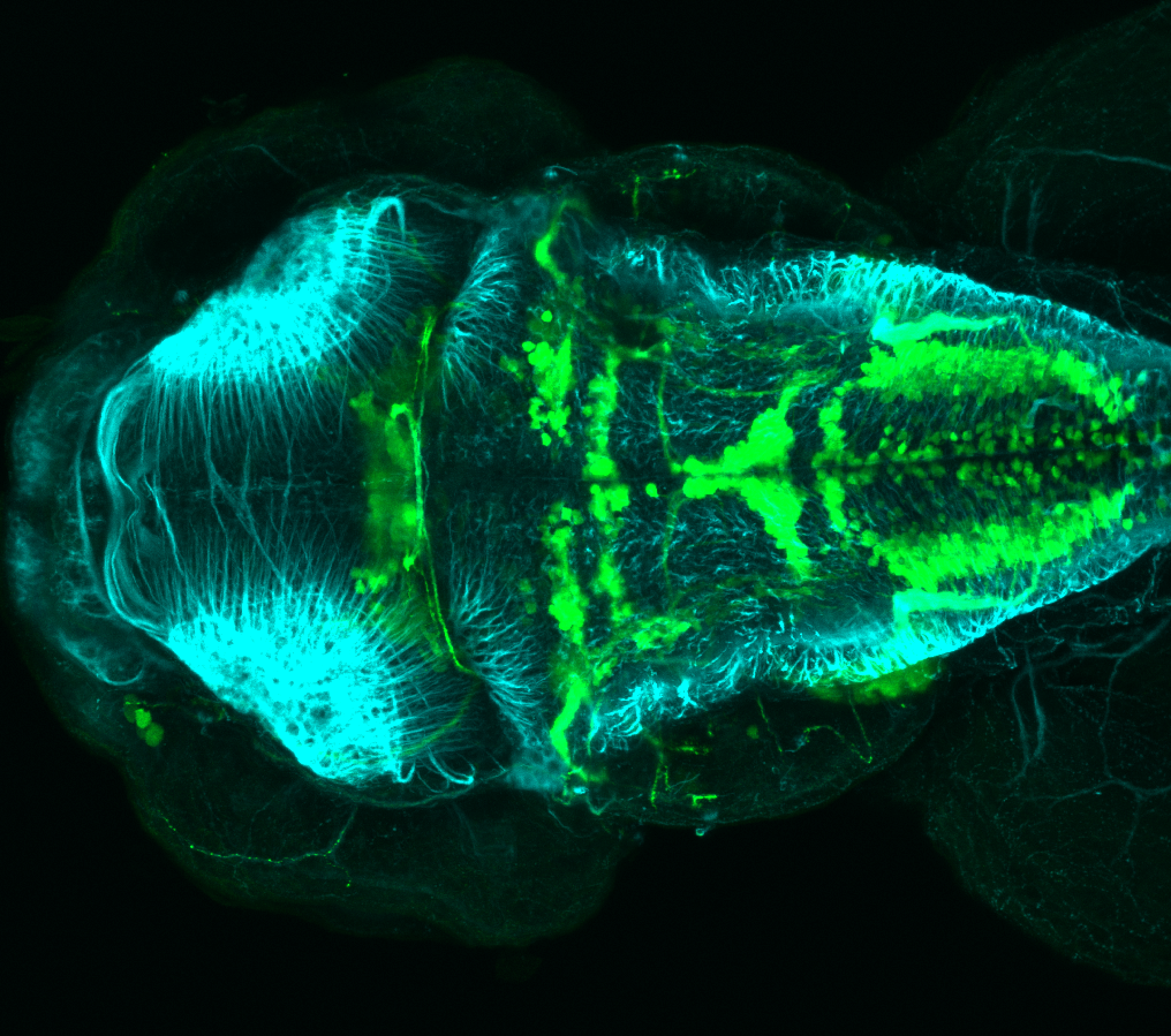
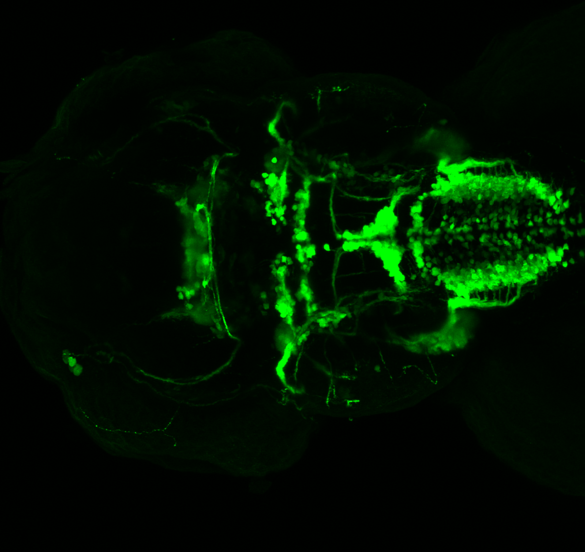
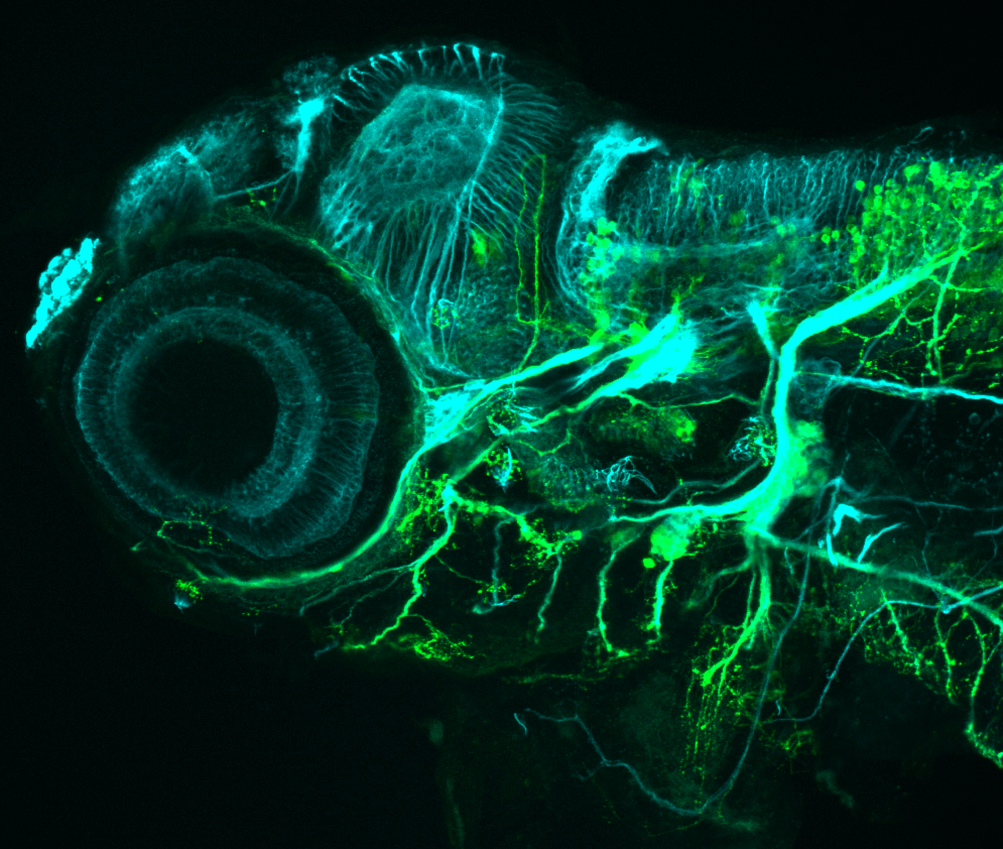
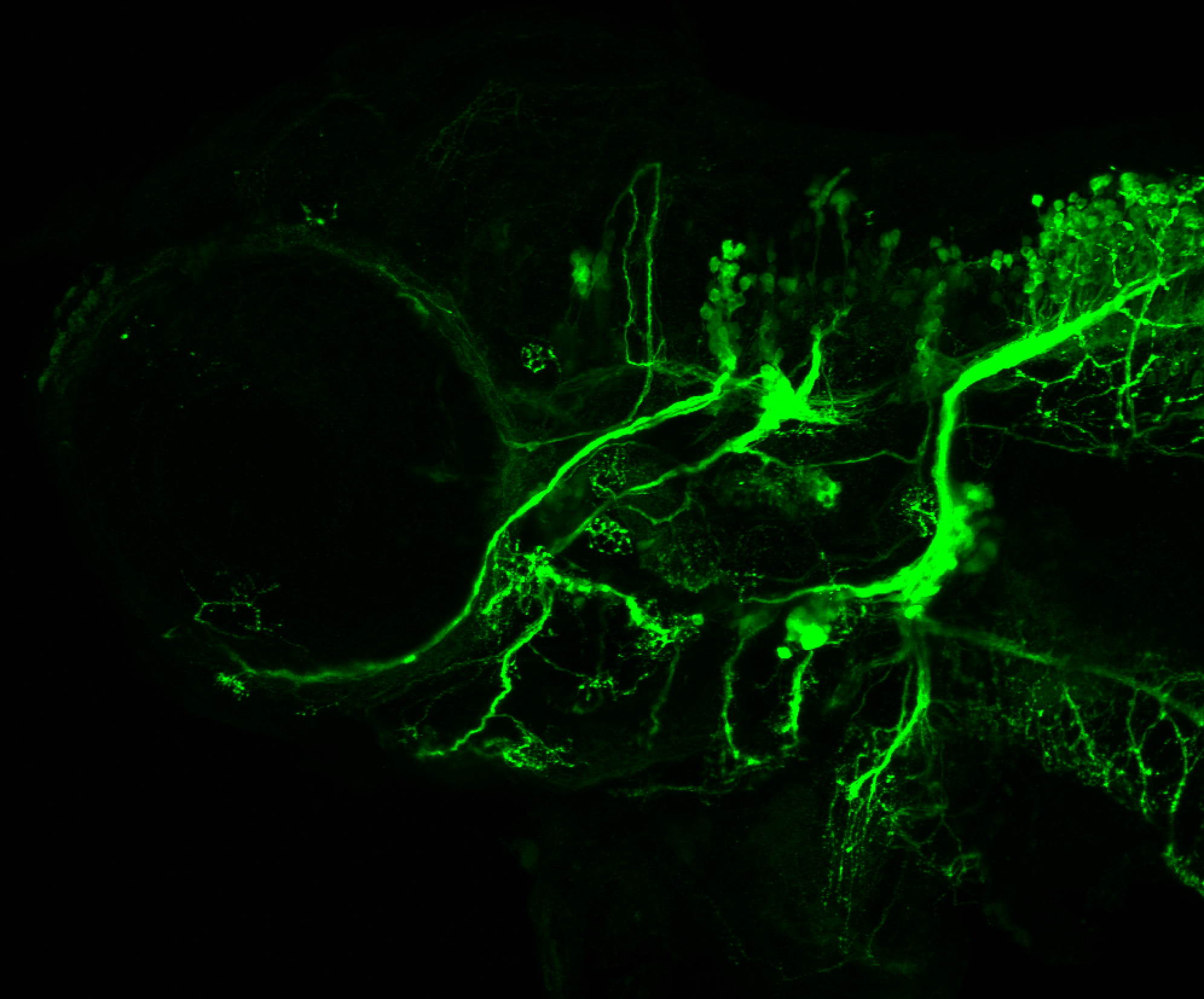
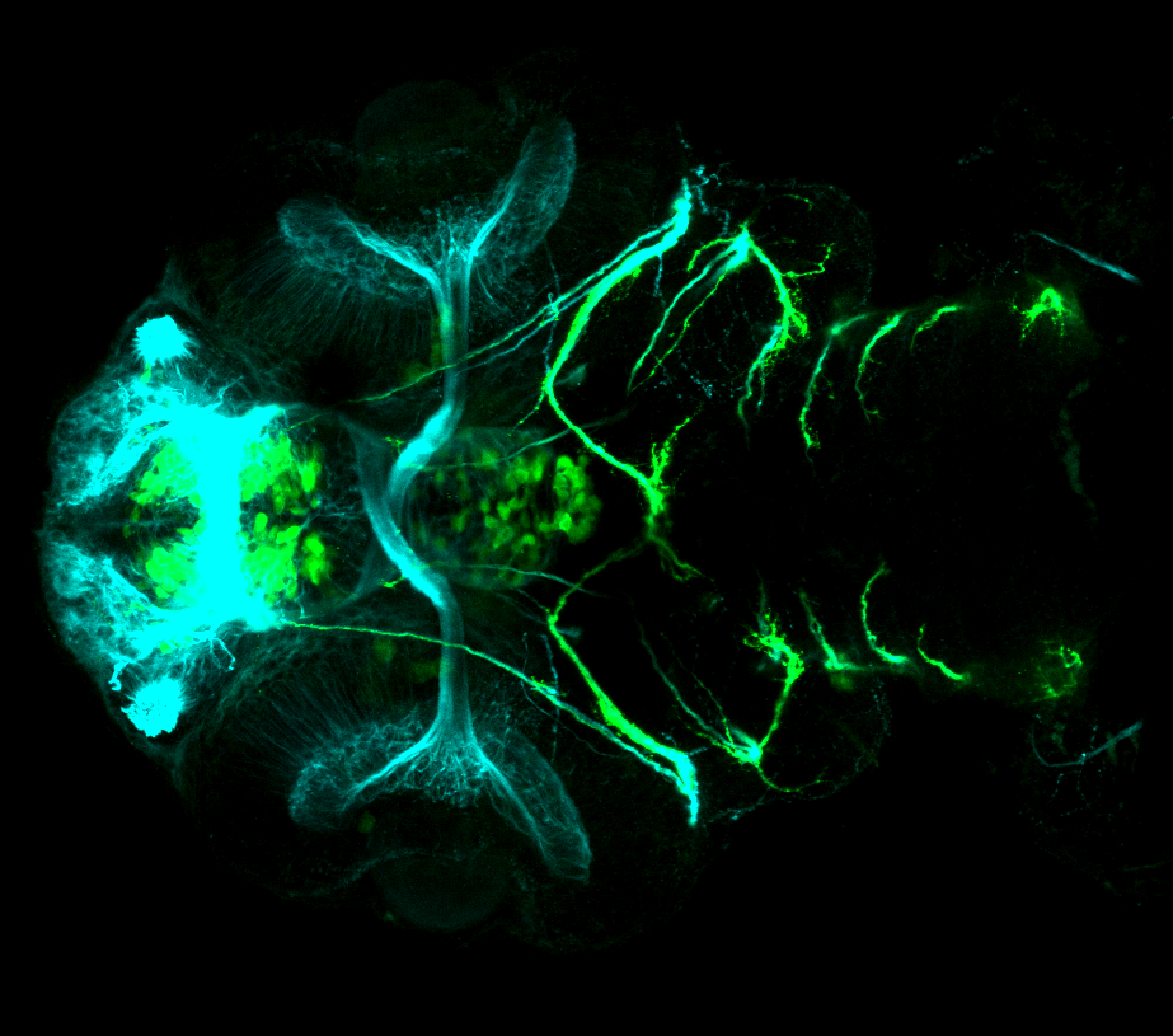
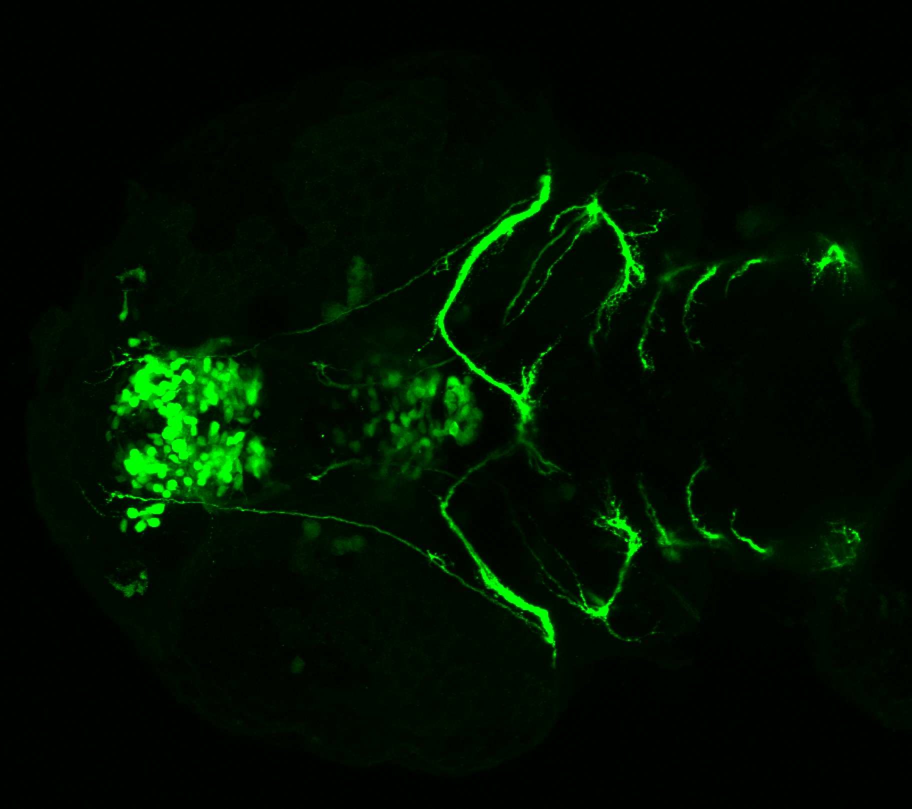
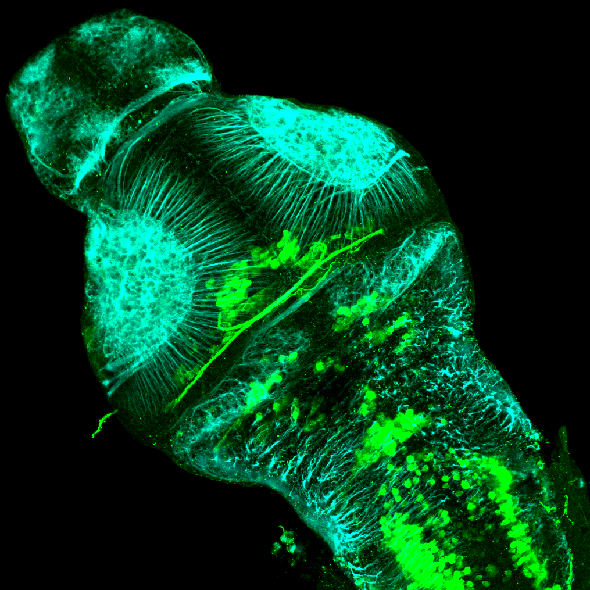
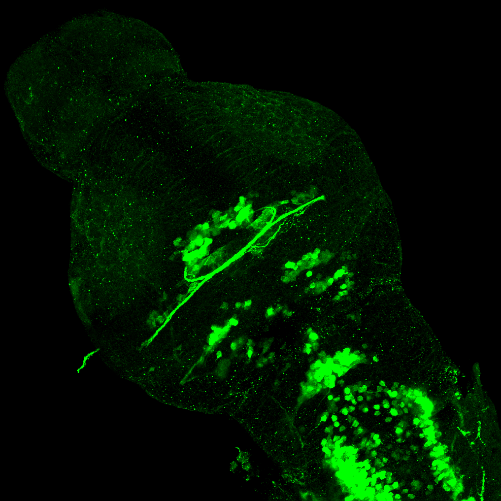
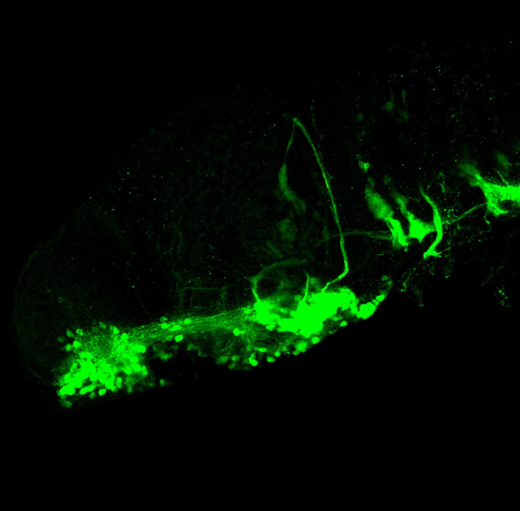
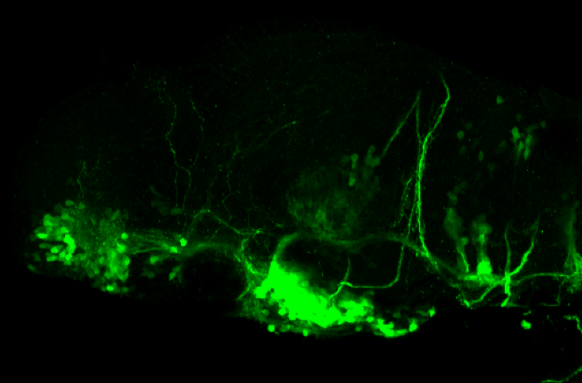
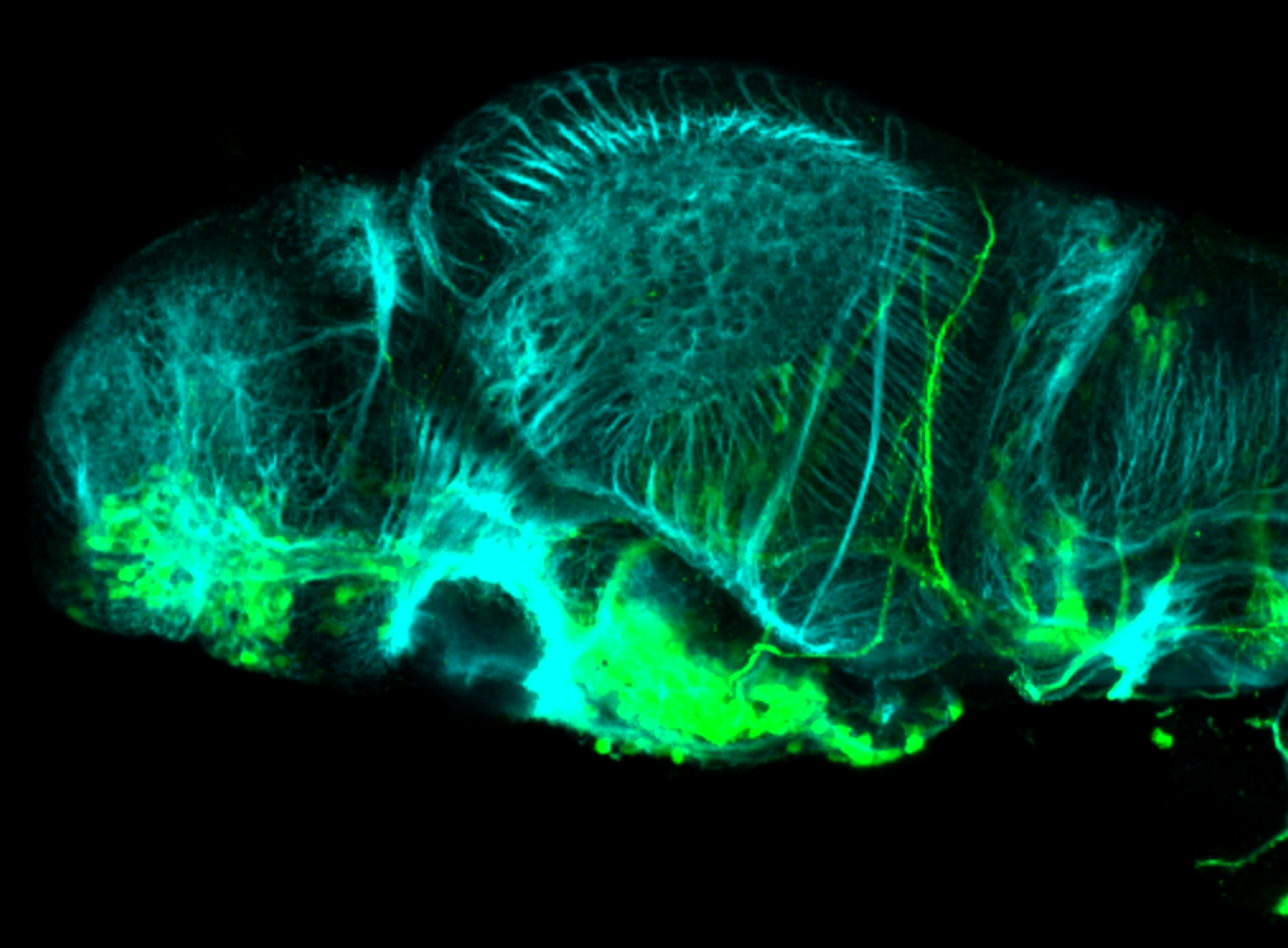
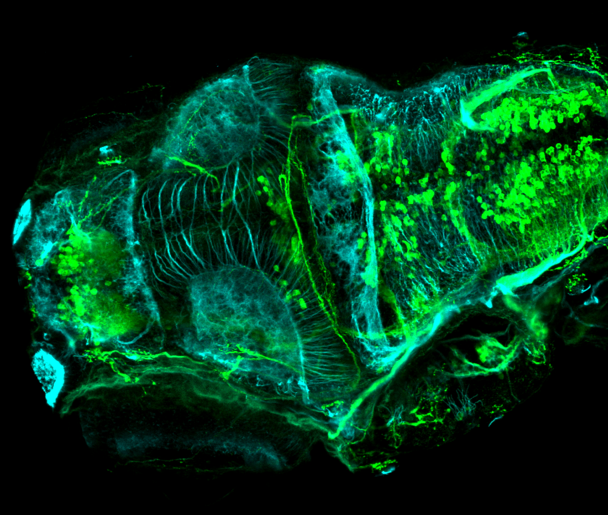
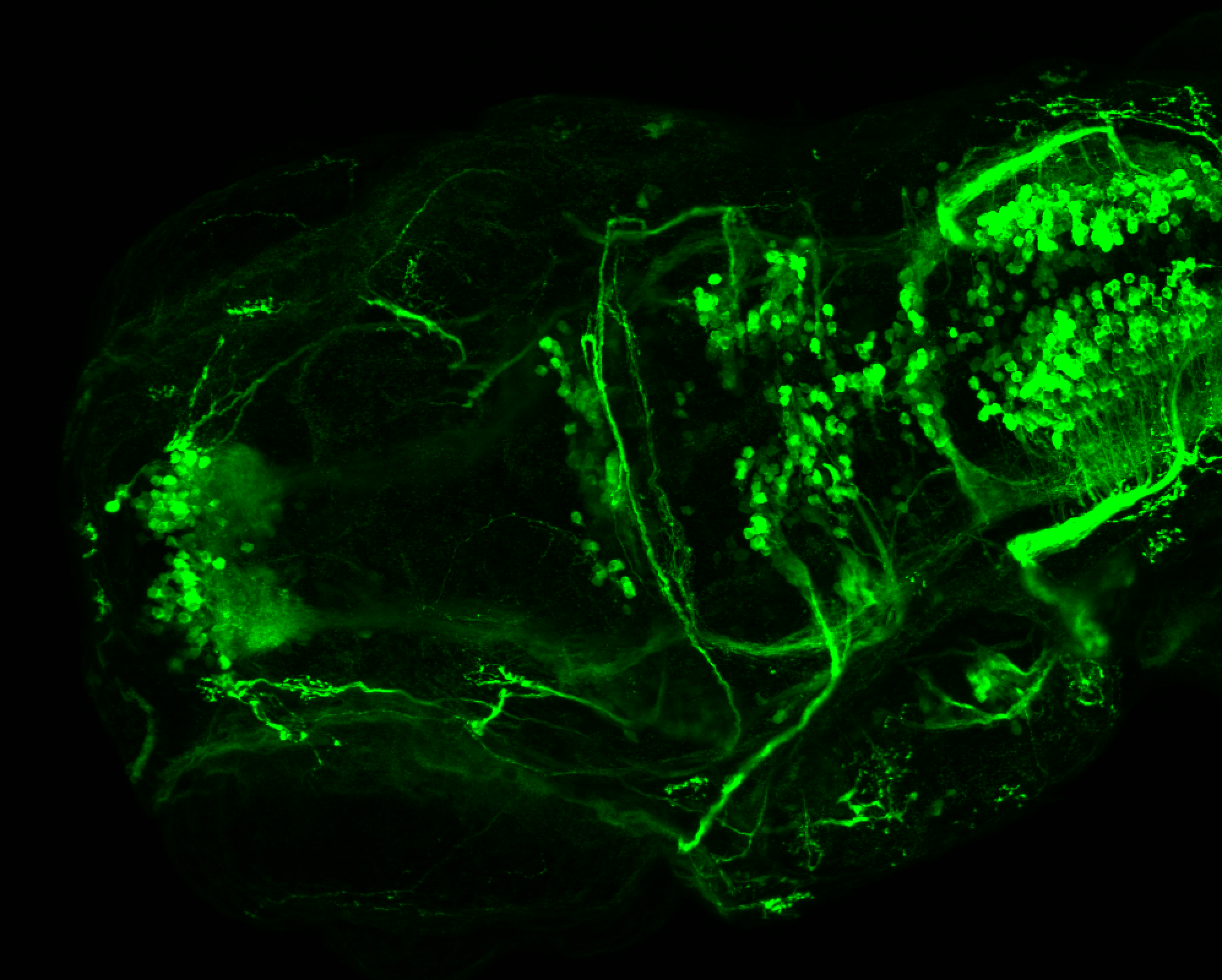
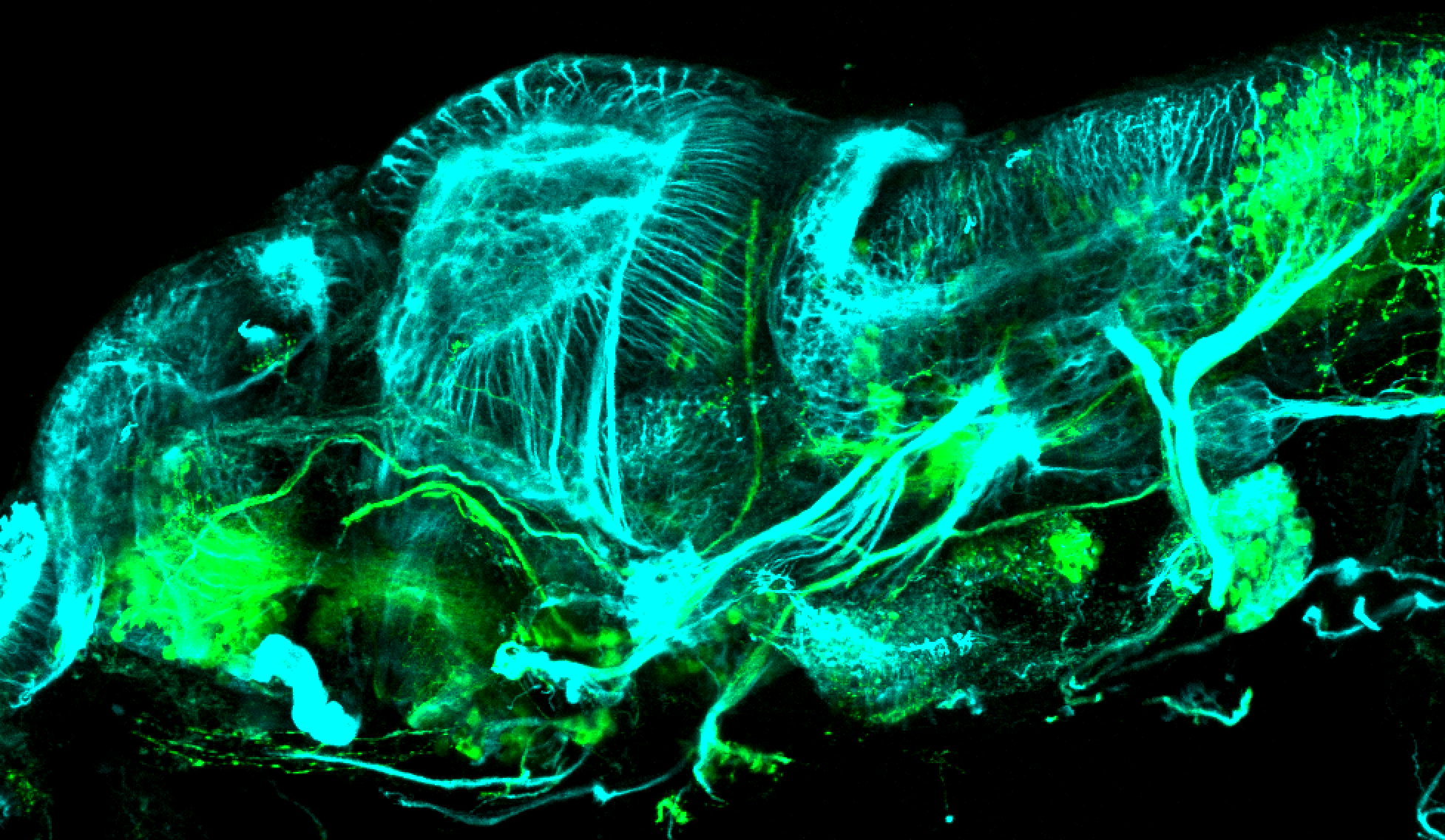
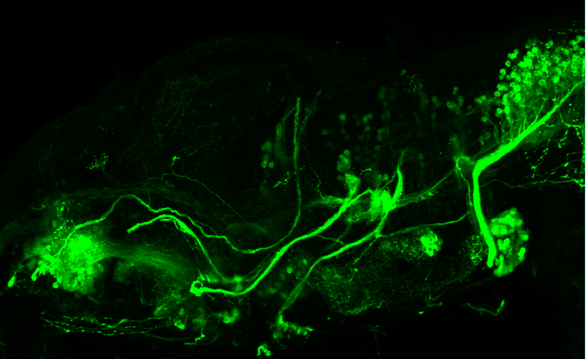
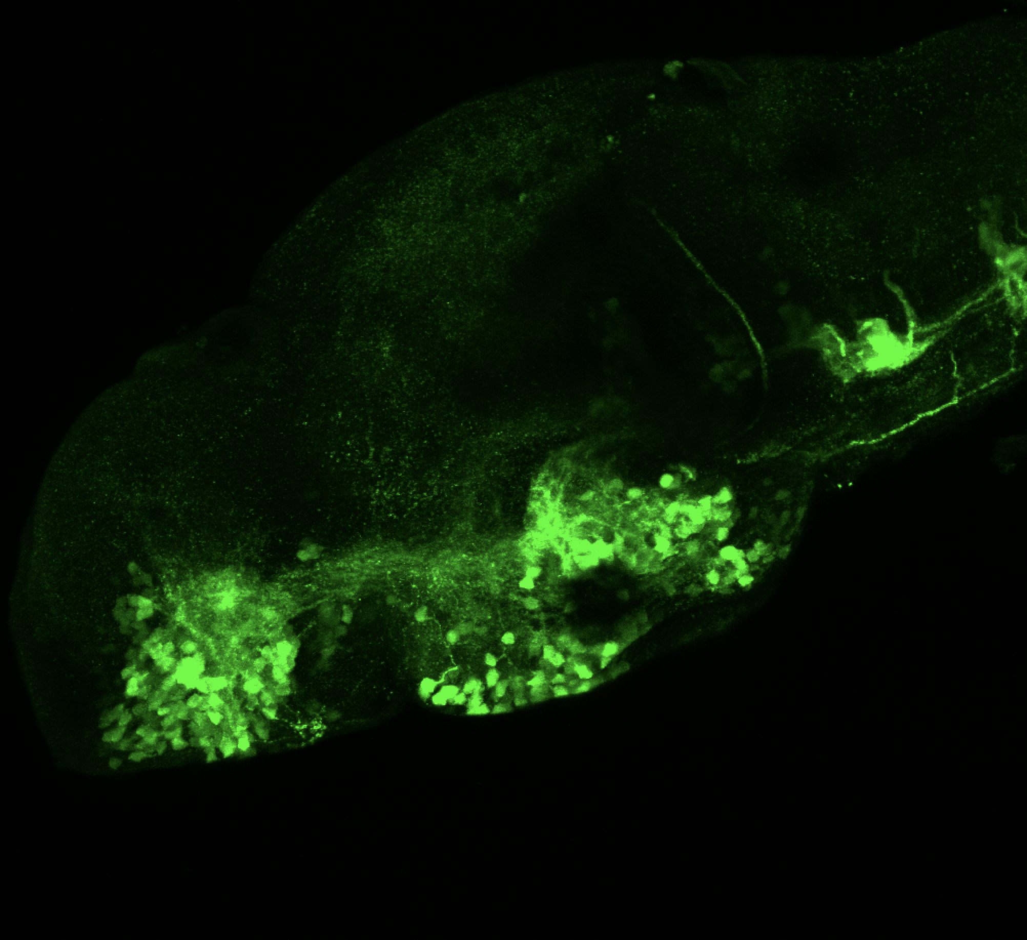
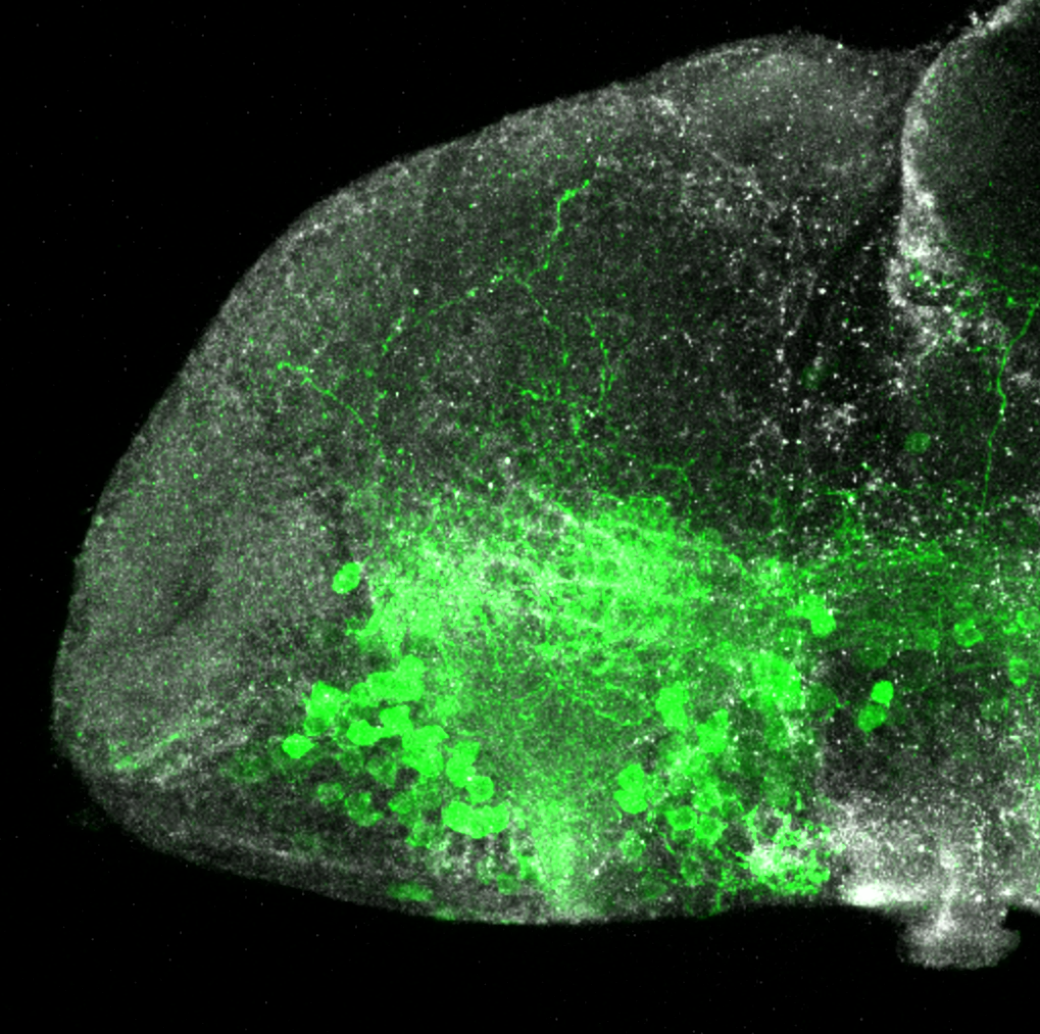
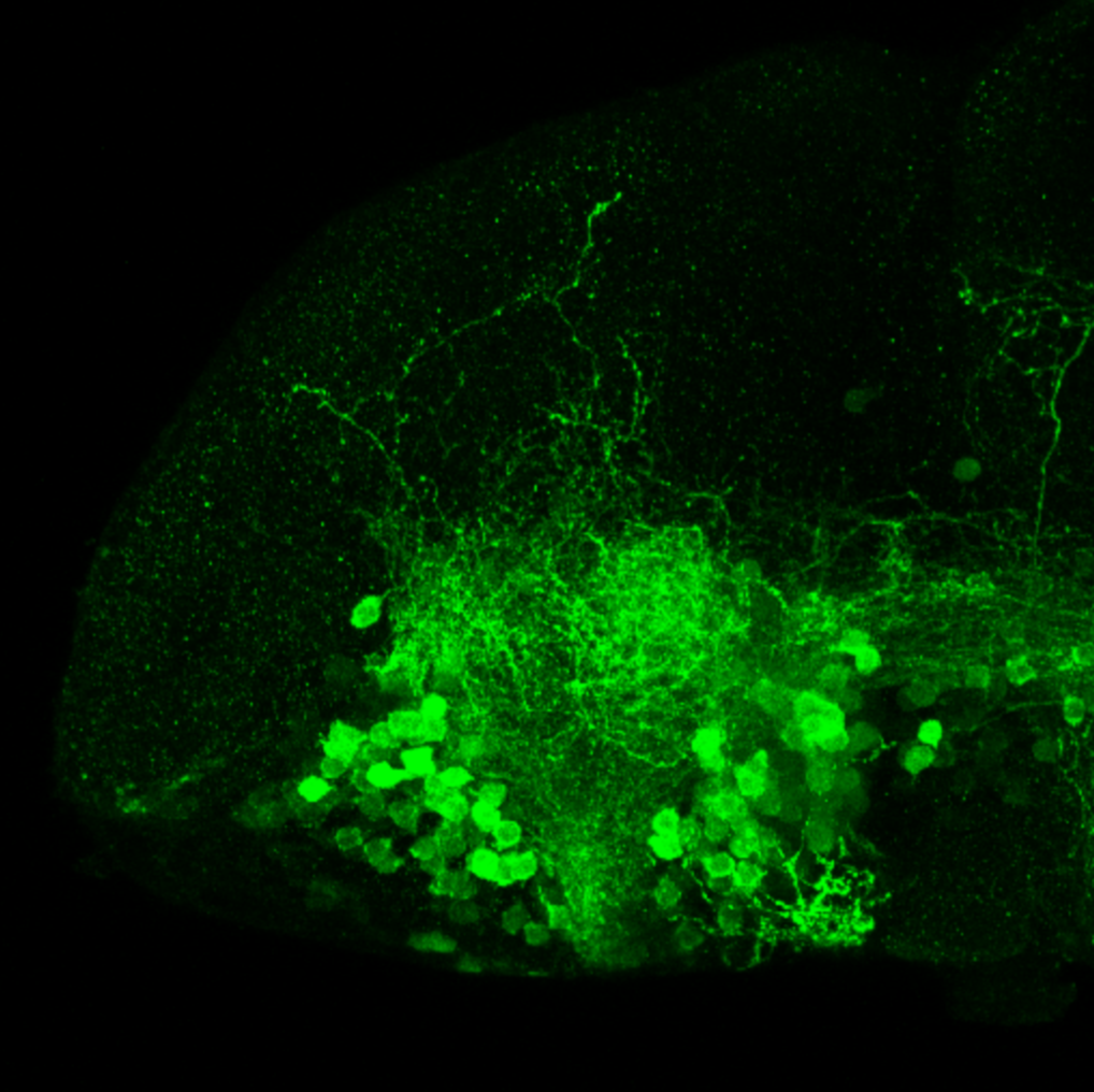
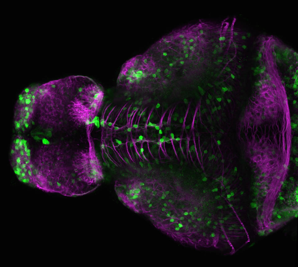
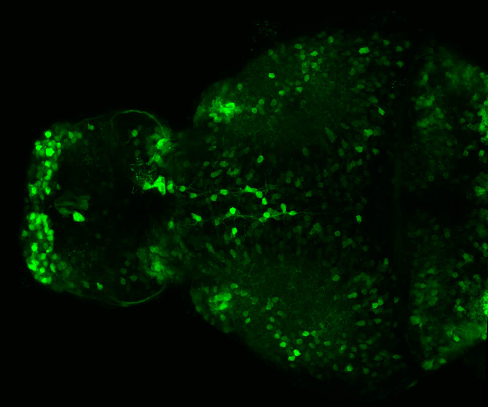
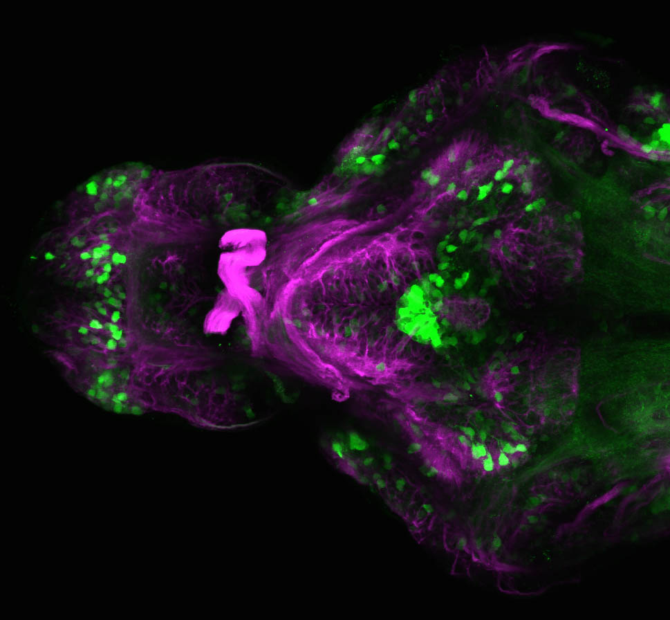
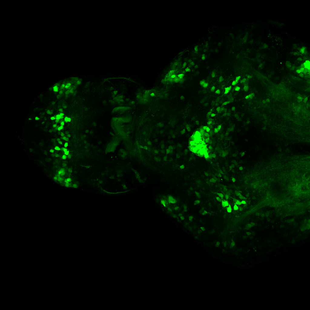
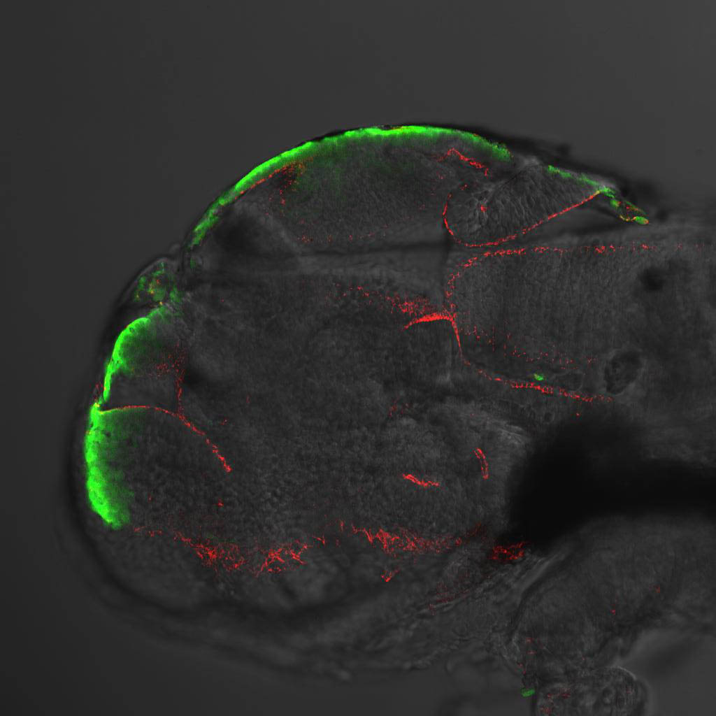
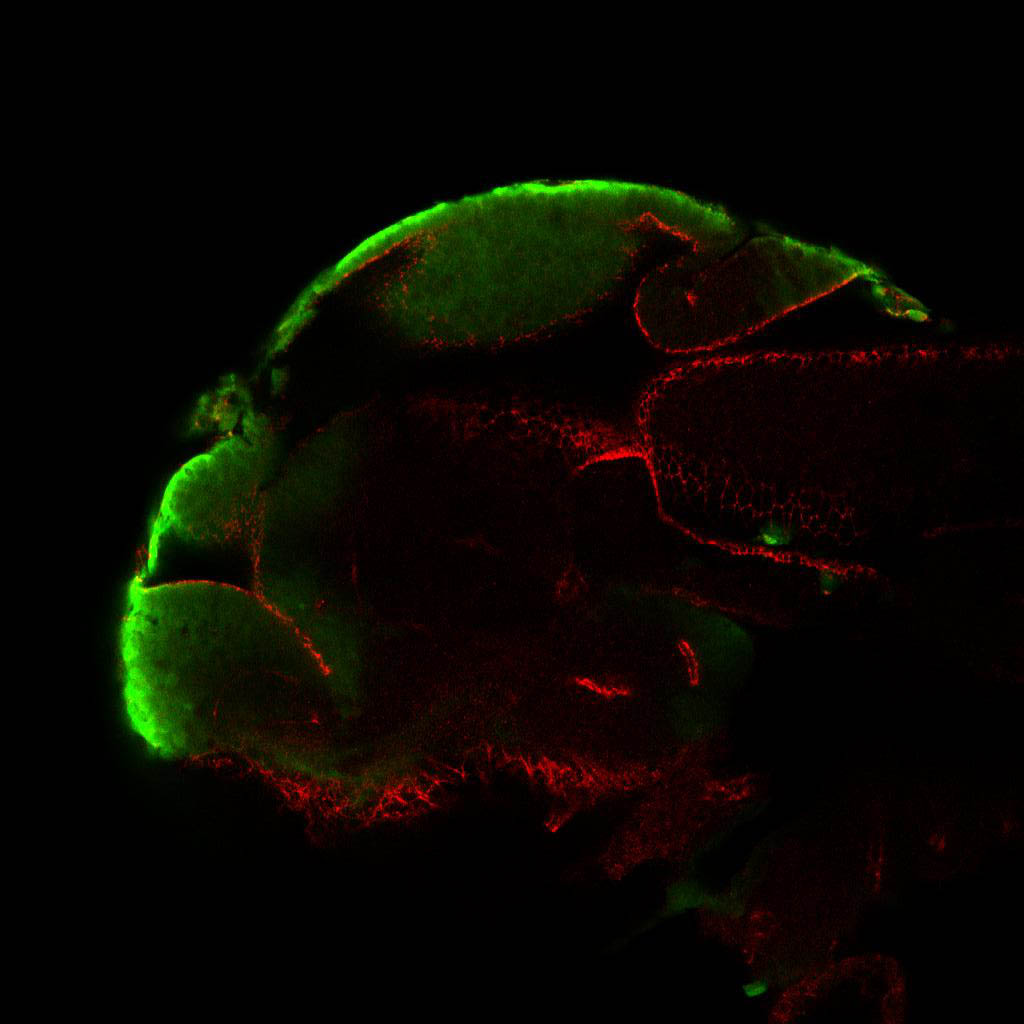
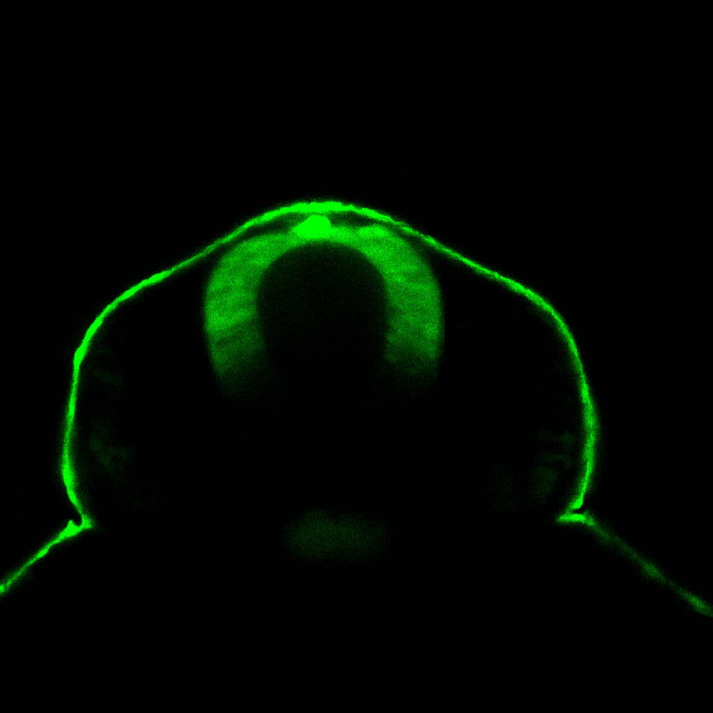
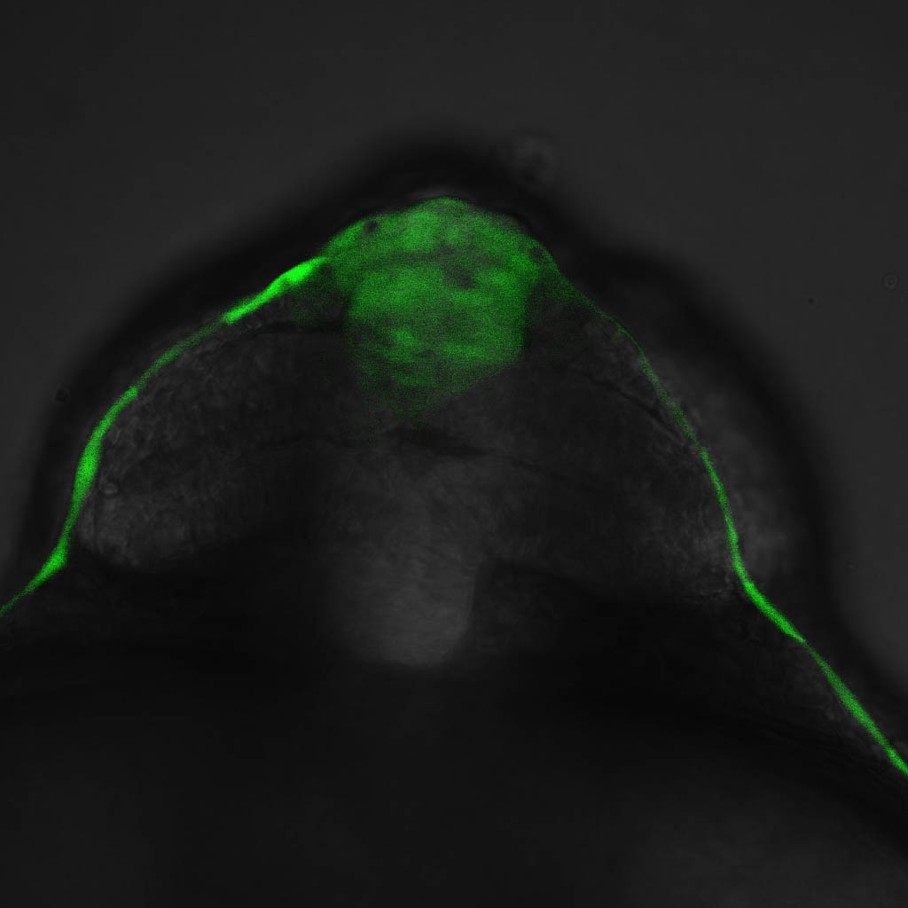
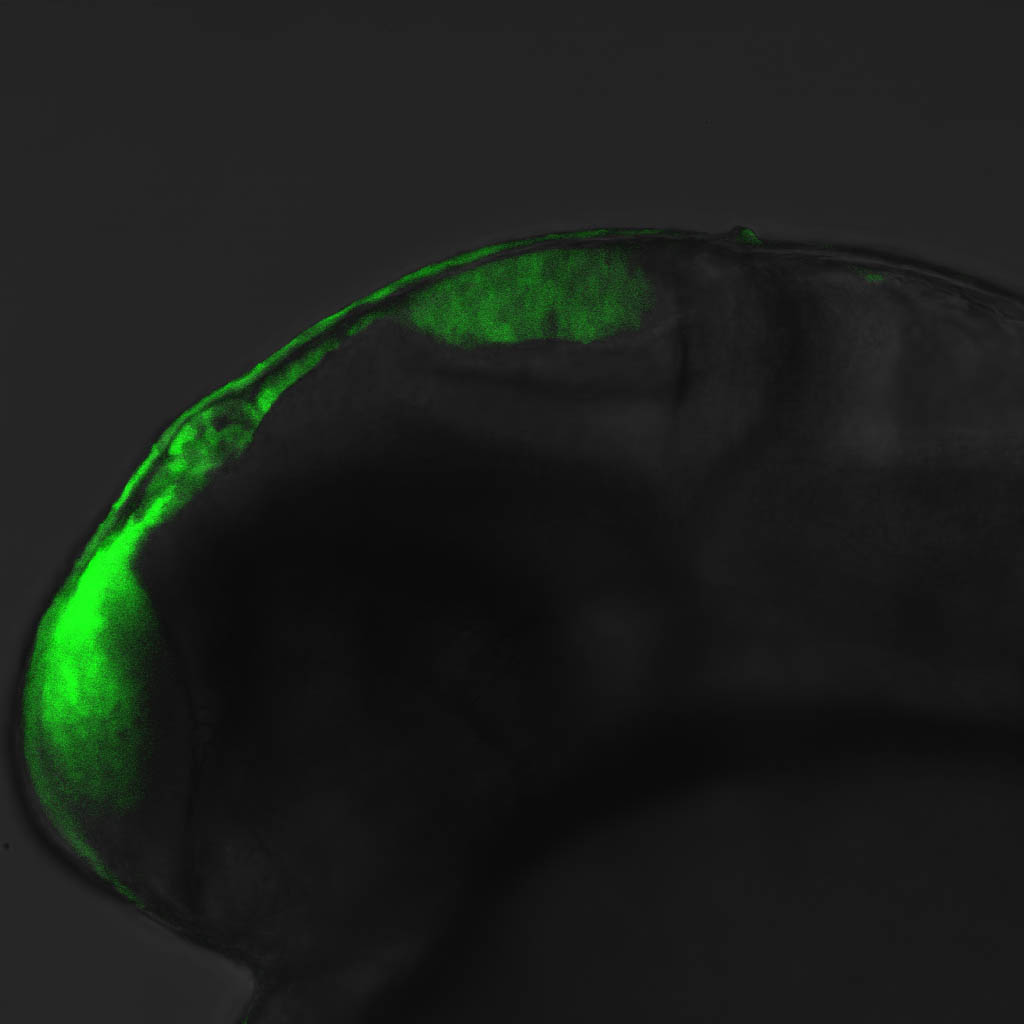
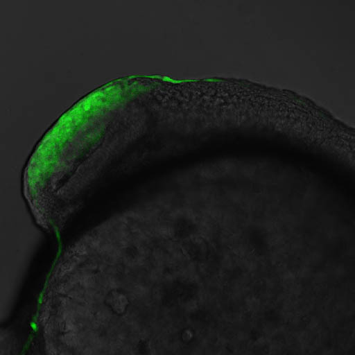
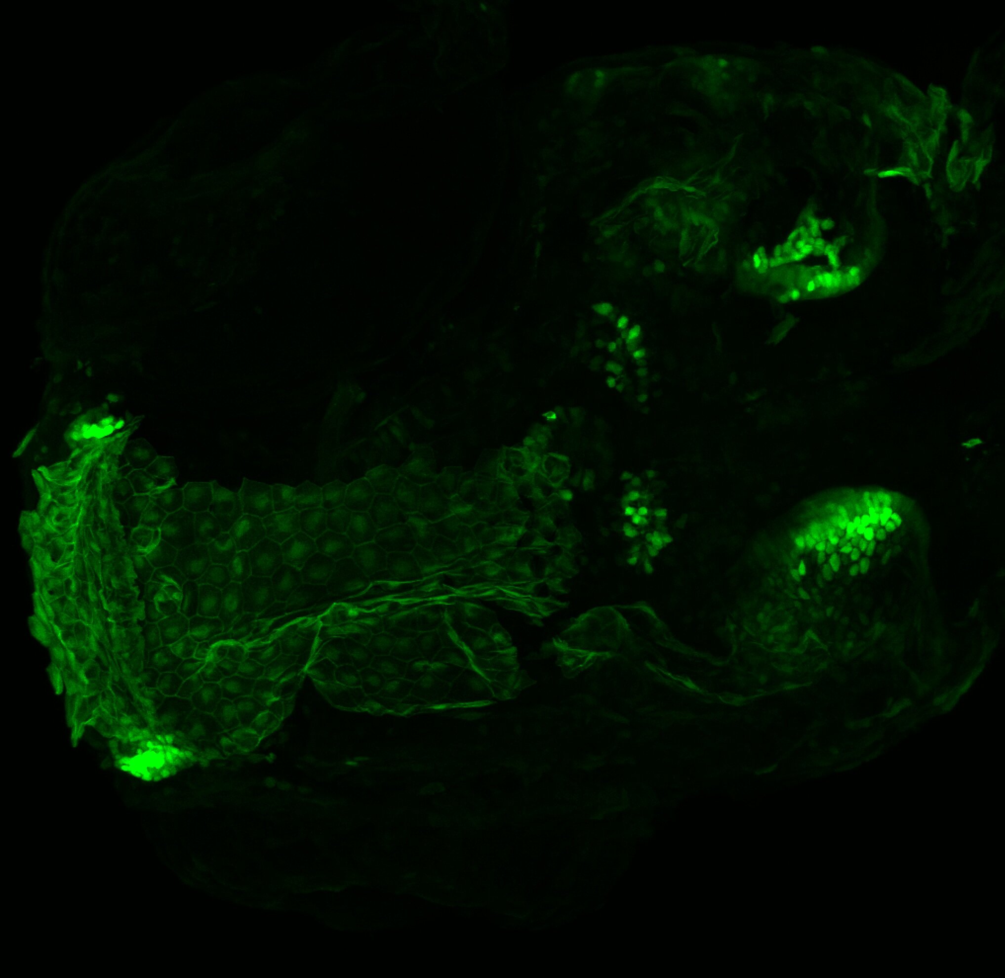
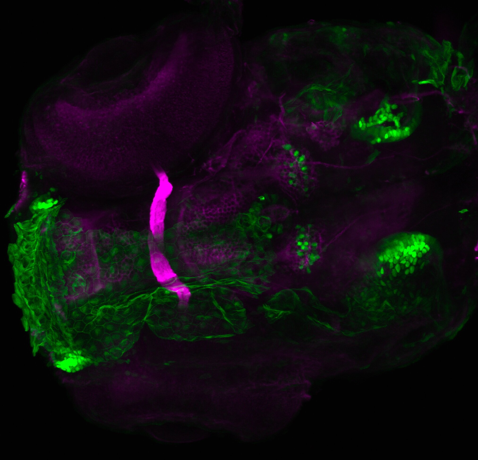
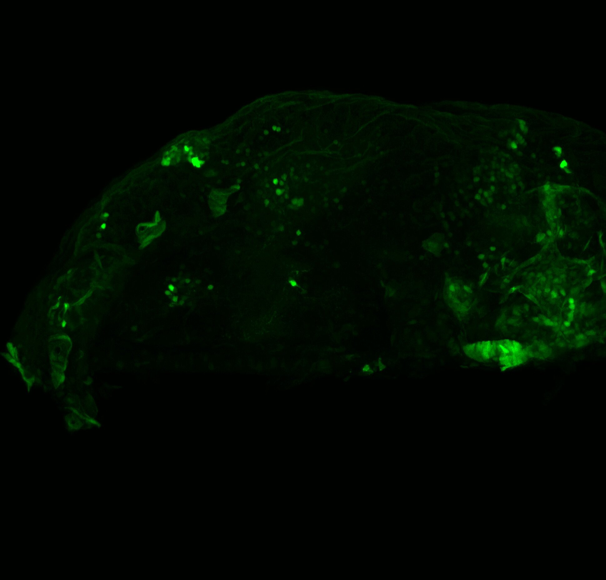
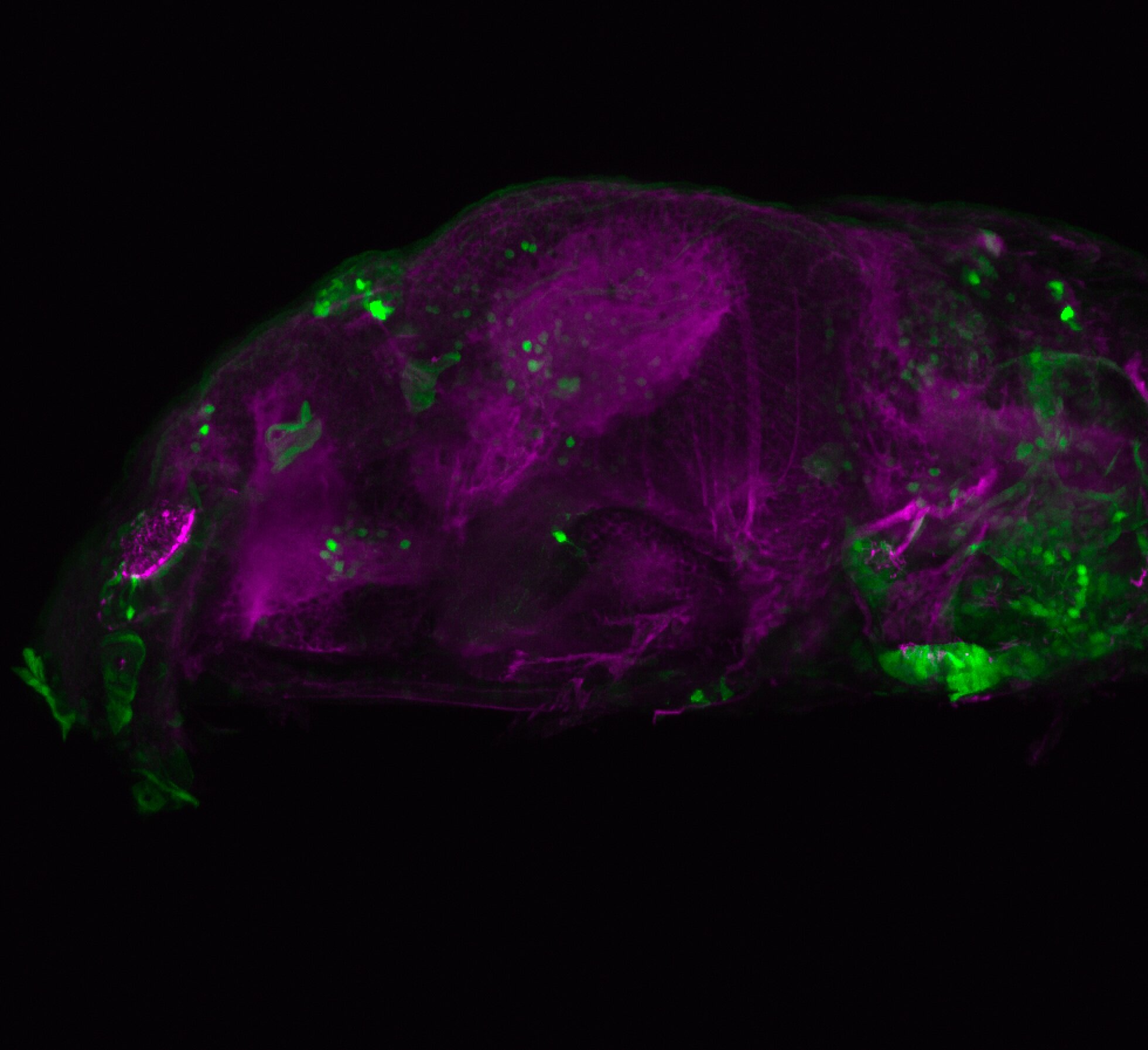
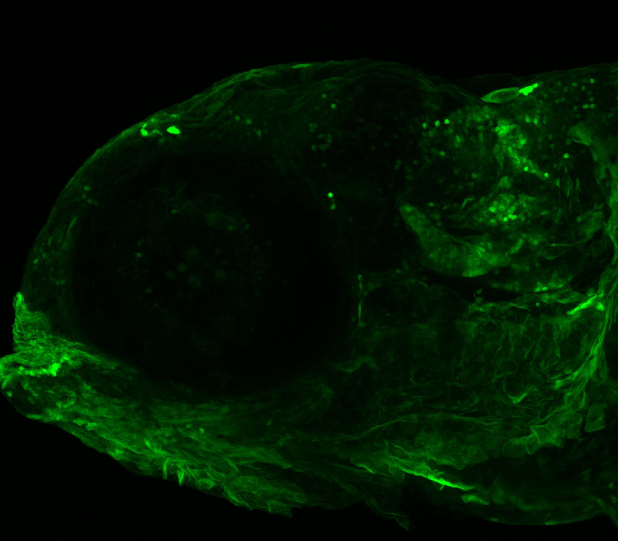
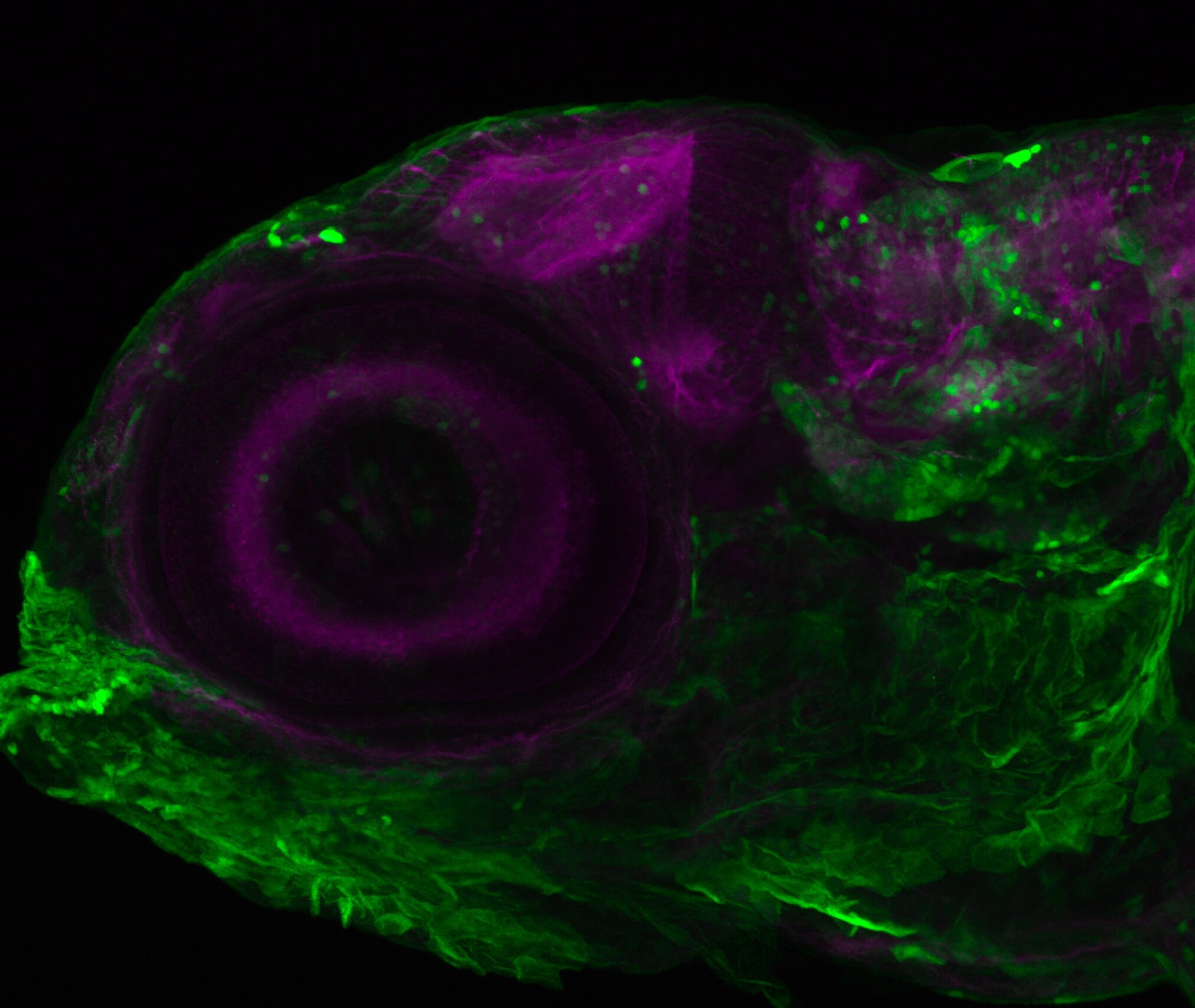
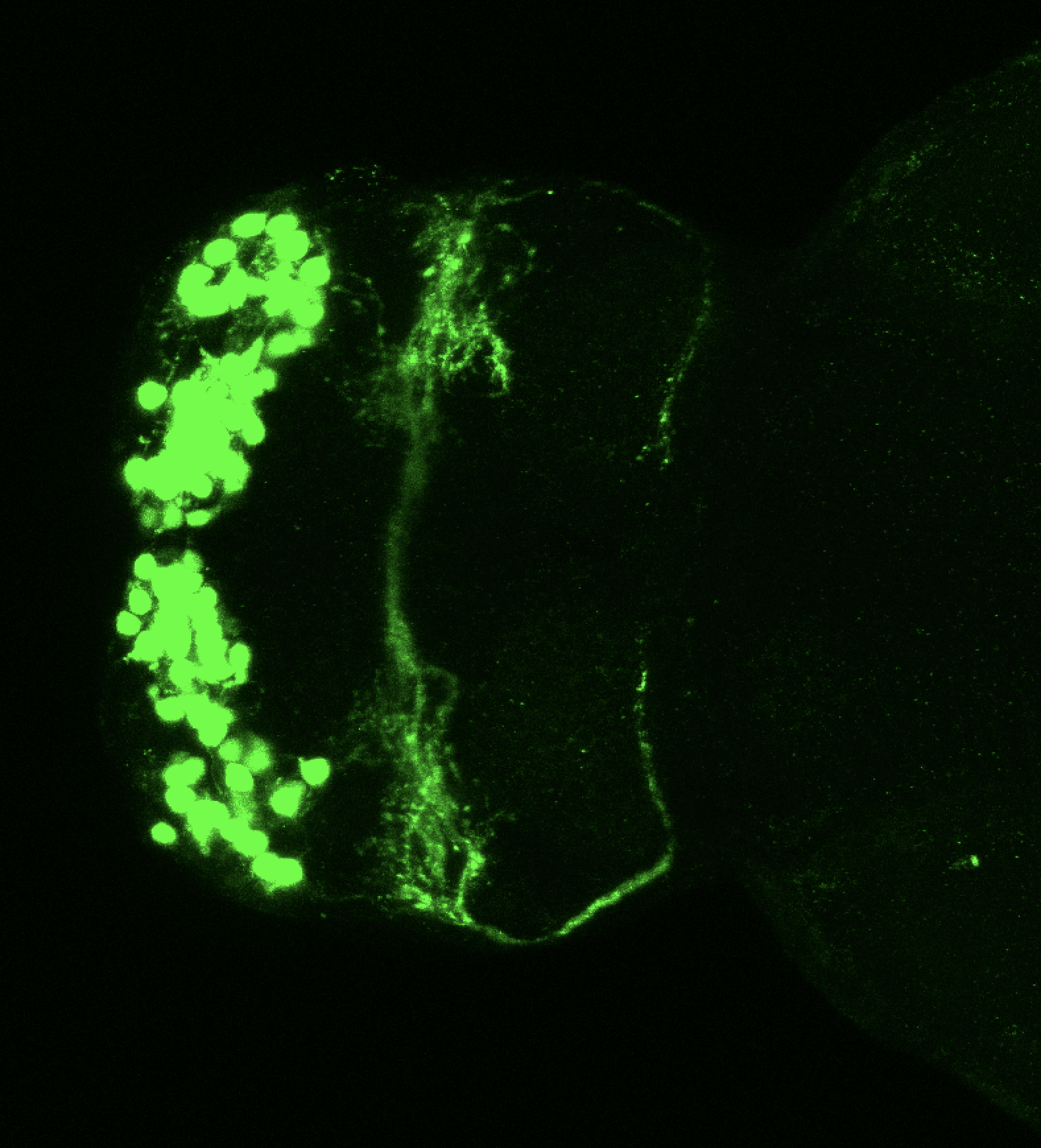
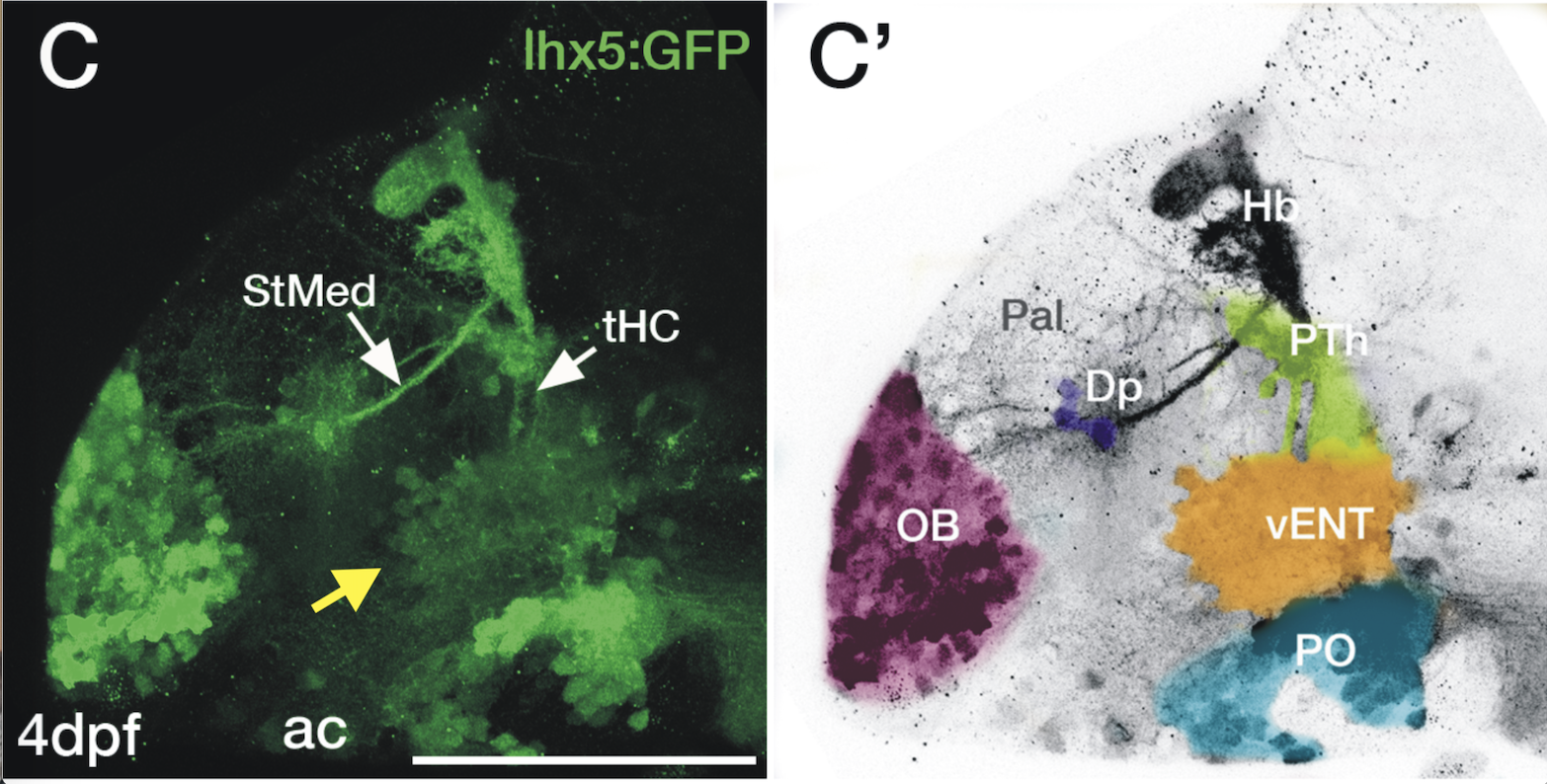
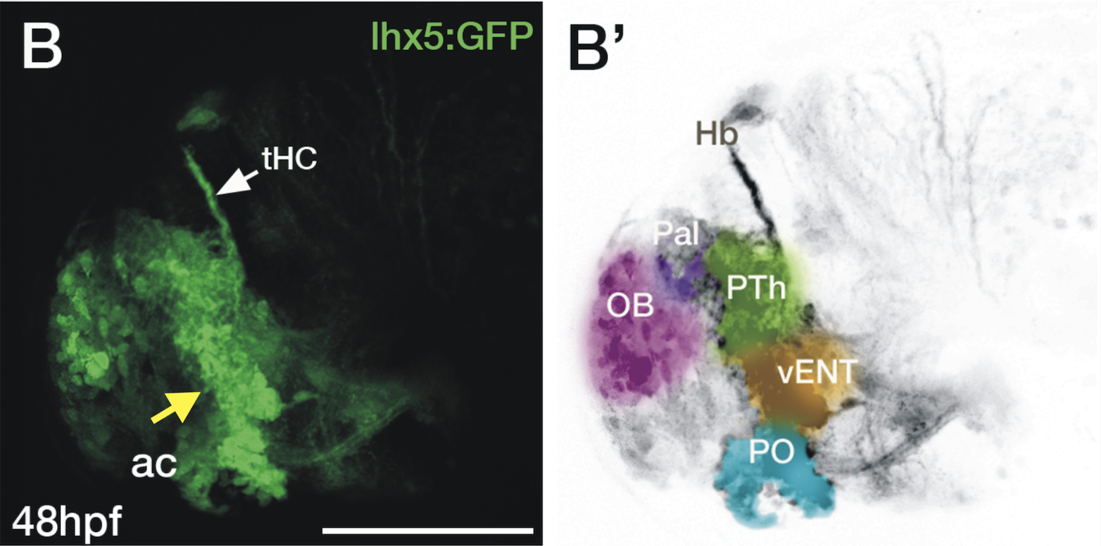
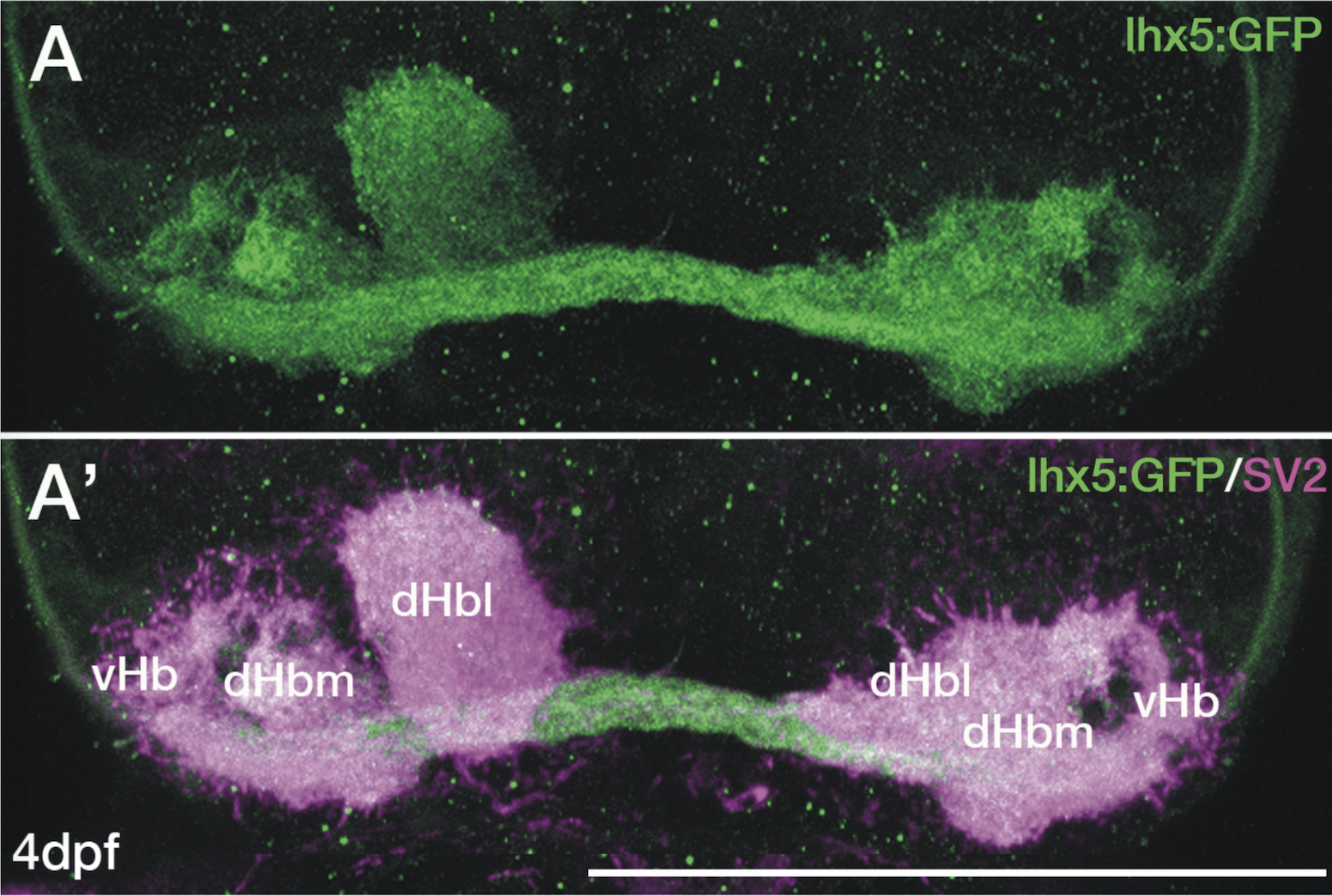
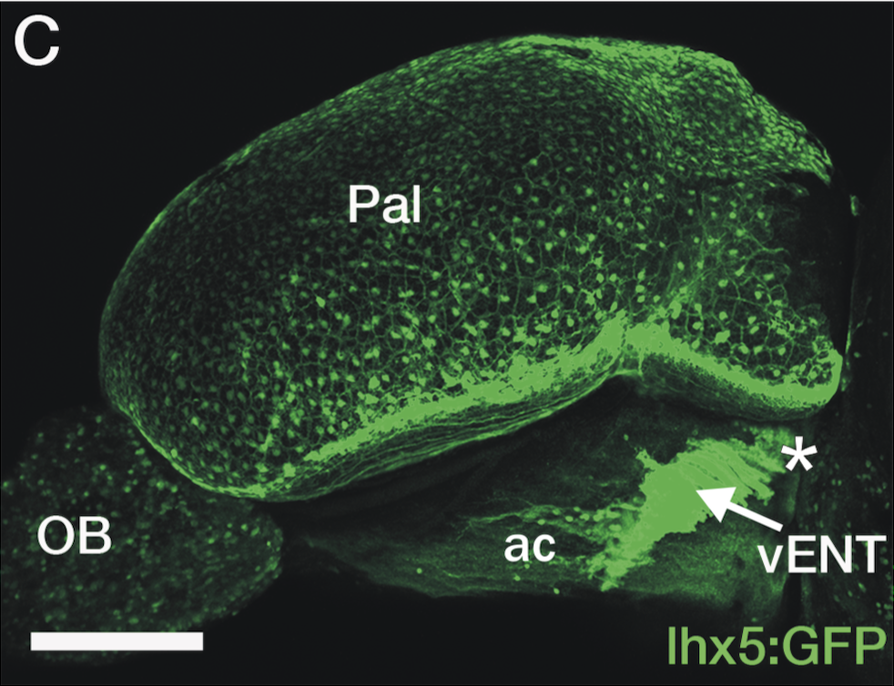
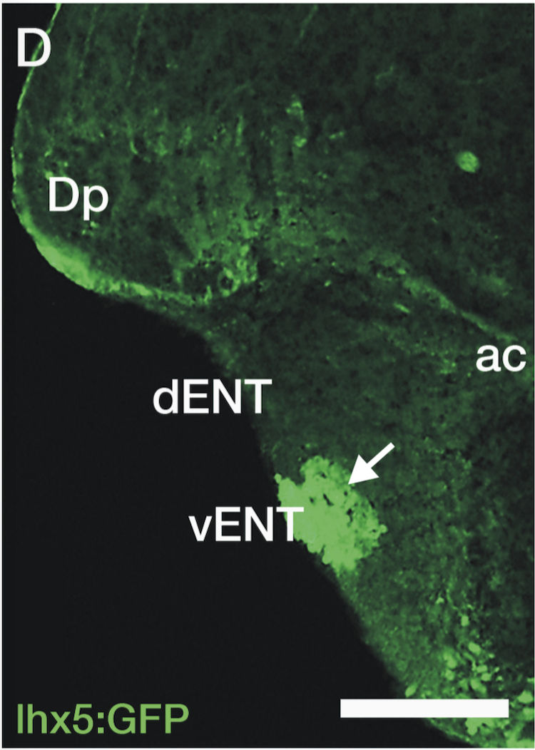
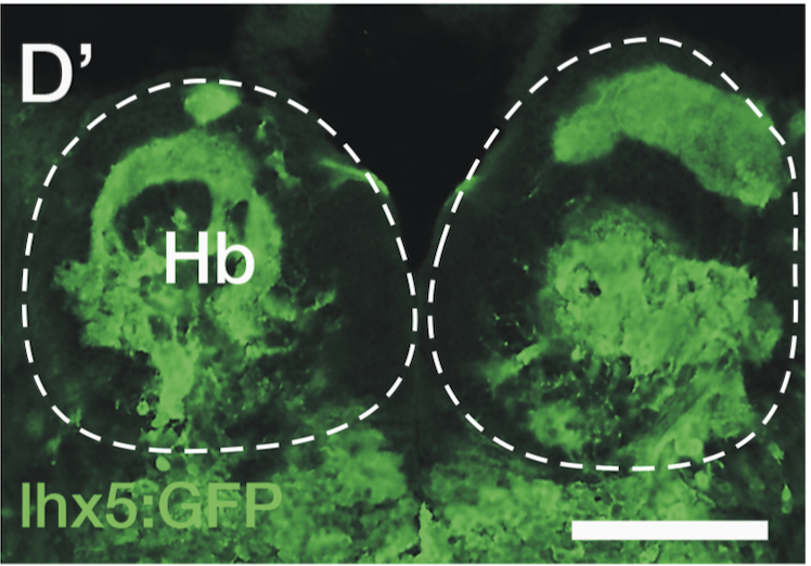
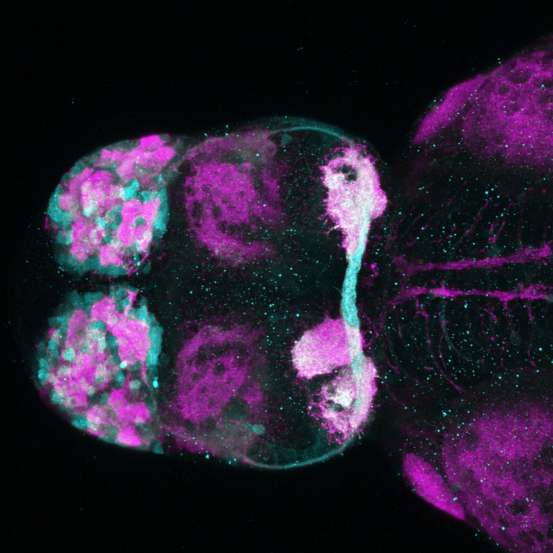
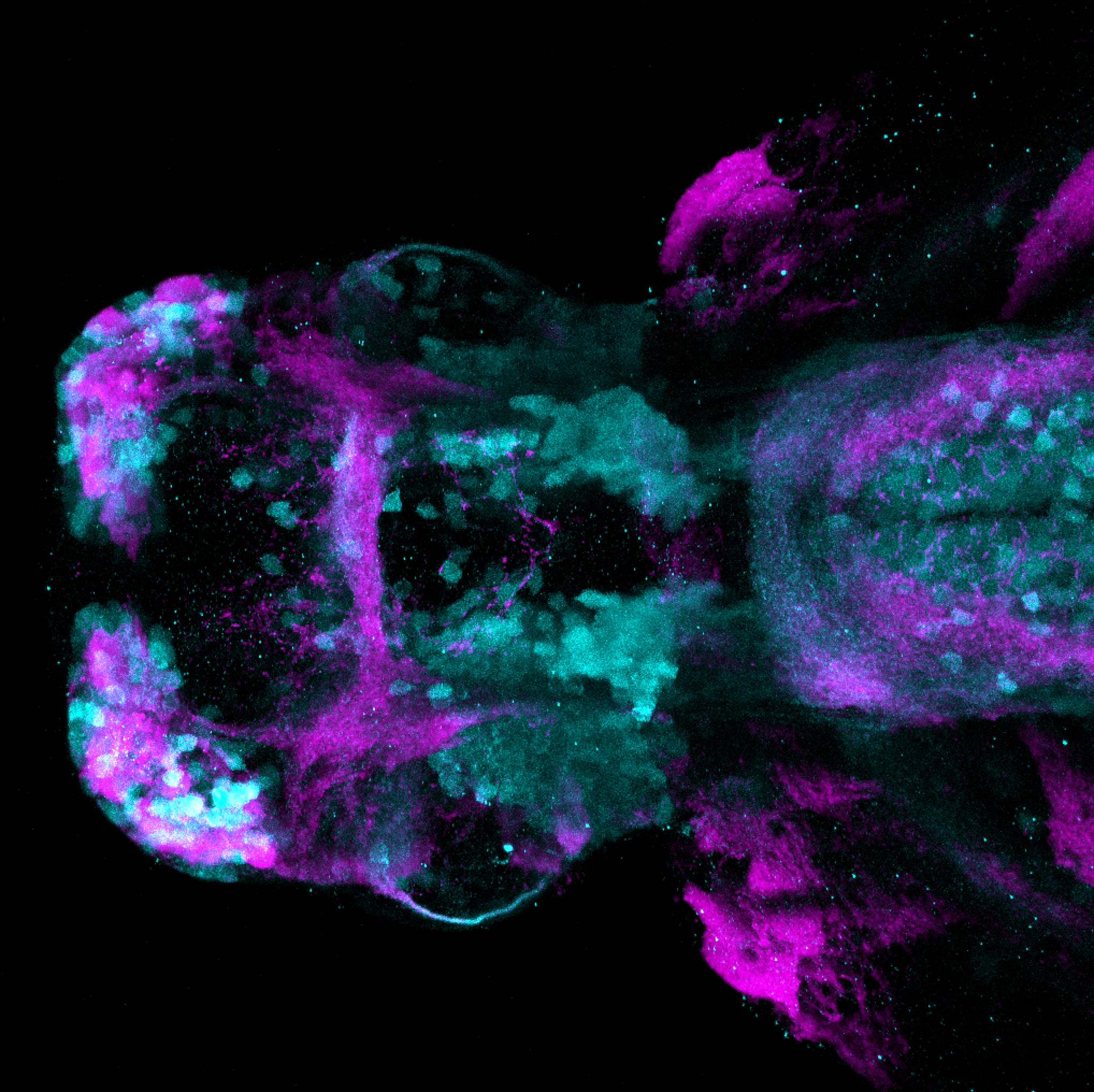
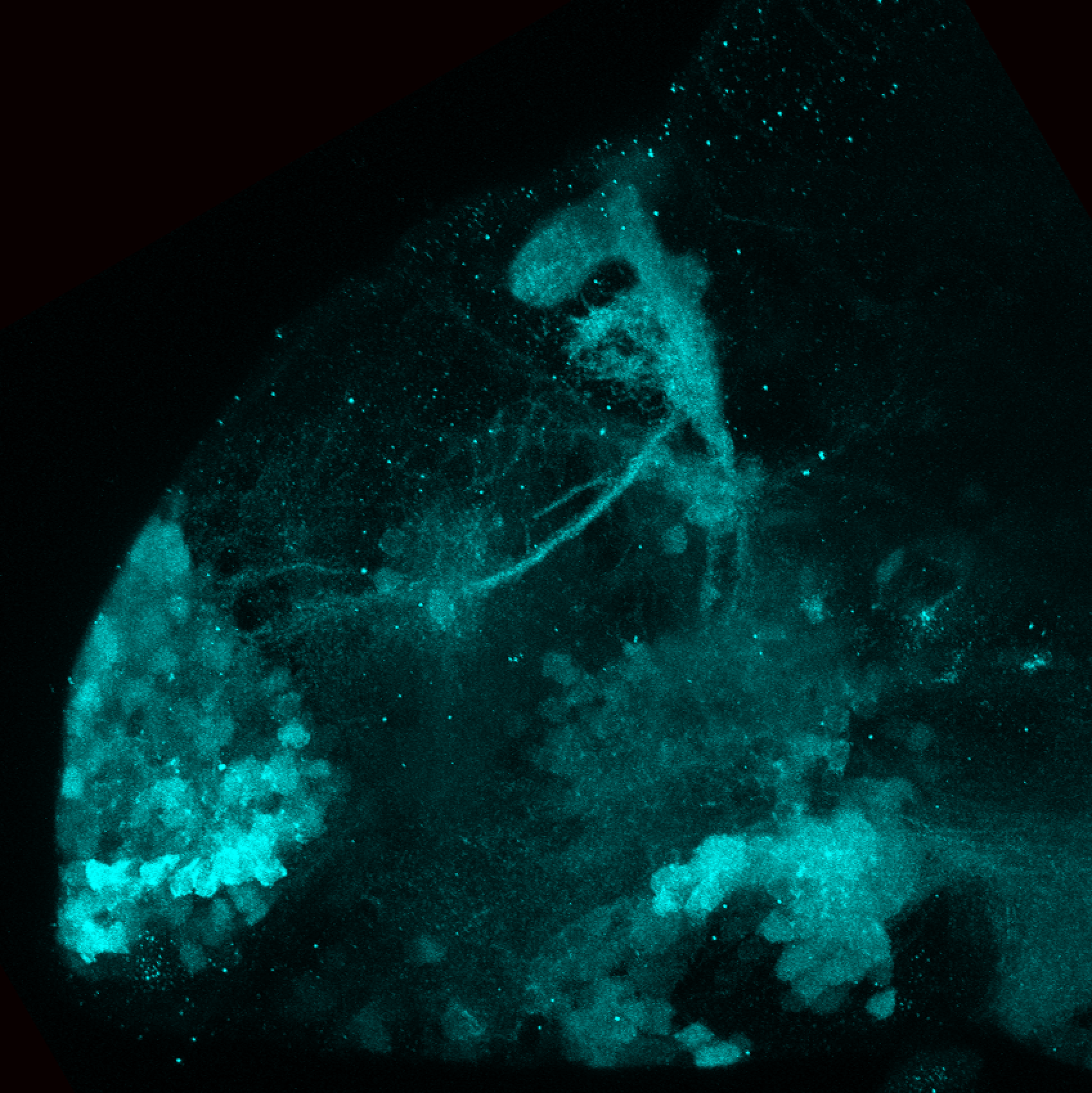
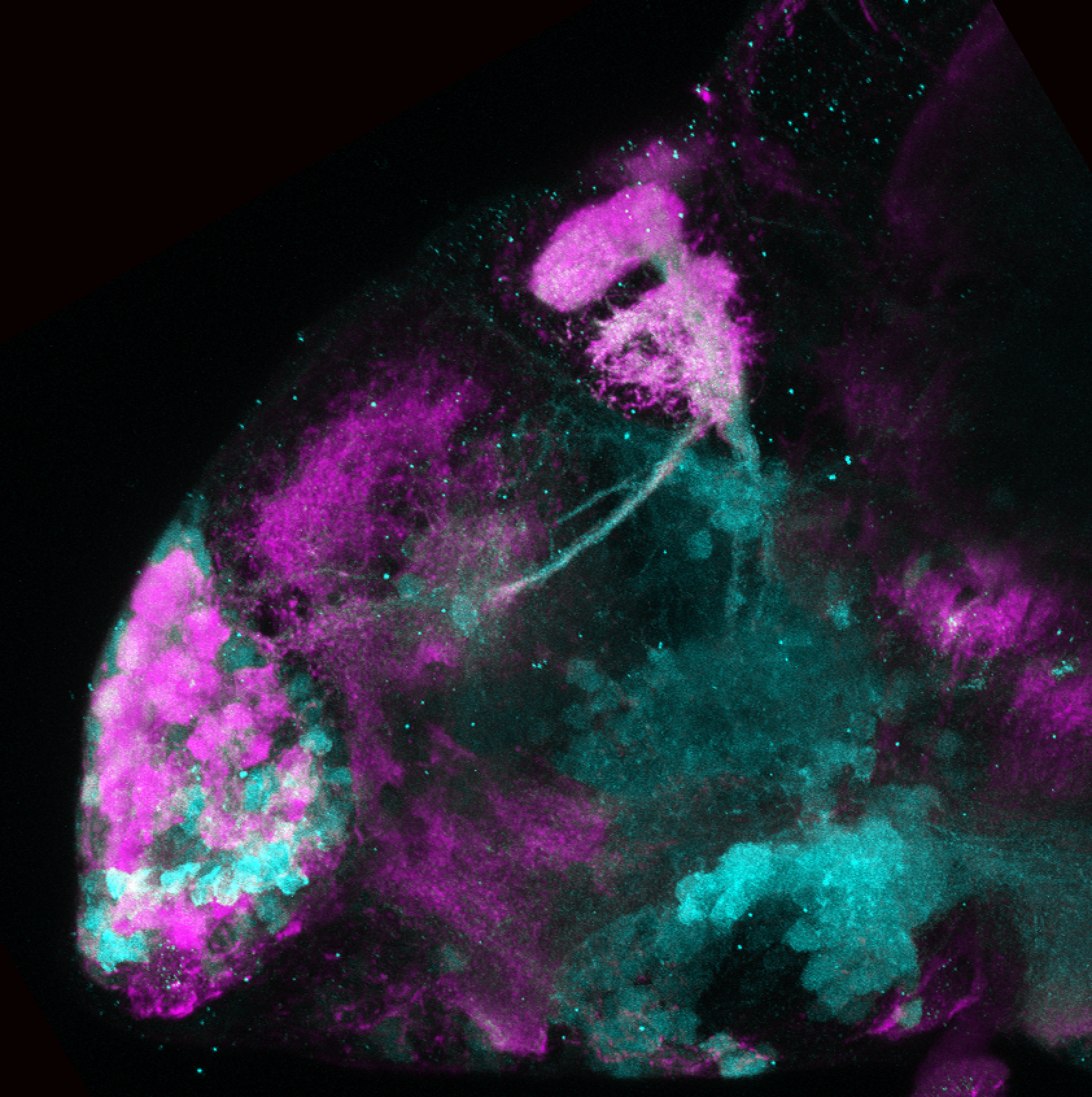
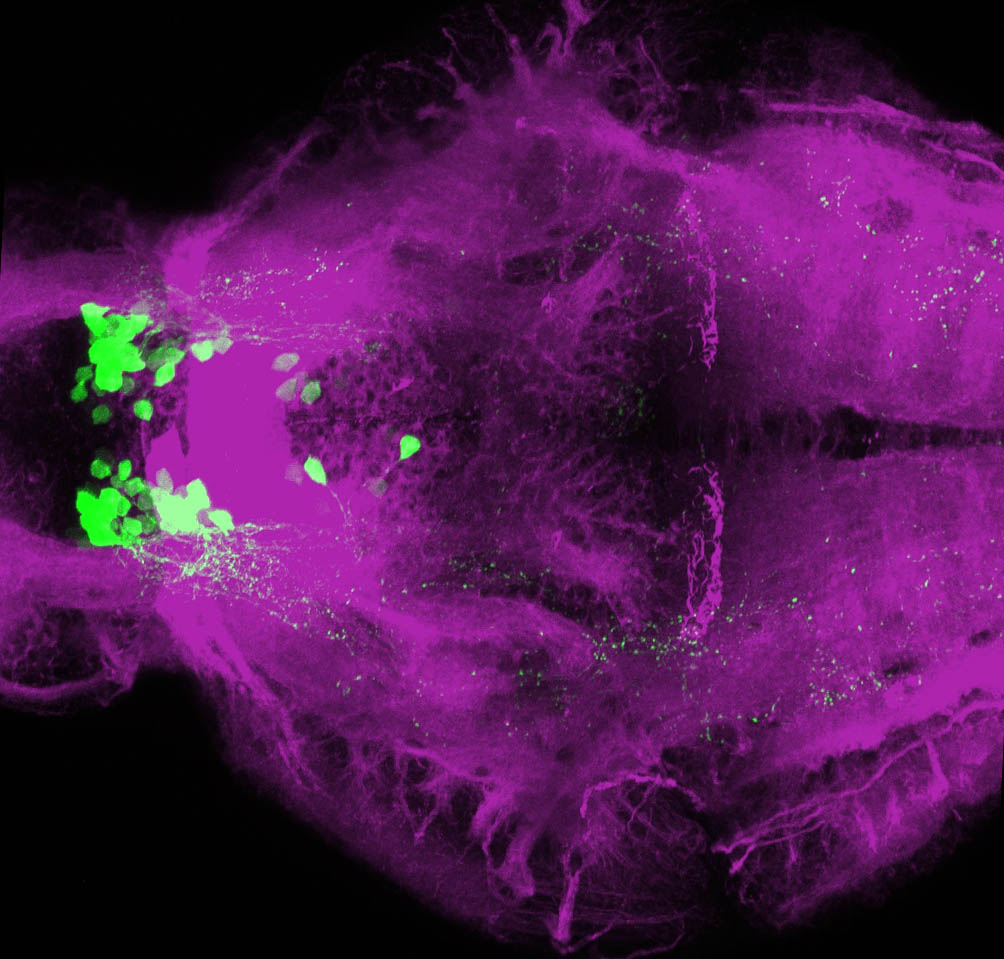
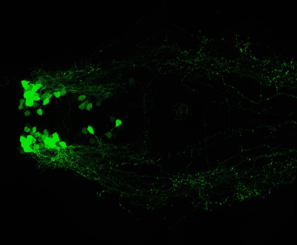
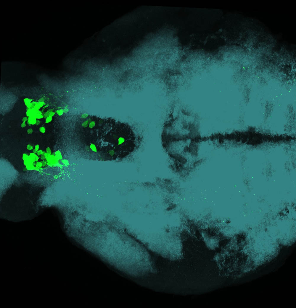
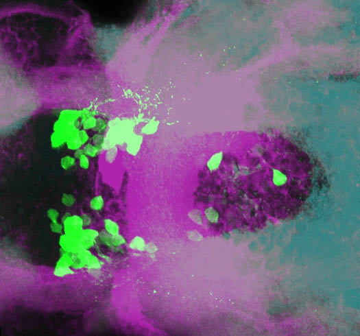
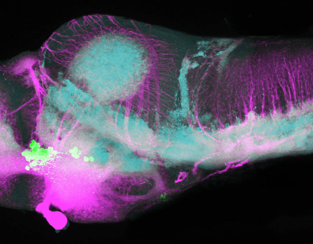
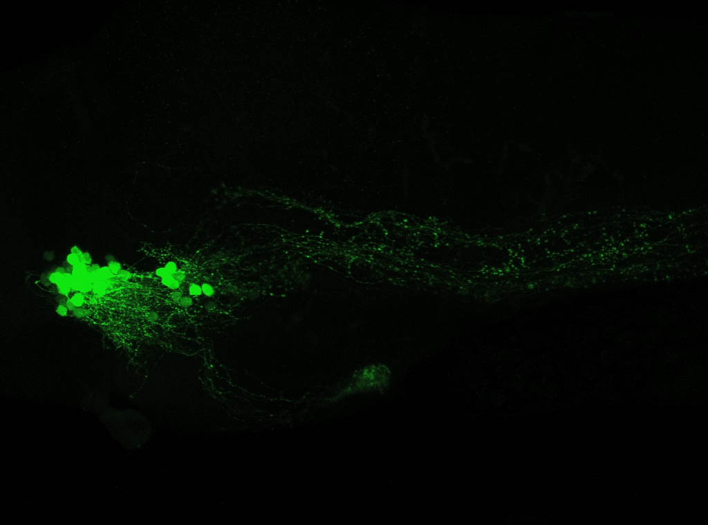
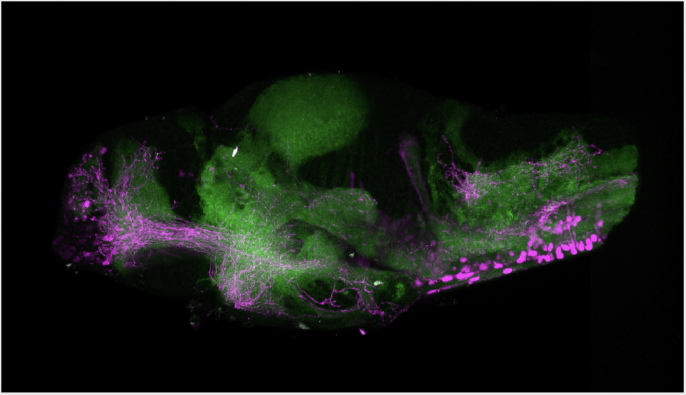
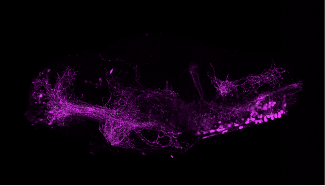
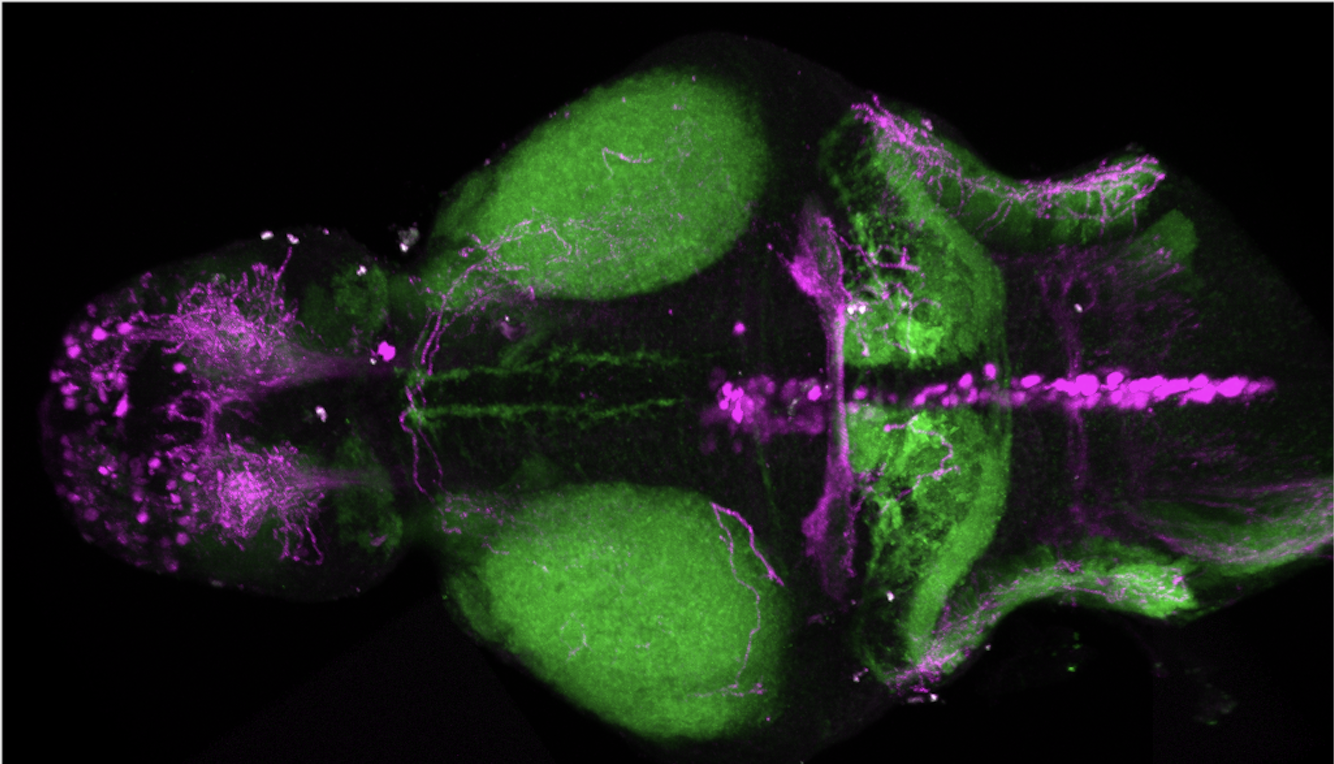
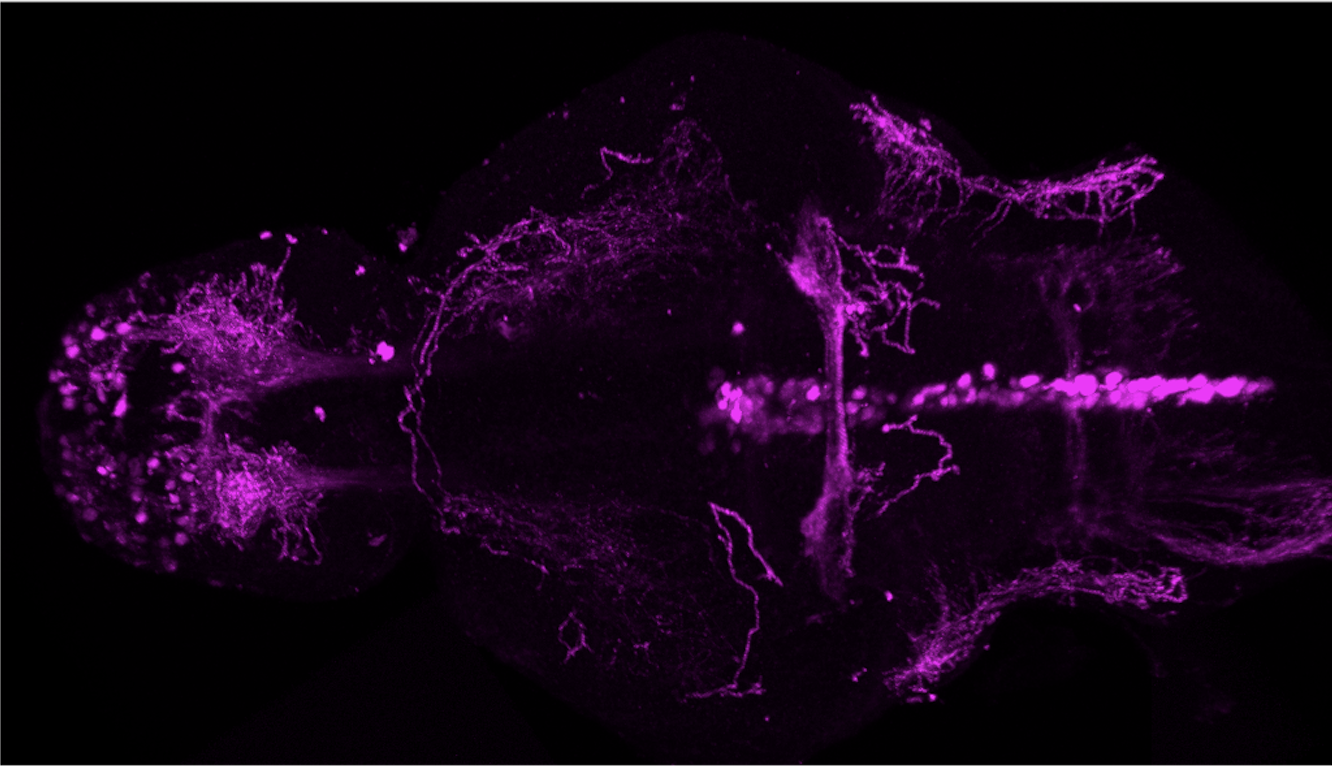
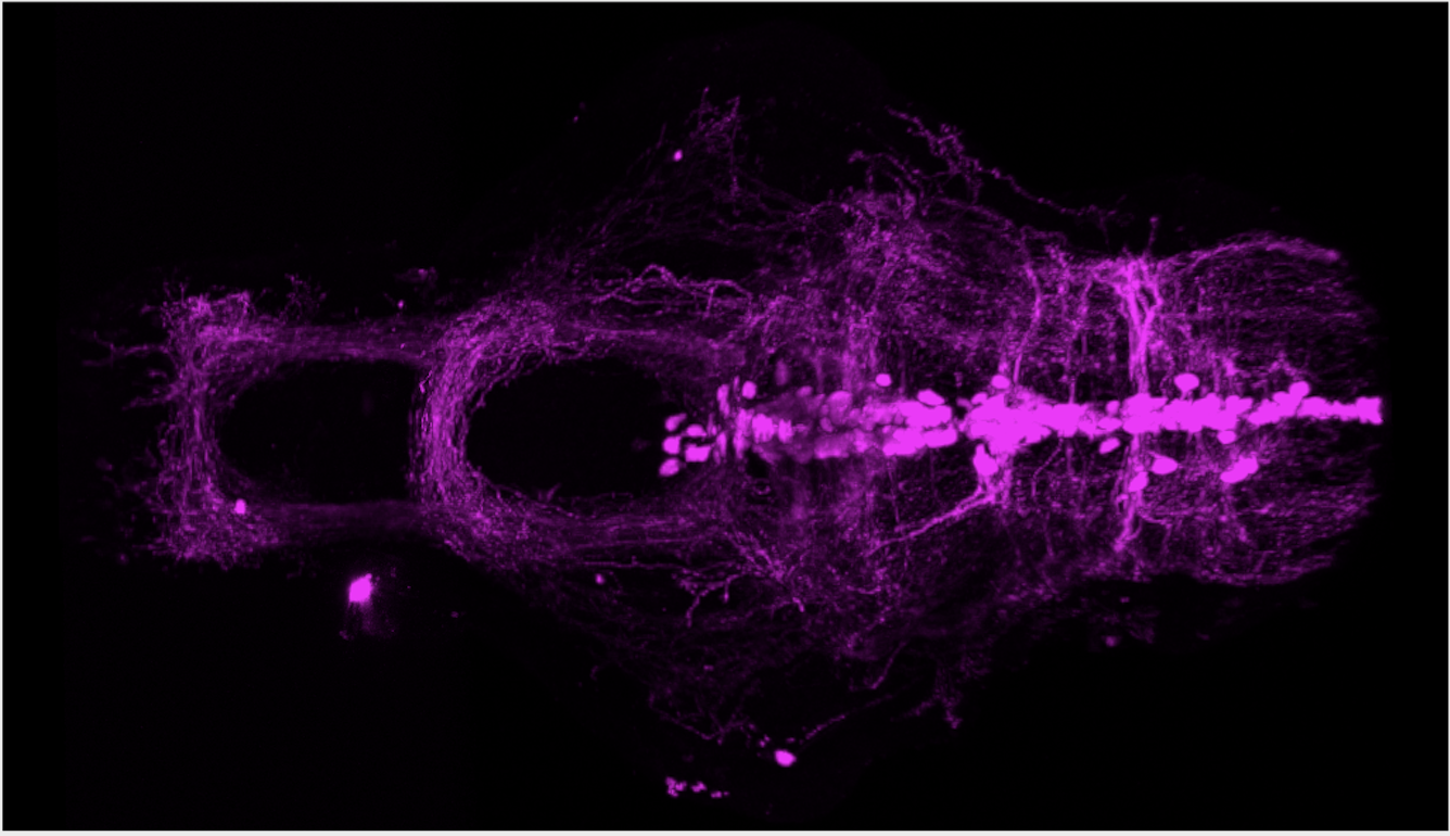
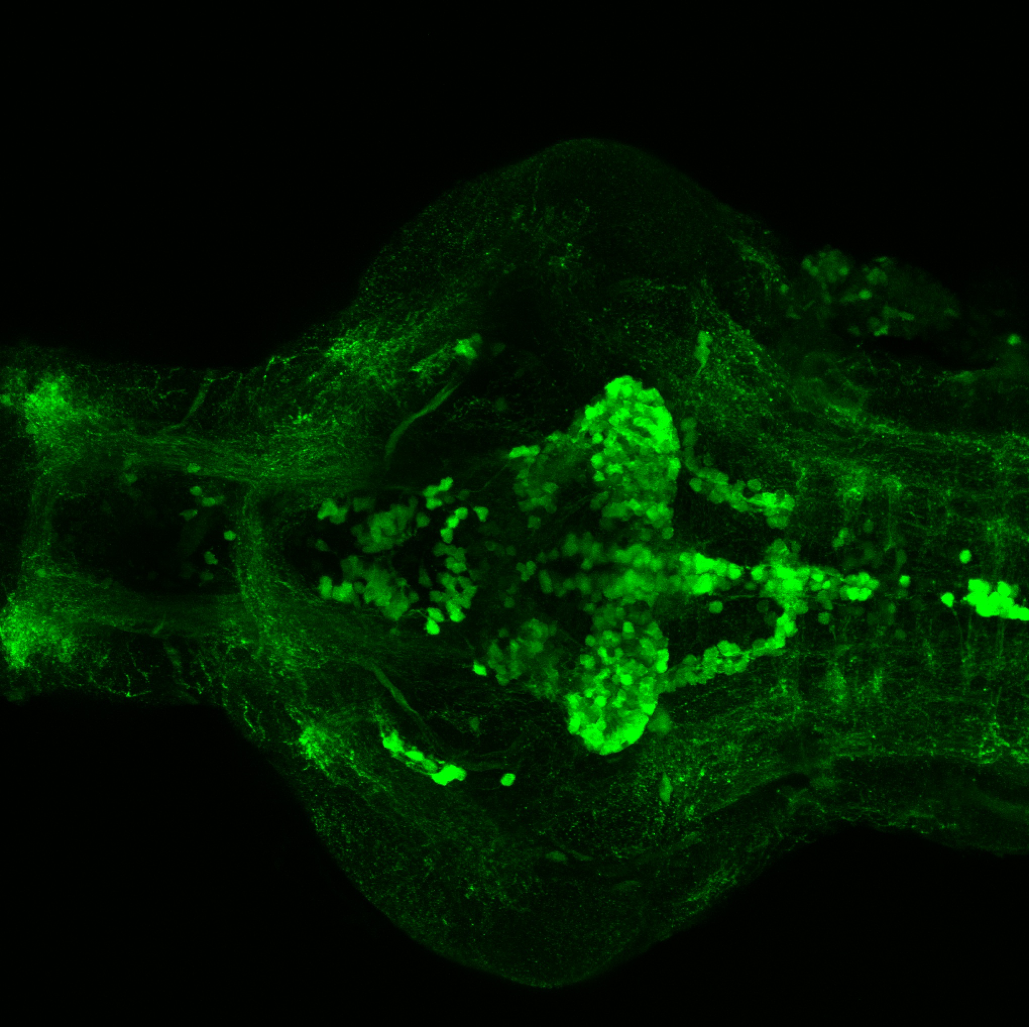
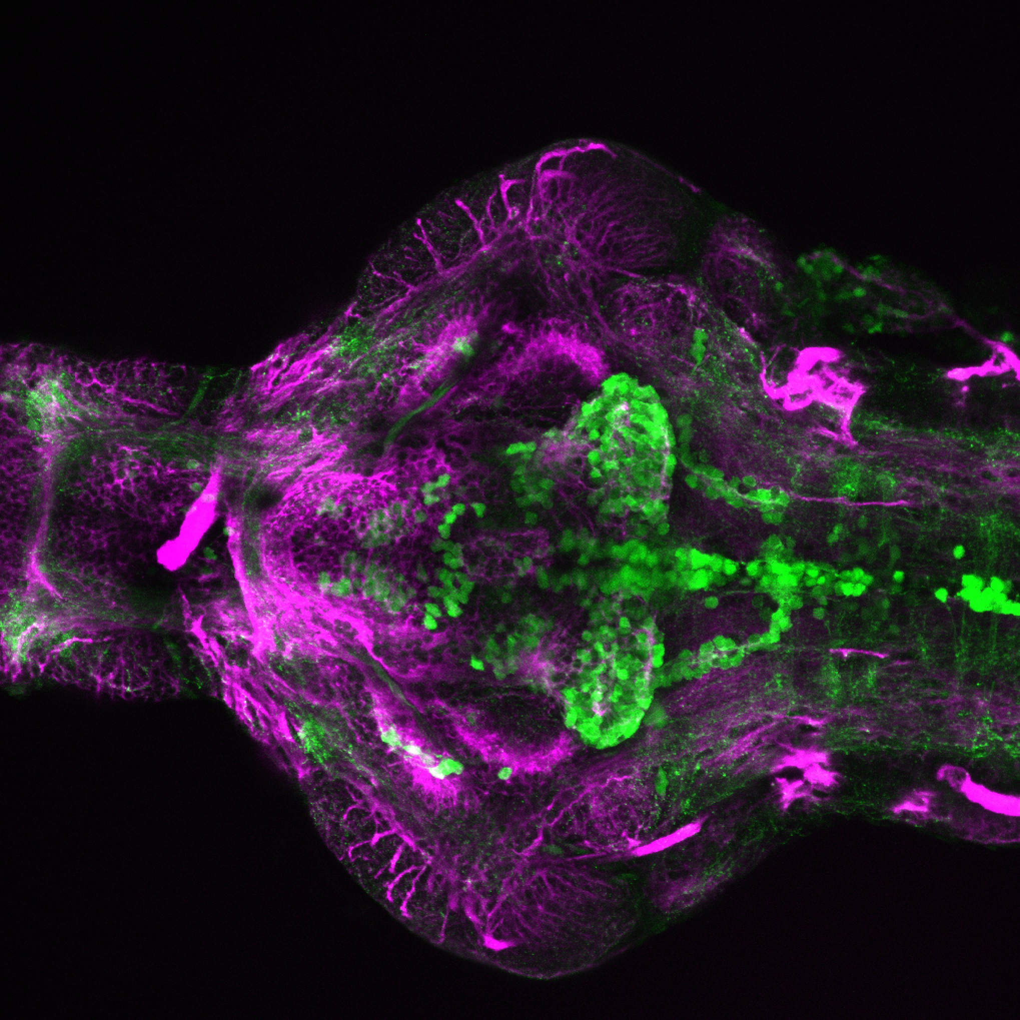
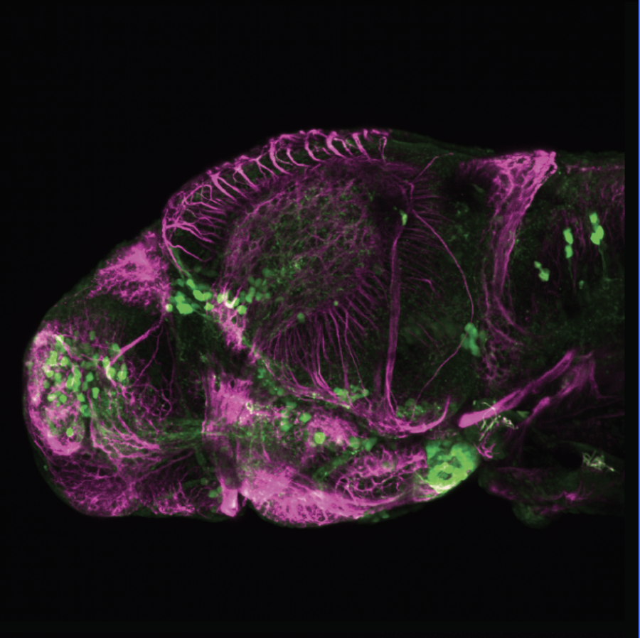
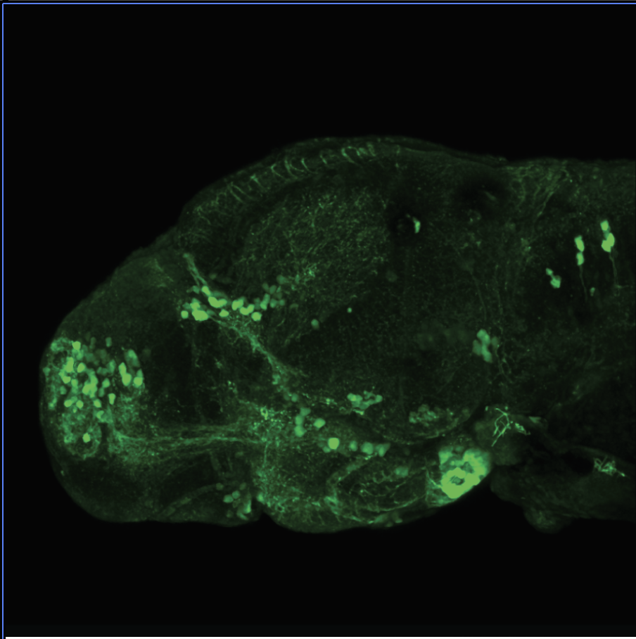
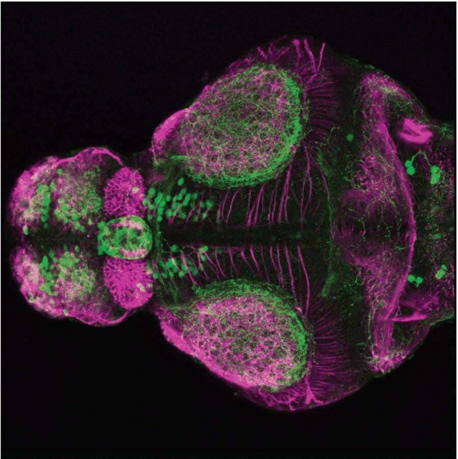
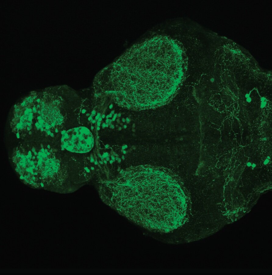
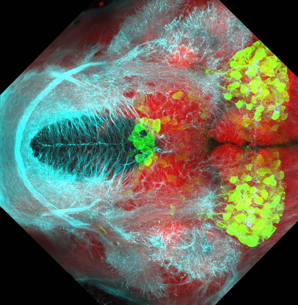
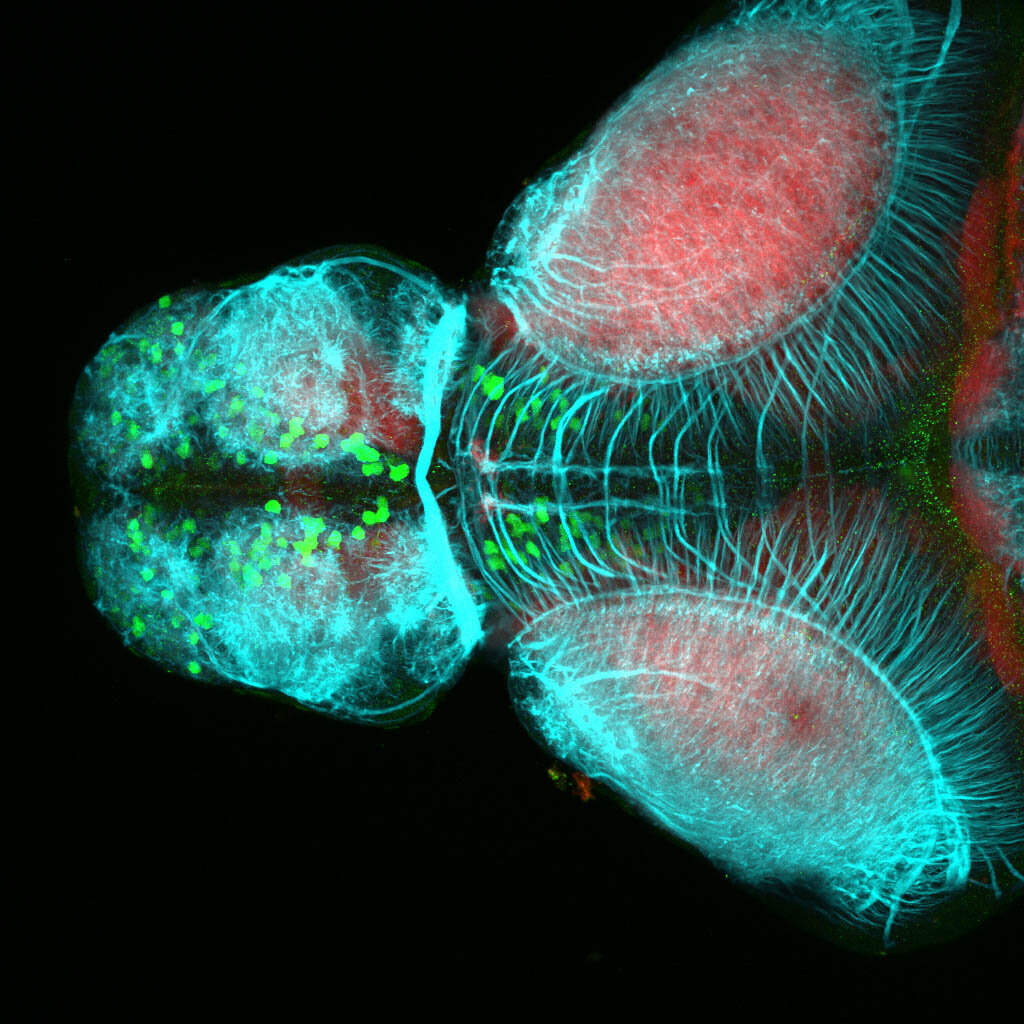
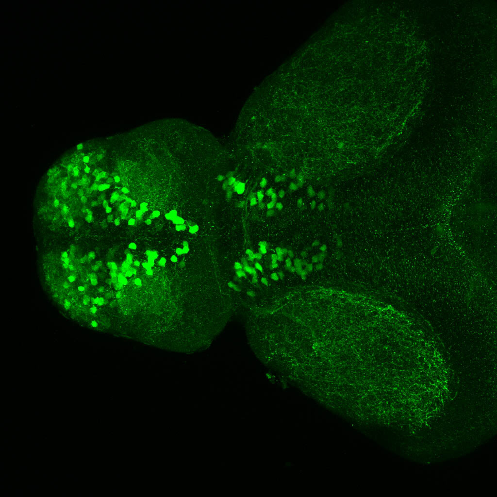
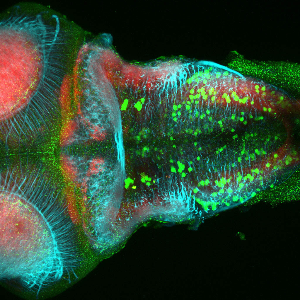

External Links:

 ZFIN

Lab or Origin: Skarmeta Lab


Expressed in: 

olfactory bulb, pineal, torus longitudinalis, optic tectum, hypothalamus, hindbrain, cerebellum


Key Publications

 Royo JL, Hidalgo C, Roncero Y, Seda MA, Akalin A, Lenhard B, et al. (2011) Dissecting the Transcriptional Regulatory Properties of Human Chromosome 16 Highly Conserved Non-Coding Regions. PLoS ONE 6(9): e24824. https://doi.org/10.1371/journal.pone.0024824

Tg(atoh7 :gapRFP)cu2Tg

Tg(atoh7 :gapRFP)cu2Tg

About

 atonal bHLH transcription factor 7 is turned on in retinal ganglion cells as they transition from proliferating neuroblast to differentiated neuron. The atoh7 promoter in this transgenic line drives the expression of a membrane tagged version of RFP so the axons of the RGCs are also labelled so the optic nerve and tract projecting from the retina to the optic tectum can be distinguished.


External Links:

 ZFIN

Lab or Origin: Bill Harris Lab


Expressed in: 

Retinal ganglion cells, cilliary marginal zone, retina, optic nerve. optic tract, optic tectum.  


Key Publications

 Zolessi, F.R., Poggi, L., Wilkinson, C.J., Chien, C.B., and Harris, W.A. (2006)
Polarization and orientation of retinal ganglion cells in vivo.
Neural Development. 1:2.

Tg(-8.0cldnb:lynGFP)zf106

Tg(-8.0cldnb:lynGFP)zf106

About

construct:
Tg(-8.0cldnb:LY-EGFP)

This transgenic line was created by the Gilmore lab “ Eight kilobases of sequence directly upstream of the Claudin B start codon were amplified from BAC zK241F11 by using the Expand Long Template PCR System (Roche). The resultant fragment was cloned into a vector containing lynEGFPpA (Koster and Fraser, 2001) flanked by sites for I-SceI, and the resultant construct was injected into one-cell zebrafish embryos by following the meganuclease transgenesis protocol (Thermes et al., 2002).”(Haas & Gilmore, 2006).

The Gilmore lab wanted to label the lateral line and neuromasts of the lateral line system, one allele of this transgenic also had EGFP expression in the nasal retina and telencephalon and has been used by other labs to study eye and telencephalic morphogenesis.


External Links:

 ZFIN

Lab or Origin: Gilmore Lab


Expressed in: 

neuromasts, lateral line, olfactory epithelium, olfactory bulb, , pallium, subpallium, tract of the habenula commissure, nasal retina.


Key Publications

 Haas, P., and Gilmour, D. (2006)
Chemokine signaling mediates self-organizing tissue migration in the zebrafish lateral line.
Developmental Cell. 10(5):673-680.

Folgueira, M., Bayley, P., Navratilova, P., Becker, T.S., Wilson, S.W., and Clarke, J.D. (2012)
Morphogenesis underlying the development of the everted teleost telencephalon.
Neural Development. 7(1):32.

Valdivia, L.E., Young, R.M., Hawkins, T.A., Stickney, H.L., Cavodeassi, F., Schwarz, Q., Pullin, L.M., Villegas, R., Moro, E., Argenton, F., Allende, M.L., and Wilson, S.W. (2011)
Lef1-dependent Wnt/β-catenin signalling drives the proliferative engine that maintains tissue homeostasis during lateral line development.
Development (Cambridge, England). 138(18):3931-3941.

Valentin, G., Haas, P., and Gilmour, D. (2007)
The chemokine SDF1a coordinates tissue migration through the spatially restricted activation of Cxcr7 and Cxcr4b.
Current biology : CB. 17(12):1026-1031.

Picker, A., Cavodeassi, F., Machate, A., Bernauer, S., Hans, S., Abe, G., Kawakami, K., Wilson, S.W., and Brand, M. (2009)
Dynamic coupling of pattern formation and morphogenesis in the developing vertebrate retina.
PLoS Biology. 7(10):e1000214.


Et(CLG-YFP)smb750

Et(CLG-YFP)smb750

About

 synonyms: clgy750, smb750Et.

This enhancer trap line was generated as part of a large enhancer screen performed by the Becker Lab. This enhancer trap transgenic line shows expression of YFP in the  olfactory bulb, telencephalon, pretectum, torus longitudinalis, cerebellum.


External Links:

 ZFIN

Lab or Origin: Becker Lab


Expressed in: 

 olfactory bulb, telencephalon, pretectum, torus longitudinalis, cerebellum.


Key Publications

 Kikuta, H., Laplante, M., Navratilova, P., Komisarczuk, A.Z., Engstrom, P.G., Fredman, D., Akalin, A., Caccamo, M., Sealy, I., Howe, K., Ghislain, J., Pezeron, G., Mourrain, P., Ellingsen, S., Oates, A.C., Thisse, C., Thisse, B., Foucher, I., Adolf, B., Geling, A., Lenhard, B., and Becker, T.S. (2007)
Genomic regulatory blocks encompass multiple neighboring genes and maintain conserved synteny in vertebrates.
Genome research. 17(5):545-555.

Tg(1.4dlx5a-dlx6a:GFP)ot1

Tg(1.4dlx5a-dlx6a:GFP)ot1

About

Tg(1.4dlx5a-dlx6a:GFP)ot1 larvae express GFP in subpallial neurons γ-aminobutyric acid (GABA)-expressing neurons. Several other brain regions also show GFP expression in this transgenic line such as the optic tectum and cerebellum.
 

 Dlx homeobox genes play essential roles in the differentiation, migration and survival of subpallial precursor cells that will later give rise to diverse subtypes of γ-aminobutyric acid (GABA)-expressing neurons. They also participate in the regulation of the Gad genes encoding the enzymes necessary for GABA synthesis (Yu et al., 2011).


Mouse over the different areas of GFP expression in the interactive images below to see the name of the brain area. 


More images

External Links:

 ZFIN

Lab of Origin: Mark Ekker Lab


Expressed in: 

olfactory bulb, subpallium, pallium, preoptic area, prethalamus, posterior tuberculum, hypothalamus, optic tectum, cerebellum.


Key Publications

Zerucha, T., Stuhmer, T., Hatch, G., Park, B.K., Long, Q., Yu, G., Gambarotta, A., Schultz, J.R., Rubenstein, J.L., and Ekker, M. (2000) 
A highly conserved enhancer in the Dlx5/Dlx6 intergenic region is the site of cross-regulatory interactions between dlx genes in the embryonic forebrain. 
The Journal of neuroscience : the official journal of the Society for Neuroscience. 20(2):709-721.

Yu, M., Xi, Y., Pollack, J., Debiais-Thibaud, M., Macdonald, R.B., and Ekker, M. (2011) 
Activity of dlx5a/dlx6a regulatory elements during zebrafish GABAergic neuron development. 
Int. J. Dev. Neurosci.. 29(7):681-91.

Et(fos:Gal4-VP16)s1168t

Et(fos:Gal4-VP16)s1168t

About

 This transgenic originates from Herwig Baier’s laboratory and is one of many enhancer trap Gal4 lines created by them. Driving Kaede expression in the posterior tuberculum,hypothalamus and cerebellum. There is a very interesting tract labelled that projects between the midbrain tegmentum and posterior tuberculum. The tract skirts the tectal neuropil. The insertion of this transgene is currently unmapped.


External Links:

 ZFIN

Lab or Origin: Baier Lab


Expressed in: 

tectum, posterior tuberculum, tegmentum, hypothalamus, cerebellum. 


Key Publications

Scott, E.K., and Baier, H. (2009) The cellular architecture of the larval zebrafish tectum, as revealed by gal4 enhancer trap lines. Frontiers in neural circuits. 3:13.

Heap, L.A., Goh, C.C., Kassahn, K.S., and Scott, E.K. (2013) Cerebellar output in zebrafish: an analysis of spatial patterns and topography in eurydendroid cell projections. Frontiers in neural circuits. 7:53.

Heap, L.A., Vanwalleghem, G.C., Thompson, A.W., Favre-Bulle, I., Rubinsztein-Dunlop, H., Scott, E.K. (2018) Hypothalamic Projections to the Optic Tectum in Larval Zebrafish. Frontiers in Neuroanatomy. 11:135.

Kani, S., Bae, Y.K., Shimizu, T., Tanabe, K., Satou, C., Parsons, M.J., Scott, E., Higashijima, S.I., and Hibi, M. (2010) Proneural gene-linked neurogenesis in zebrafish cerebellum. Developmental Biology. 343(1-2):1-17. 

Et(fos:Gal4-VP16)s1026t

Et(fos:Gal4-VP16)s1026t

About

 This transgenic originates from Herwig Baier’s laboratory and is one of many enhancer trap Gal4 lines created by them. It labels prethalamus, thalamus and posterior tubercular regions. It also has retinal, habenular, pineal, optic tectum and subpallial expression.


External Links:

 ZFIN

Lab or Origin:


Expressed in: 

 subpallium, habenulae, pineal, emminentia thalami, prethalamus, thalamus, posterior tuberculum, retina, optic tectum, hypothalamus, preglomerular complex.


Key Publications

 Scott, E.K., and Baier, H. (2009) The cellular architecture of the larval zebrafish tectum, as revealed by gal4 enhancer trap lines. Frontiers in neural circuits. 3:13.

Et(fos:Gal4-VP16)s1137t

Et(fos:Gal4-VP16)s1137t

About

 This transgenic originates from Herwig Baier’s laboratory and is one of many enhancer trap Gal4 lines created by them. It shows dense expression of kaede in the telencephalon and throughout all the habenula subnuclei. The insertion of this transgene is currently unmapped.


External Links:

 ZFIN

Lab or Origin: Baier Laboratory


Expressed in: 

 telencephalon, posterior tuberculum, habenula, hypothalamus


Key Publications

Mason, L., Scott, E.K., Staub, W., Finger-Baier, K., and Baier, H. (2009) Expression patterns from GAL4 enhancer trap screen. ZFIN Direct Data Submission. . (http://zfin.org).

Scott, E.K., and Baier, H. (2009) The cellular architecture of the larval zebrafish tectum, as revealed by gal4 enhancer trap lines. Frontiers in neural circuits. 3:13. 

Tg(gata2:eGFP)bi105

Tg(gata2:eGFP)bi105

About

The Tg(gata2:eGFP)bi105 enhancer trap transgenic line was generated as part of an enhancer trap screen by the Becker Lab using the Tol2-transposase system. It has GFP expression throughout the pallium, in a small subpallial nucleus, parapineal organ, optic tectum, pretectum, posterior tuberculum and lateral hypothalamus. In the hindbrain, several cell bodies are labelled in the superior raphe and areas of the reticular formation and caudally in areas of the medulla oblongata


External Links:

 ZFIN

Lab of Origin: Tom Becker Lab


Transgene expressed in: 

Key Publications


Folgueira, M., Bayley, P., Navratilova, P., Becker, T.S., Wilson, S.W., and Clarke, J.D. (2012)
Morphogenesis underlying the development of the everted teleost telencephalon.
Neural Development. 7(1):32


Ragvin, A., Moro, E., Fredman, D., Navratilova, P., Drivenes, O., Engström, P.G., Alonso, M.E., Mustienes, E.D., Gomez Skarmeta, J.L., Tavares, M.J., Casares, F., Manzanares, M., van Heyningen, V., Molven, A., Njølstad, P.R., Argenton, F., Lenhard, B., and Becker, T.S. (2010)
Long-range gene regulation links genomic type 2 diabetes and obesity risk regions to HHEX, SOX4, and IRX3.
Proceedings of the National Academy of Sciences of the United States of America. 107(2):775-780

Turner, K.J., Hawkins, T.A., Yáñez, J., Anadón, R., Wilson, S.W., Folgueira, M. (2016)
Afferent Connectivity of the Zebrafish Habenulae.
Frontiers in neural circuits. 10:30


Tg(glyt2:GFP)/Tg(slc6a5:GFP)

Tg(glyt2:GFP)/Tg(slc6a5:GFP)

About

 Synonyms: Tg(slc6a5:GFP), Tg(slc6a5:GFP)cf3, cf3Tg

Previously called glycine transporter 2 now named solute carrier family 6 member 5(slc6a5), this trangenic line labels glycinergic neruons.


External Links:

 ZFIN

Lab or Origin: Joe Fetcho Lab


Expressed in: 

 glycinergic neurons, spinal cord, pineal, hindbrain.


Key Publications

McLean, D.L., Fan, J., Higashijima, S., Hale, M.E., and Fetcho, J.R. (2007)
A topographic map of recruitment in spinal cord.
Nature. 446(7131):71-75.

Barreiro-Iglesias, A., Mysiak, K.S., Adrio, F., Rodicio, M.C., Becker, C.G., Becker, T., and Anadón, R. (2013)
Distribution of glycinergic neurons in the brain of glycine transporter-2 Tg(glyt2:gfp) transgenic adult zebrafish: Relation with brain-spinal descending systems.
The Journal of comparative neurology. 521(2):389-425.
 

Tg(isl1:GFP)rw0

Tg(isl1:GFP)rw0

About

 This transgenic line expresses GFP in cranial motor neurons. It was made using a construct that fuses Islet-1 promotor/enhancer sequences to GFP.

isl1 cranial nerves expanded-01.png

isl1 cranial nerves expanded-02.png

More images of this transgenic

External Links:

 ZFIN

Lab or Origin: Okomoto Lab


Expressed in: 

cranial motor neurons, hindbrain, subpallium, preoptic area. 


Key Publications

Higashijima, S., Hotta, Y., and Okamoto, H. (2000)
Visualization of cranial motor neurons in live transgenic zebrafish expressing green fluorescent protein under the control of the islet-1 promoter/enhancer.
The Journal of neuroscience. 20(1):206-218

Suli, A., Mortimer, N., Shepherd, I., and Chien, C.B. (2006)
Netrin/DCC signaling controls contralateral dendrites of octavolateralis efferent neurons.
The Journal of neuroscience. 26(51):13328-13337.

Schoppik, D., Bianco, I.H., Prober, D.A., Douglass, A.D., Robson, D.N., Li, J.M.B., Greenwood, J.S.F., Soucy, E., Engert, F., Schier, A.F. (2017)
Gaze-stabilizing central vestibular neurons project asymmetrically to extraocular motoneuron pools. The Journal of neuroscience. 37(47):11353-11365.

Rebman, J.K., Kirchoff, K.E., Walsh, G.S. (2016)
Cadherin-2 Is Required Cell Autonomously for Collective Migration of Facial Branchiomotor Neurons.
PLoS One. 11:e0164433.

Barsh, G.R., Isabella, A.J., Moens, C.B. (2017)
Vagus Motor Neuron Topographic Map Determined by Parallel Mechanisms of hox5 Expression and Time of Axon Initiation.
Current biology : CB. 27(24):3812-3825.e3.

Cox, J.A., Lamora, A., Johnson, S.L., and Voigt, M.M. (2011)
Diverse mechanisms for assembly of branchiomeric nerves.
Developmental Biology. 357(2):305-17.

Et(krt4:EGFP)sqet11

Et(krt4:EGFP)sqet11

About

This enhancer trap construct carries the EGFP gene controlled by a partial epithelial promoter from the keratin8 gene.


External Links:

 ZFIN

Lab or Origin: Korzh Lab


Expressed in: 

 olfactory bulb, subpllium, parapineal, torus longitudinlis, optic tectum, posterior tuberal region, hypothalamus, pituitary


Key Publications

 Parinov, S., Kondrichin, I., Korzh, V., and Emelyanov, A. (2004) Tol2 transposon-mediated enhancer trap to identify developmentally regulated zebrafish genes in vivo. Developmental dynamics : an official publication of the American Association of Anatomists. 231(2):449-459.

Et(krt4:EGFP)sqet33

Et(krt4:EGFP)sqet33

About

 Enhancer trap line from Vladimir Korzh lab that has EGFP expression in the roof plate.


External Links:

 ZFIN

Lab or Origin: Korzh Lab


Expressed in: 

 roof plate


Key Publications

 Parinov, S., Kondrichin, I., Korzh, V., and Emelyanov, A. (2004) Tol2 transposon-mediated enhancer trap to identify developmentally regulated zebrafish genes in vivo. Developmental dynamics : an official publication of the American Association of Anatomists. 231(2):449-459.

Kondrychyn, I., Teh, C., Sin, M., and Korzh, V. (2013) Stretching morphogenesis of the roof plate and formation of the central canal. PLoS One. 8(2):e56219.

Et(krt4:EGFP)sqet24

Et(krt4:EGFP)sqet24

About

 Enhancer trap line from Vladimir Korzh lab that has EGFP expression in the epithalamus, thalamic eminence, hypothalamic lobes, cerebellum, medulla oblongata.


External Links:

 ZFIN

Lab or Origin: Korzh Lab


Expressed in: 

epithalamus, thalamic eminence, hypothalamus, cerebellum, medulla oblongata


Key Publications

Parinov, S., Kondrichin, I., Korzh, V., and Emelyanov, A. (2004)
Tol2 transposon-mediated enhancer trap to identify developmentally regulated zebrafish genes in vivo.
Developmental dynamics : an official publication of the American Association of Anatomists. 231(2):449-459.

Tg(lhx5:GFP)b1205

Tg(lhx5:GFP)b1205

About

 This transgenic line recapitulates the expression of lhx5 transcription factor. Generated by the Westerfield lab using BAC transgenesis. A kaede version of this line is also available. This transgenic has been used to study pathfinding in early telencephalic development (Guo et al., 2012; Zhang et al., 2012, Turner et al., 2019) and also to look at areas afferent to the habenula at later stages of development (Turner et al., 2016).


External Links:

 ZFIN

Lab or Origin: Westerfield Lab


Expressed in: 

 olfactory bulb, pallium, ventral entopeduncular nucleus, preoptic area, prethalamus, stria medularis, tract of the habenula commissure, habenula commisure, habenula neuropil.


Key Publications

Gao, J., Zhang, C., Yang, B., Sun, L., Zhang, C., Westerfield, M., and Peng, G. (2012)
Dcc Regulates Asymmetric Outgrowth of Forebrain Neurons in Zebrafish.
PLoS One. 7(5):e36516.

Zhang, C., Gao, J., Zhang, H., Sun, L., and Peng, G. (2012)
Robo2-Slit and Dcc-Netrin1 Coordinate Neuron Axonal Pathfinding within the Embryonic Axon Tracts.
The Journal of neuroscience : the official journal of the Society for Neuroscience. 32(36):12589-12602.

Turner, K.J., Hawkins, T.A., Yáñez, J., Anadón, R., Wilson, S.W., Folgueira, M. (2016)
Afferent Connectivity of the Zebrafish Habenulae.
Frontiers in neural circuits. 10:30.

Turner, K.J., Hoyle, J., Valdivia, L.E., Cerveny, K.L., Hart, W., Mangoli, M., Geisler, R., Rees, M., Houart, C., Poole, R.J., Wilson, S.W., Gestri, G. (2019)
Abrogation of Stem Loop Binding Protein (Slbp) function leads to a failure of cells to transition from proliferation to differentiation, retinal coloboma and midline axon guidance deficits.
PLoS One. 14:e0211073.

Tg(oxt:EGFP)

Tg(oxt:EGFP)

About

 This transgenic construct drives GFP expression in oxytocin expressing neurons in the neuro-secretary pre-optic area(NPO) part of the neuroendocrine system.


External Links:

 ZFIN

Lab or Origin: Gil Levkowitz Lab


Expressed in: 

preoptic area, neurosecretory preoptic area (NPO).  


Key Publications

Gutnick, A., Blechman, J., Kaslin, J., Herwig, L., Belting, H.G., Affolter, M., Bonkowsky, J.L., and Levkowitz, G. (2011)
The hypothalamic neuropeptide oxytocin is required for formation of the neurovascular interface of the pituitary.
Developmental Cell. 21(4):642-654.


Machluf, Y., Gutnick, A., and Levkowitz, G. (2011)
Development of the zebrafish hypothalamus.
Annals of the New York Academy of Sciences. 1220(1):93-105.

Tg(Pet1:KalTA4)

Tg(Pet1:KalTA4)

About

 This transgenic line was made in the Wilson Lab by Kate Turner, using the pet1 promoter cloned by Lillesaar et al., 2009. The promoter drives KalTA4 and was cloned into a tol2 backbone using the Gateway cloning method (Kwan et al., 2007).

Tg(Pet1:KalTA4) drives expression in serotinergic raphe neurons. In the images below, this driver line was crossed to Tg(14XUAS:mRFP,Xla.Cryg:GFP)tpl2(Auer et al., 2014) and larvae were labelled with anti-RFP and anti-SV2 antibodies to visualise serotinergic raphe neurons and their projections. For a detailed description of the serotinergic system in zebrafish read the excellent Lillesaar et al., 2009 paper. For an overview of the serotinergic system look at our neurotransmitters section.


External Links:

 ZFIN

Lab or Origin: Wilson Lab


Expressed in: 

superior raphe, inferior raphe,  serotinergic neurons


Key Publications

Lillesaar, C., Stigloher, C., Tannhäuser, B., Wullimann, M.F., and Bally-Cuif, L. (2009)
Axonal projections originating from raphe serotonergic neurons in the developing and adult zebrafish, Danio rerio, using transgenics to visualize raphe-specific pet1 expression.
The Journal of comparative neurology. 512(2):158-182. 

pku2Et/ ETvmat2:GFP

pku2Et/ ETvmat2:GFP

About

 Synonyms: Et(gata2a:EGFP)pku2, Et(gata2a:GFP)zf81, pku2Et, ETvmat2:GFP

The ETvmat2:GFP transgenic zebrafish line was identified from a large scale enhancer trap screen (unpublished data (Wen et al., 2008)) using a Tol2 vector containing a 249 bp zebrafish gata2 minimal promoter linked to a GFP reporter gene. (Wen et al., 2008). The enhancer trap vector was inserted in the second intron of vmat2 gene with the transcription direction of the GFP reporter opposite to that of the vmat2 gene (Wen et al., 2008).

This enhancer trap line recapitulates the expression pattern of vmat2 and labels most monaminergic neurons in the zebrafish. Vesicular monoamine transporter 2 (Vmat2) is a monoamine transporter VMAT2 is a membrane protein that transports monoamines, including neurotransmitters such as dopamine, norepinephrine, serotonin, and histamine from the cytosol into synaptic vesicles.


External Links:

 ZFIN

Lab or Origin: PKU Zebrafish Functional Genomics Group


Expressed in: 

 monoaminergic neurons, olfactory bulb, telencephalon, pretectum, pineal, posterior tuberculum, torus longitudinalis, raphe, ventral lateral group of serotinergic neurons in hindbrain(VL) , hypothalamus, locus coerulus.


Key Publications

Wen, L., Wei, W., Gu, W., Huang, P., Ren, X., Zhang, Z., Zhu, Z., Lin, S., and Zhang, B. (2008)
Visualization of monoaminergic neurons and neurotoxicity of MPTP in live transgenic zebrafish. Developmental Biology. 314(1):84-92. 

Farrar, M.J., Kolkman, K.E., Fetcho, J.R. (2018)
Features of the structure, development and activity of the Zebrafish Noradrenergic System explored in new CRISPR transgenic lines.
The Journal of comparative neurology. 526(15):2493-2508.

 Tg(pou4f1-hsp70l:GFP)

Tg(pou4f1-hsp70l:GFP)

About

Tg(pou4f1-hsp70l:GFP) formally Tg(brn3a-hsp70:GFP) expresses green fluorescent protein (GFP) under the control of enhancer elements of brn3a, a POU-domain transcription-factor-encoding gene expressed in the habenula, retina, optic tectum, torus semicircularis and cranial sensory ganglia including the neuromasts of the lateral line and inner ear.

GFP is expressed in retinal ganglion cells and amacrine cells. Projections from RGCs can be seen traversing the optic nerve and innervating the superficial neuropil layer of the optic tectum. In addition, there were neurons expressing GFP in the deeper layers of the tectum. Given that the tectum sends motor outputs from the deeper layers it is likely that these neurons were involved in sending motor outputs to the hindbrain.

The tectobulbar tract descends from the deeper layers of the optic tectum ipsilaterally to connect with the reticulospinal neurons in the hindbrain (Sato et al., 2007).

This transgenic expresses GFP in the dorsal medial habenula subnuclei. Projections from dHbM neurons to the IPN can be seen traversing the fasiculus retroflexus and terminating in the Interpeduncular nuclei. Habenula axons exhibit a spiralling terminal morphology when innervating the IPN (Aizawa et al., 2005).


External Links:

 ZFIN

Lab of Origin: Hitoshi Okamoto Lab


Transgene expressed in: 

dorsal habenula, retina, optic tectum, tecto-bulbar tract, fasiculus retroflexus, neuromasts of lateral line, cranial nerve ganglia, hair cells of inner ear.

Key Publications

Sato, T., Hamaoka, T., Aizawa, H., Hosoya, T., and Okamoto, H. (2007)
Genetic single-cell mosaic analysis implicates ephrinB2 reverse signaling in projections from the posterior tectum to the hindbrain in zebrafish.
The Journal of Neuroscience 27(20):5271-5279.

Aizawa, H., Bianco, I.H., Hamaoka, T., Miyashita, T., Uemura, O., Concha, M.L., Russell, C., Wilson, S.W., and Okamoto, H. (2005)
Laterotopic Representation of Left-Right Information onto the Dorso-Ventral Axis of a Zebrafish Midbrain Target Nucleus.
Current biology. 15(3):238-243.

deCarvalho, T.N., Subedi, A., Rock, J., Harfe, B.D., Thisse, C., Thisse, B., Halpern, M.E., Hong, E. (2014) Neurotransmitter map of the asymmetric dorsal habenular nuclei of zebrafish.
Genesis (New York, N.Y. : 2000). 52(6):636-55.


Tg(-2.7shh:GFP)

Tg(-2.7shh:GFP)

About

The hedgehog family of genes encodes a group of morphogenic preoteins that have a critical role in the development and patterning of midline brain structures and other systems. In zebrafish  two of the three vertebrate hh genes are duplicated shha and shhb. This trangenic from the Nüsslein-Volhard lab has been used to study the role that Shh plays in patterning both the retina and the diencephalon.


All images of shh:GFP were produced by Monica Folgueira.

External Links:

 ZFIN

Lab or Origin: Nüsslein-Volhard, Christiane


Expressed in: 

basal plate midbrain, zona limitans intrathalamica, prethalamus, retina.


Key Publications

 Neumann, C.J. and Nüsslein-Volhard, C. (2000) Patterning of the zebrafish retina by a wave of sonic hedgehog activity. Science (New York, N.Y.). 289(5487):2137-2139.

Scholpp, S., Foucher, I., Staudt, N., Peukert, D., Lumsden, A., and Houart, C. (2007) Otx1l, Otx2 and Irx1b establish and position the ZLI in the diencephalon. Development (Cambridge, England). 134(17):3167-3176