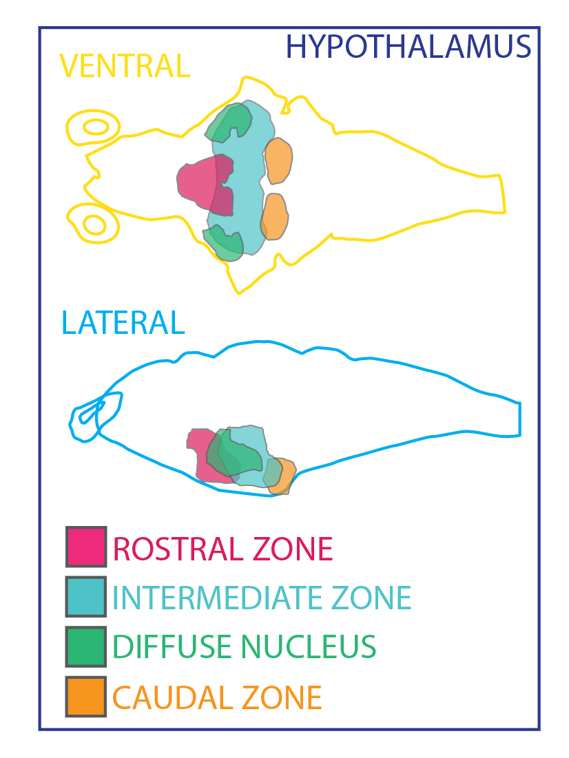
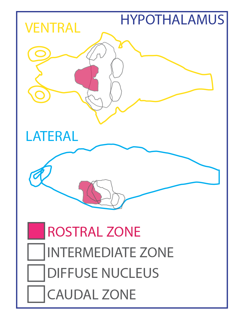
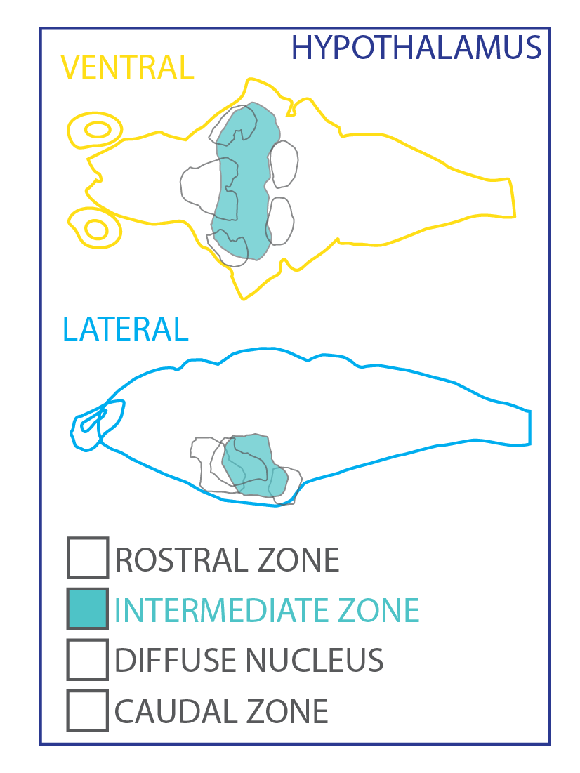
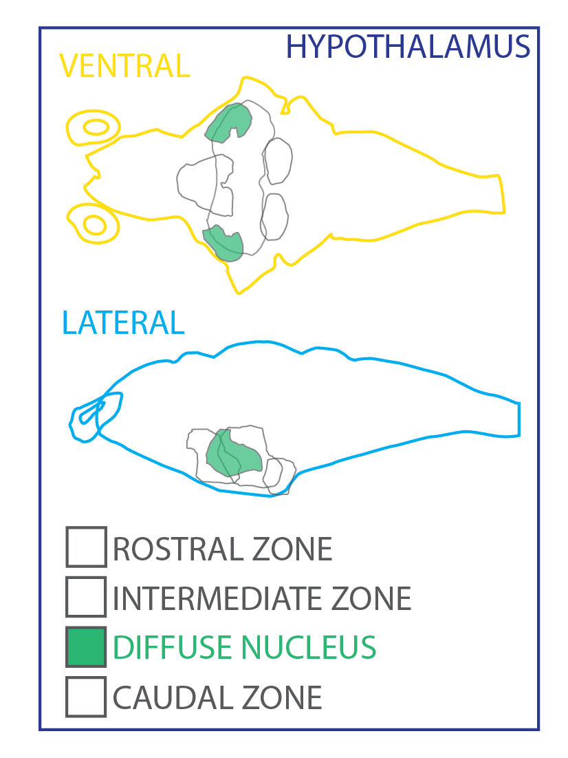
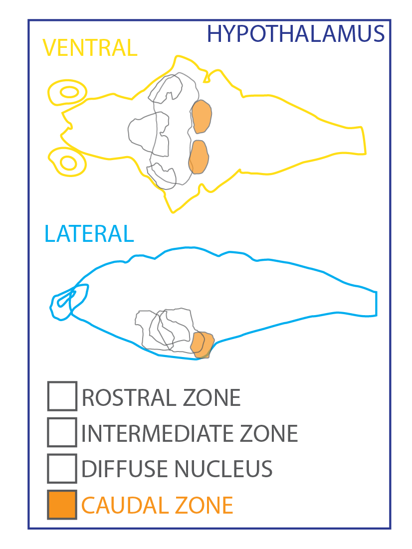
Forebrain > Diencephalon > Preglomerular Complex

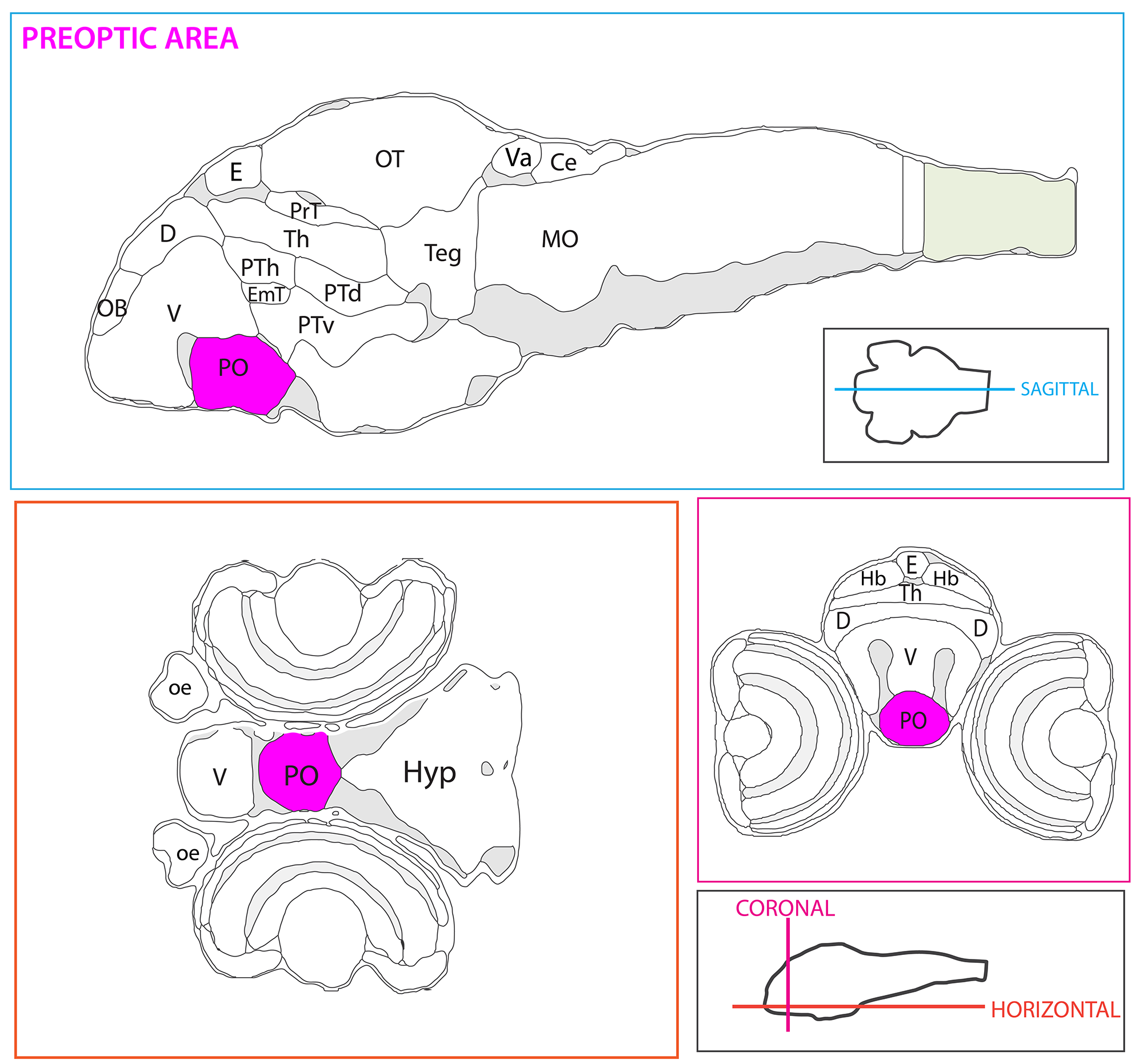
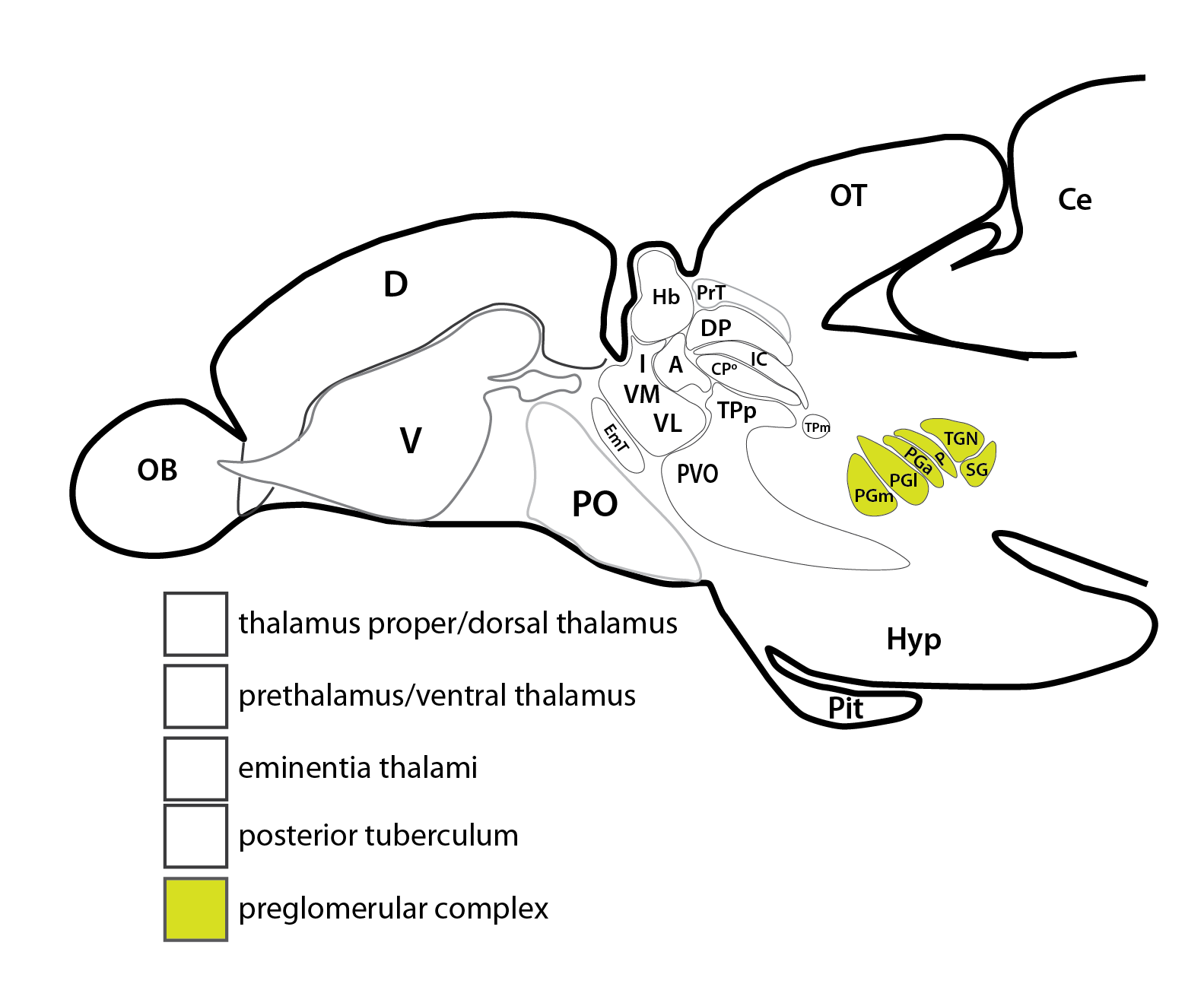
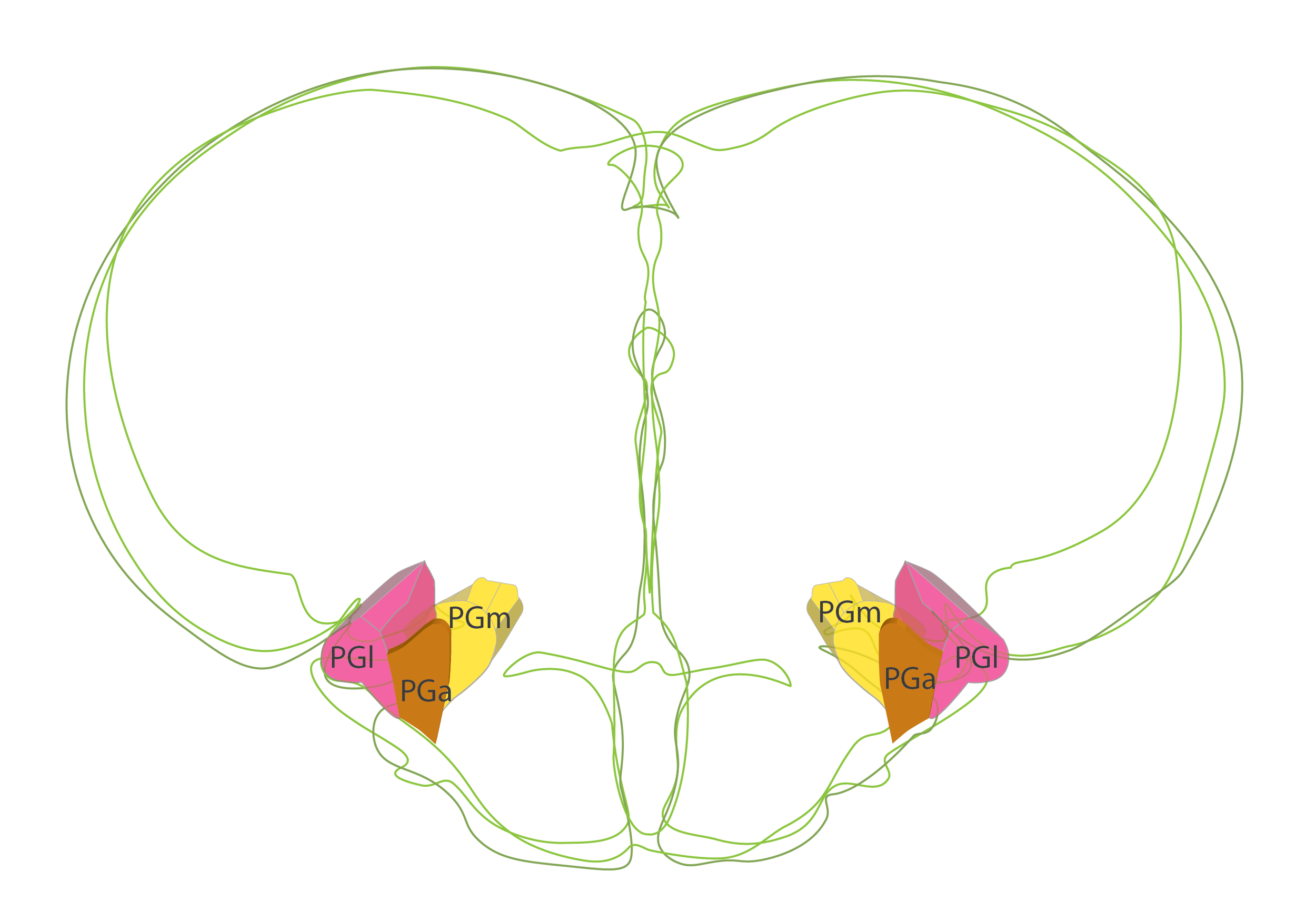
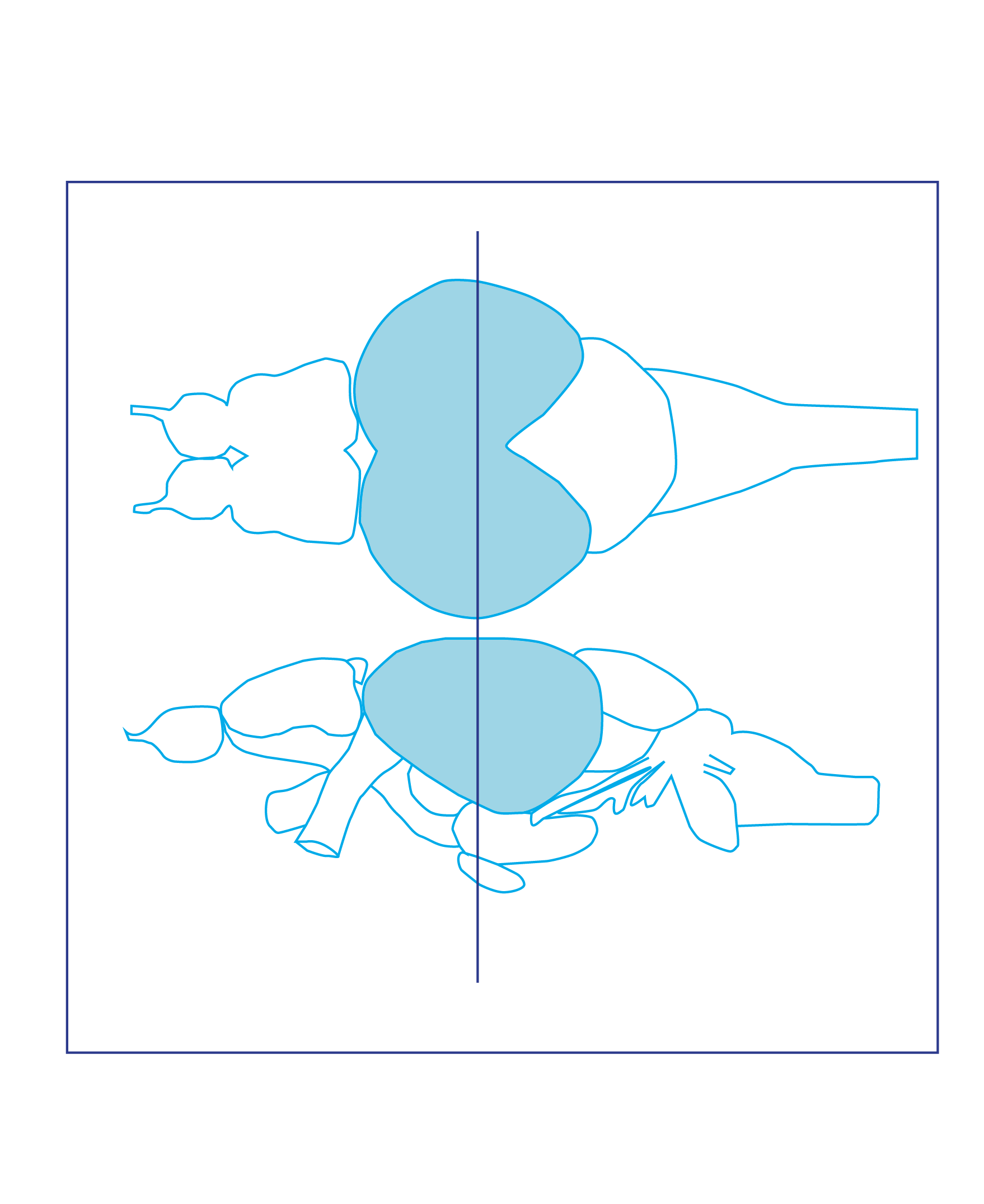
Schematic showing the position of the preglomerular complex in sagittal, horizontal and coronal sections through the zebrafish brain.
Based on the anatomical segmentation of 3 day old zebrafish larval brain by Thomas Müller, Olaf Ronneberger, Wolfgang Driever and colleagues. For details see Ronneberger et al., Nat. Meth. 2012 and http://vibez.informatik.uni-freiburg.de
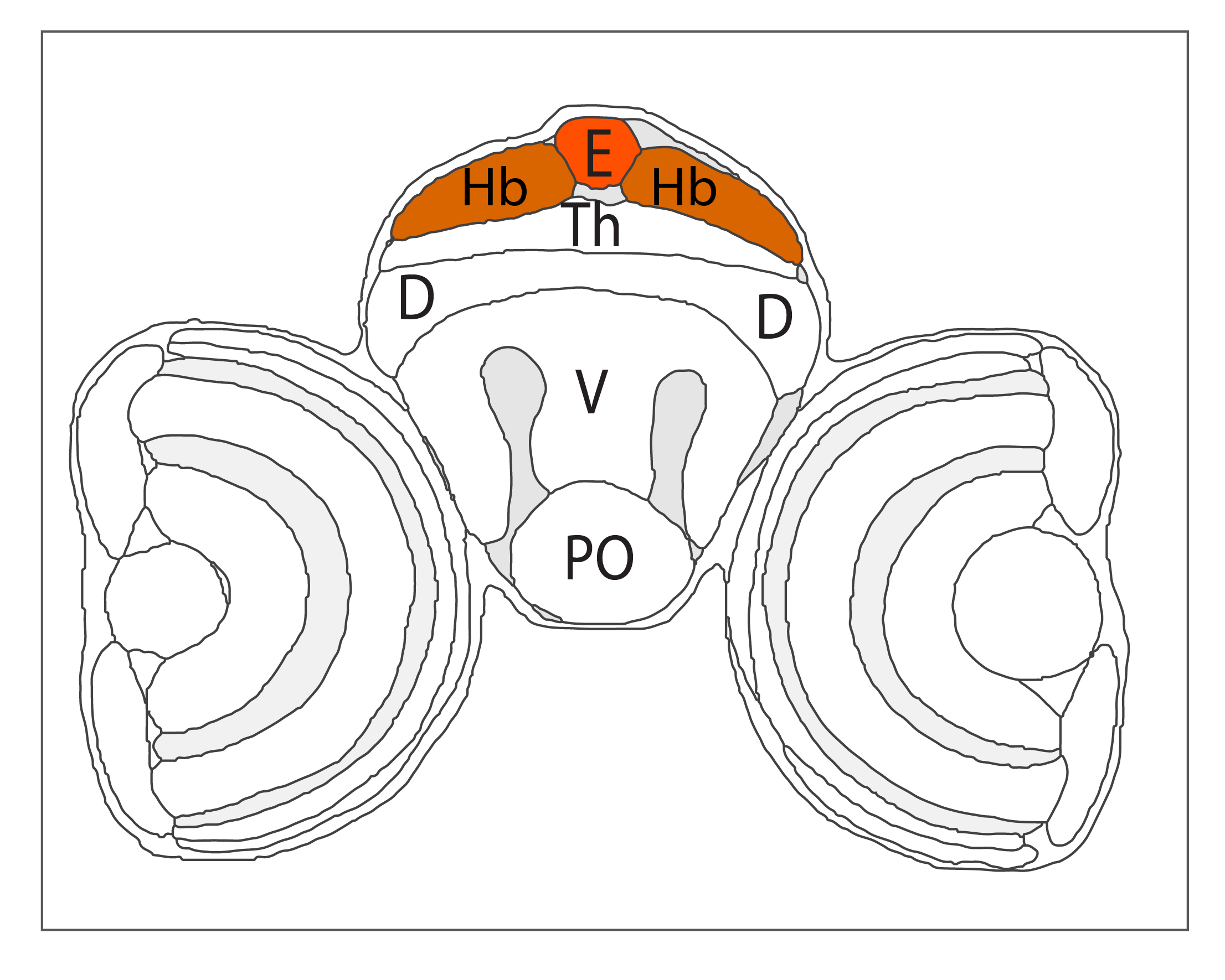
Abbreviations: Ce, cerebellar plate; D, dorsal telencephalon/pallium; E, epiphysis; EmT, eminentia thalami; Hb, habenula; Hyp, hypothalamus; MO, medulla oblongata; OB, olfactory bulb; OT, optic tectum; PG, preglomerular complex; PO, preoptic area;PrT, pretectum; PTd, posterior tuberculum dorsal part; PTh, prethalamus; PTv posterior tuberculum ventral part; Teg, tegmentum; Th, thalamus; TS, torus semicircularis; V, ventral telencephalon/subpallium; Va, valvula cerebelli.
Description
“The thalamus proper is less prominent in zebrafish than in mammals. This is the case because teleosts possess a preglomerular complex, an elaborated migrated agglomeration of nuclei related to the posterior tuberculum absent in mammals. In fact, the preglomerular complex serves as a predominant sensory relay station in the teleostean diencephalon (Wullimann and Northcutt, 1990; Northcutt, 2008; Yamamoto and Ito, 2008).”(Mueller, 2012). The PG complex in teleosts receives sensory input from several sensory modalities including visual, auditoy, lateral line, gustatory and potentially somatosensory (Bloch et al., 2020).
"The preglomerular complex consists of the anterior, the lateral, the medial, and the caudal preglomerular nuclei (PGa, PGl, PGm, PGc) and additionally the tertiary gustatory nucleus (TGN), the posterior thalamic (P), and the subglomerular (SG) nucleus. These nuclei have been interpreted as migrated derivatives of the embryonic posterior tuberculum (Braford and Northcutt, 1983; Northcutt, 2008). They serve as sensory relay stations projecting to different parts of the pallium, comparable to sensory thalamic nuclei in mammals. Based on their projection patterns to pallial parts of the adult zebrafish telencephalon and on expression patterns of pax6 in larval zebrafish, these nuclei have been interpreted as homologous to thalamic nuclei of mammals (Yamamoto and Ito, 2008)." (Mueller., 2012)
The lateral preglomerular nucleus receives visual information from the optic tectum and projects to the pallium. This ascending visual circuit has been compared to the ascending thalamocortical visual pathway in mammals and other vertebrates. Some new work on the development of the preglomerular complex has shown that the majority of PG cells are derived from the midbrain rather than from the forebrain as the thalamus is in amniotes suggesting that the PG complex may not be homologous to the thalamus of amniotes (Bloch et al., 2020).
In the adult
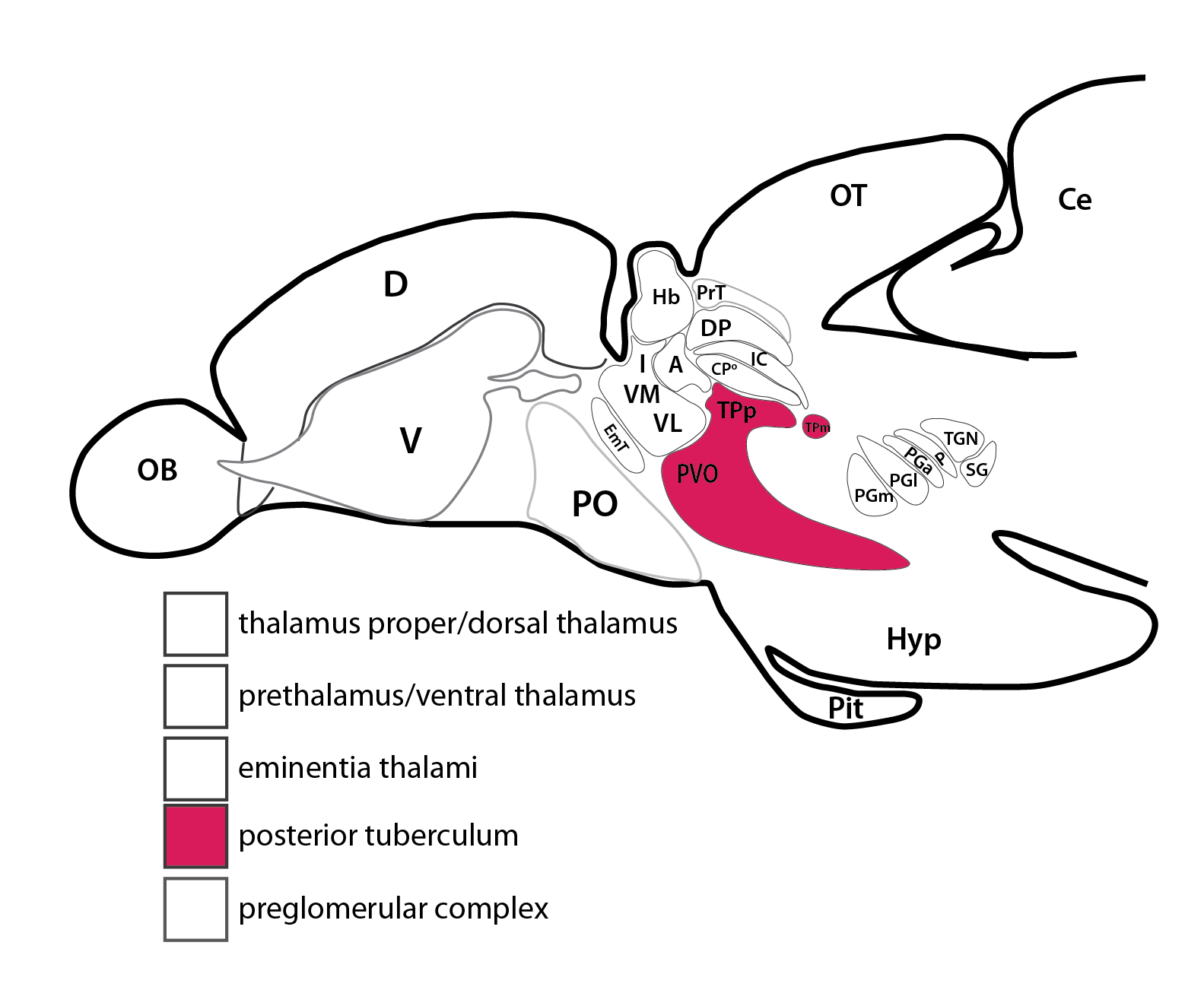
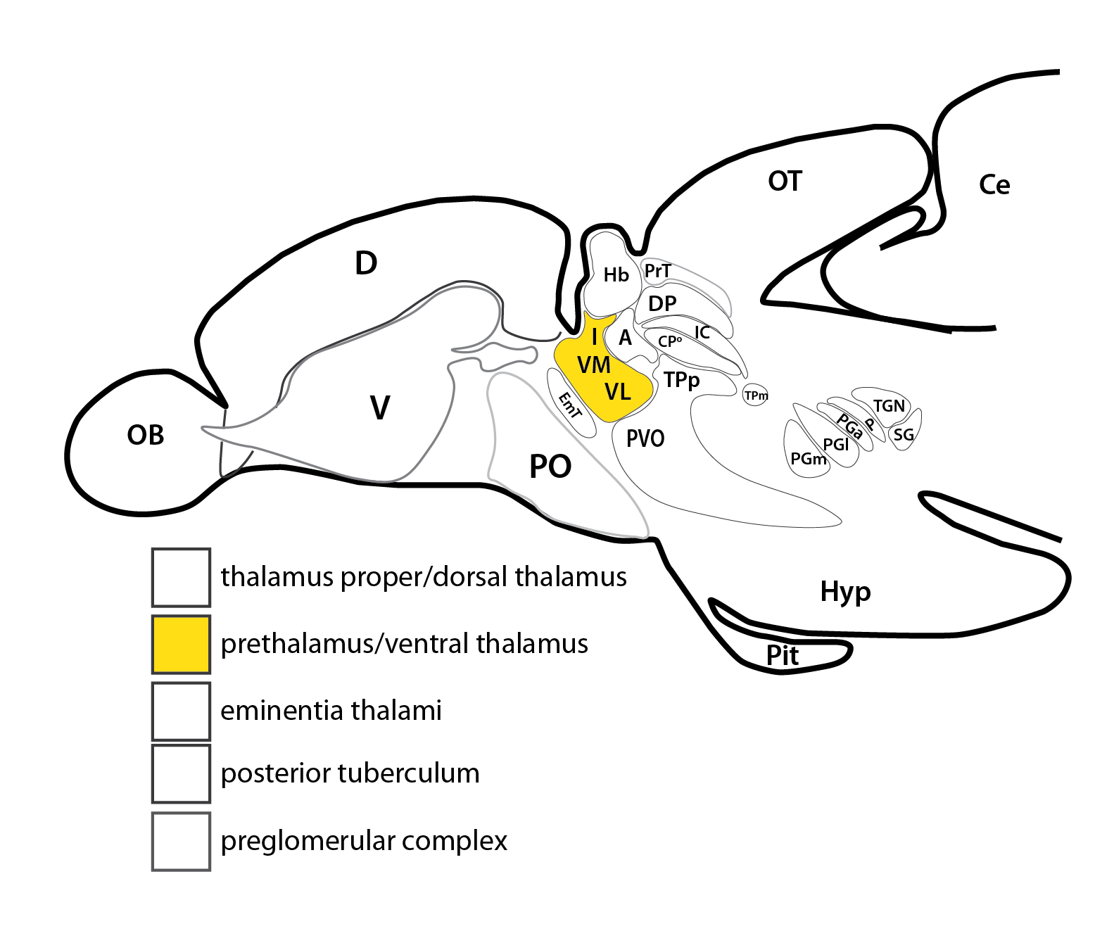
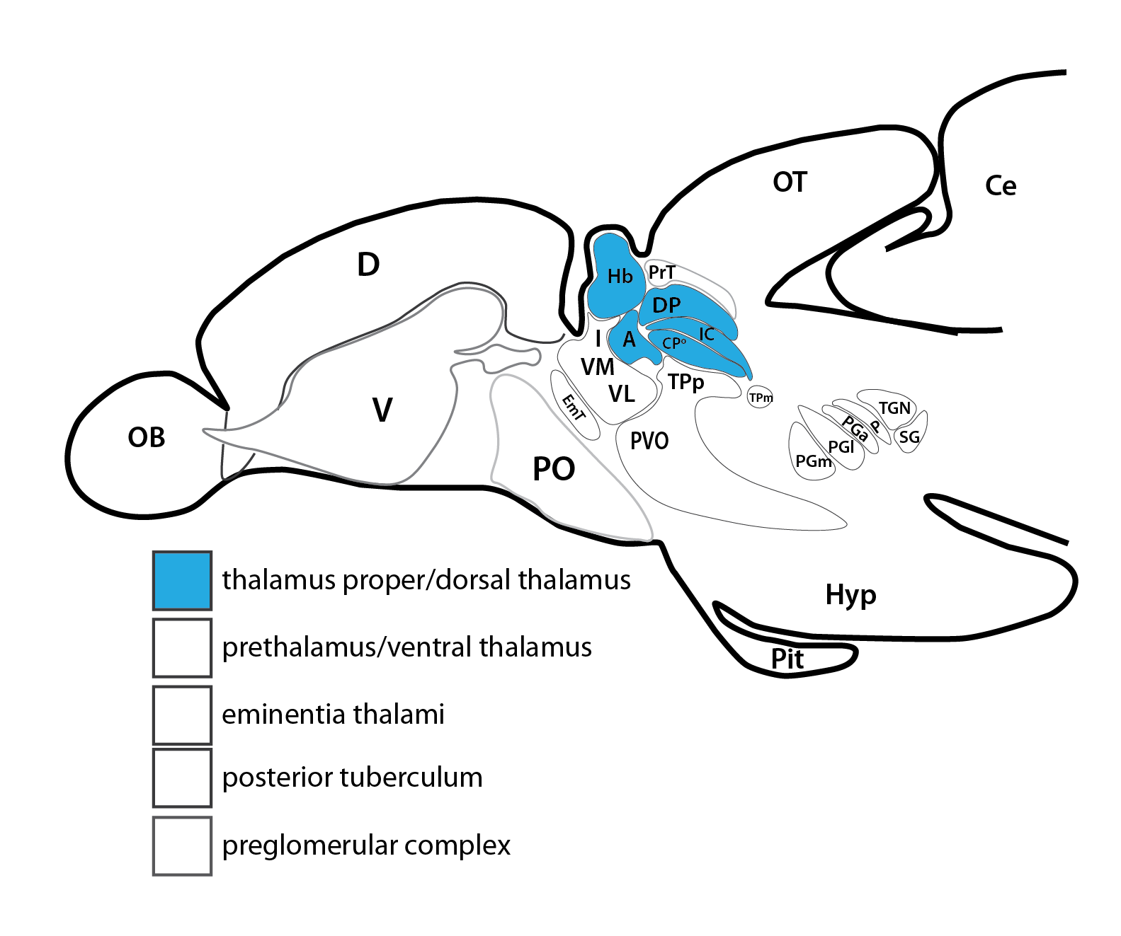
The preglomerular complex is part of the wider thalamus.
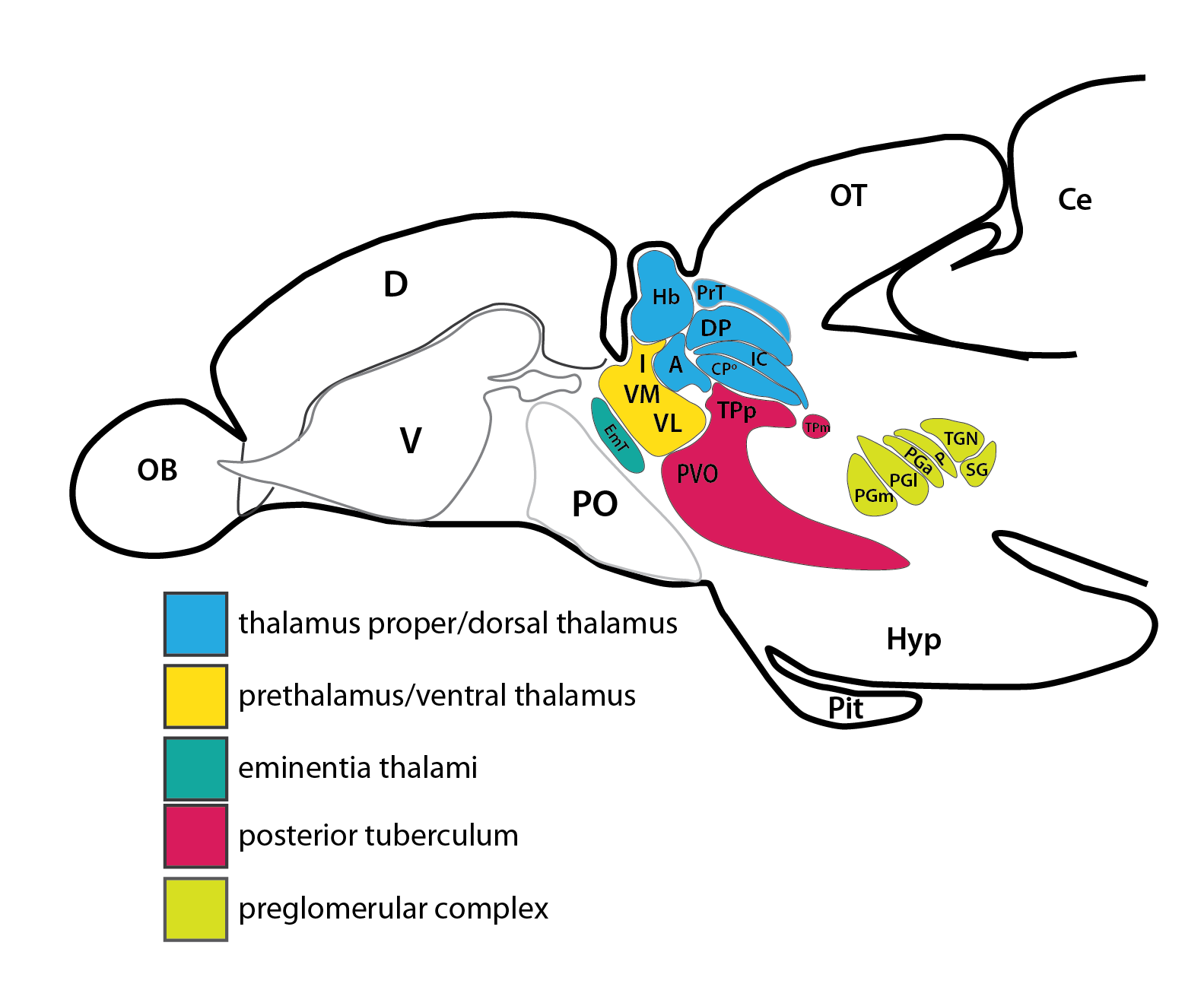
Schematic adapted from Mueller 2012 showing the nuclei that comprise the wider thalamus.
Abbreviations: A, anterior thalamic nucleus; CPo, centroposterior thalamic nucleus; D, dorsal telencephalon (pallium); DP, dorsoposterior thalamic nucleus; EmT, eminentia thalami; Hyp, hypothalamus; Hb, habenula; I, intermediate thalamic nucleus; IC, intercalated thalamic nucleus; OB, olfactory bulb; OT, optic tectum; P, posterior thalamic (preglomerular) nucleus; PG, preglomerular complex; PGa, anterior preglomerular nucleus; PGc, caudal preglomerular nucleus; PGl, lateral preglomerular nucleus; PGm, medial preglomerular nucleus; PO, preoptic region; PrT, pretectum; PVO, paraventricular organ; SG, subglomerular nucleus; TGN, tertiary gustatory nucleus; TPp, periventricular nucleus of posterior tuberculum; TPm, migrated nucleus of posterior tuberculum; V,ventral telencephalon (subpallium); VL, ventrolateral thalamic nucleus; VM, ventromedial thalamic nucleus.
The preglomerular complex is formed of 7 different nuclei. The anterior group is formed by the anterior preglomerular nucleus (PGa), lateral preglomerular nucleus (PGl) and the medial preglomerular nucleus (PGm). The posterior group includes the tertiary gustatory nucleus (TGN), subglomerular nucleus (SG), posterior thalamic (preglomerular) nucleus, (P) and the caudal preglomerular nucleus (PGc).
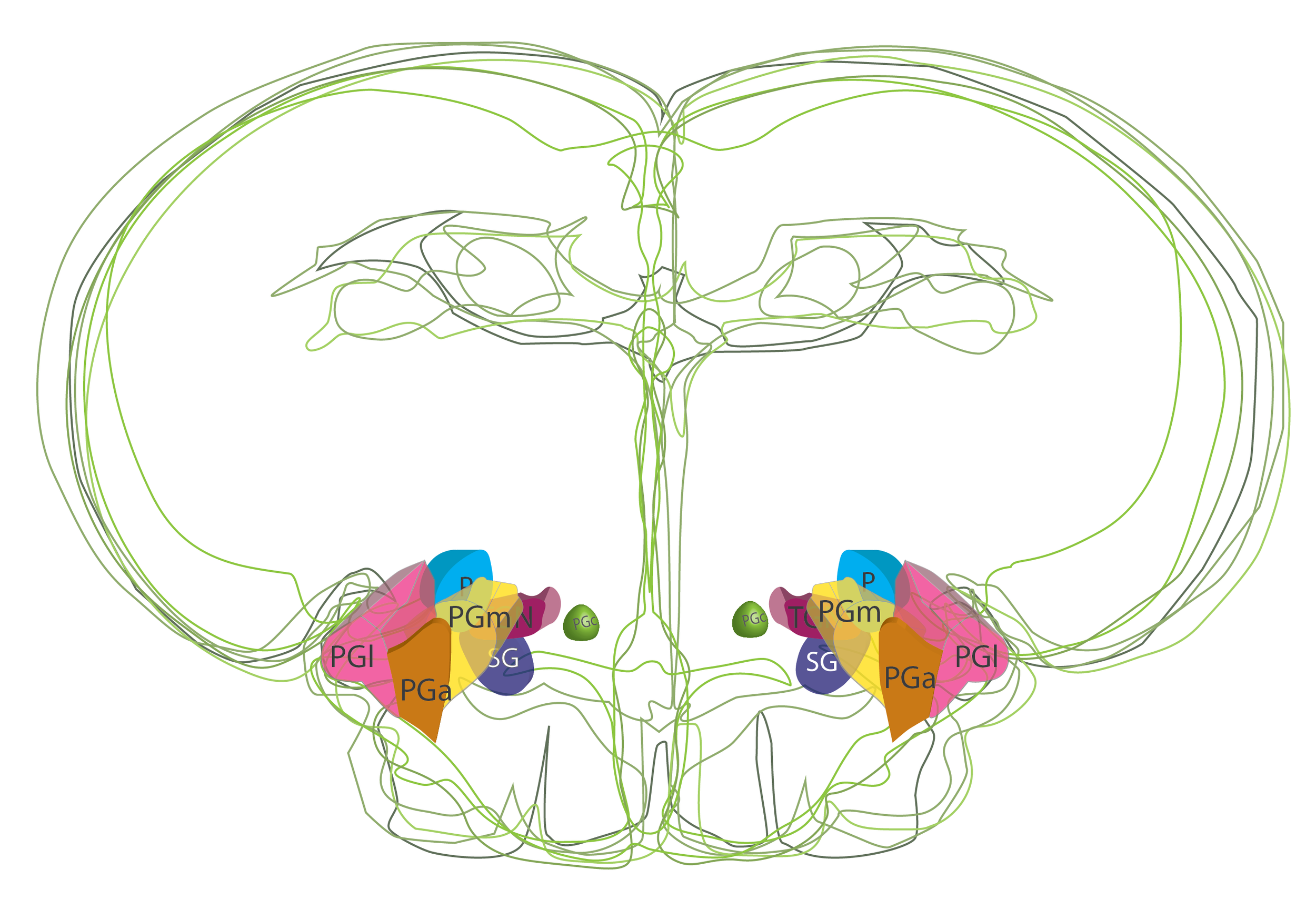

Positions of preglomerular nuclei taken from Neuroanatomy of the Zebrafish Brain (Wullimann et al., 1996).
Ontology
is part of: diencephalon, posterior tuberculum.
has parts: P, posterior thalamic(preglomerular)nucleus, PGa, anterior preglomerular nucleus; PGc, caudal preglomerular nucleus; PGl, lateral preglomerular nucleus; PGm, medial preglomerular nucleus; TGN, tertiary gustatory nucleus;SG, subglomerular nucleus.

Anatomical segmentation of 3 day old zebrafish larval brain by Thomas Müller, Olaf Ronneberger, Wolfgang Driever and colleagues. For details see Ronneberger et al., Nat. Meth. 2012 and http://vibez.informatik.uni-freiburg.de
Transgenic Lines that label this brain region

Antibodies that label this brain region
Key Publications
Bloch et al.
Non-thalamic origin of zebrafish sensory nuclei implies convergent evolution of visual pathways in amniotes and teleosts.
Elife, 2020 vol. 9 pp. 233-27
Thomas Mueller
What is the Thalamus in Zebrafish?
Front Neurosci. 2012; 6: 64.
Yamamoto, N., and Ito, H.
Visual, lateral line, and auditory ascending pathways to the dorsal telencephalic area through the rostrolateral region of the lateral preglomerular nucleus in cyprinids.
J. Comp. Neurol. 2008. 508, 615–647.