Viewing entries in
antibody

4C4

4C4

4C4

ABOUT THIS ANTIBODY

Anti-4C4 labels microglia

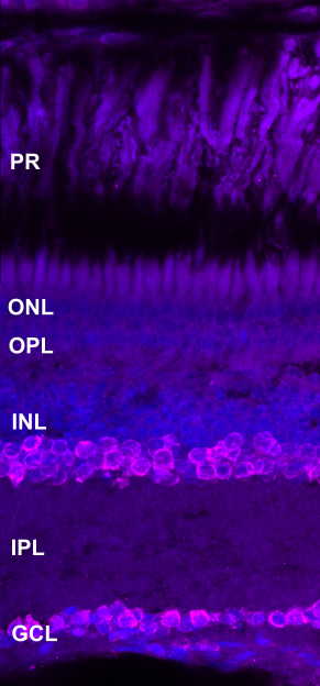
4C4 antibody has been found to bind to microglia in the zebrafish retina (Raymond et al., 2006). In the killifish 4C4 labelled Müller glia in the ganglion cell layer, particularly the endfeet. Further, staining was found in the photoreceptors.

Mouse Monoclonal anti-4C4 (Merck, Cat#92092321, dilution 1:150)
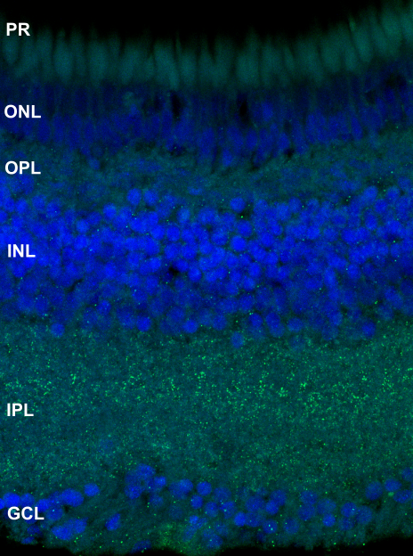
image

by Eva-Maria Breitenbach

Section of 4 mpf male killifish sodium citrate antigen retrieval anti-4C4 in magenta and DAPI in blue

PS = photoreceptors, ONL = outer nuclear layer, OPL = outer plexiform layer, INL = inner nuclear layer, IPL = inner plexiform layer, GCL = ganglion cell layer


labels these retinal cell types

Microglia


key publications

Raymond PA, Barthel LK, Bernardos RL and Perkowski JJ. 2006. Molecular characterization of retinal stem cells and their niches in adult zebrafish. BMC Developmental Biology. 6:1-17.

Calbindin

Calbindin

ABOUT THIS ANTIBODY

Rabbit Polyclonal anti-Calbindin (Swant, Cat#1CB38, dilution 1:200)

image

by Ryan MacDonald


labels these retinal cell types

Amacrine and bipolar cells


Carbonic Anhydrase 1 (CA1)

Carbonic Anhydrase 1 (CA1)

ABOUT THIS ANTIBODY

Rabbit Polyclonal anti-CA1 (Abcam, Cat#ab108367, dilution 1:200)

image

by Ryan MacDonald

labels these retinal cell types

Microglia


Caspase-3

Caspase-3

Caspase-3

ABOUT THIS ANTIBODY

Anti-Caspase-3 labels apoptosis

Caspase-3 antibody, caspases, are crucial mediators in apoptosis and caspase-3 is activated during death protease. Its antibody labels apoptosis in mice (Gashegu et al., 2007). In the killifish, we found two cells stained in the ganglion cell layer (white arrow) and some staining in the outer plexiform layer (yellow arrow).

Rabbit Polyclonal anti-Caspase-3 (BD Biosciences, Cat#559565, dilution 1:200)

image

by Eva-Maria Breitenbach

Section of 4 mpf male killifish sodium citrate antigen retrieval anti-Caspase-3 in green and DAPI in blue


labels these retinal cell types

Cells undergoing apoptosis


key publications

Gashegu J, Ladha R, Vanmuylder N, Philippson C, Bremer F, Rooze M and Louryan S. 2007. HSP110, caspase‐3 and‐9 expression in physiological apoptosis and apoptosis induced by in vivo embryonic exposition to all‐trans retinoic acid or irradiation during early mouse eye development. Journal of Anatomy. 210:532-541.

Glial fibrillary acidic protein (Gfap)

Glial fibrillary acidic protein (Gfap)

Glial fibrillary acidic protein (Gfap)

ABOUT THIS ANTIBODY

Anti-Gfap labels gliosis

Glial fibrillary acidic protein (Gfap) is an intermediate filament protein found in Müller glia and is upregulated under retinal stress. It has been found to label retinal gliosis in zebrafish (Dhakal et al., 2021). Gfap antibody showed a strong staining along the Müller glia in the killifish.

Rabbit polyclonal anti-Gfap (Proteintech, Cat#16825-1-AP, dilution 1:100)

image

by Eva-Maria Breitenbach

Section of 2 mpf female killifish sodium citrate antigen retrieval anti-Gfap (green) and DAPI (blue)

PS = photoreceptors, ONL = outer nuclear layer, OPL = outer plexiform layer, INL = inner nuclear layer, IPL = inner plexiform layer, GCL = ganglion cell layer


labels these retinal cell types

Gliosis


key publications

Dhakal S, Rotem-Bamberger S, Sejd JR, Sebbagh M, Ronin N, Frey RA, Beitsch M, Batty M, Taler K, Blackerby JF and Inbal A. 2021. Selective requirements for vascular endothelial cells and circulating factors in the regulation of retinal neurogenesis. Frontiers in Cell and Developmental Biology. 9:1-23.

Glial fibrillary acidic protein (Gfap) [ZRF-1]

Glial fibrillary acidic protein (Gfap) [ZRF-1]

ABOUT THIS ANTIBODY

Anti-Gfap [ZPR-1] labels gliosis

Glial fibrillary acidic protein (Gfap) is an intermediate filament protein found in Müller glia and is upregulated under retinal stress. It has been found to label retinal gliosis in zebrafish (Dhakal et al., 2021). Gfap antibody showed a strong staining along the Müller glia in the killifish.

Mouse monoclonal anti-Gfap [ZPR-1] (ZIRC, Cat#AB_10013806, dilution 1:50)

image

by Ryan MacDonald


labels these retinal cell types

Gliosis


key publications

Dhakal S, Rotem-Bamberger S, Sejd JR, Sebbagh M, Ronin N, Frey RA, Beitsch M, Batty M, Taler K, Blackerby JF and Inbal A. 2021. Selective requirements for vascular endothelial cells and circulating factors in the regulation of retinal neurogenesis. Frontiers in Cell and Developmental Biology. 9:1-23.

Glutamate receptor 2 (GluR2)

Glutamate receptor 2 (GluR2)

Glutamate receptor 2 (GluR2)

ABOUT THIS ANTIBODY

Anti-GluR2 labels horizontal cell dendrites

Glutamate receptor 2 (GluR2) antibody which marks GluR2 subunits of AMPA receptors. It stains horizontal cell dendrites in the monkeys (Haverkamp, Grünert and Wässle, 2000). In the killifish staining was found in the inner and outer plexiform layer which suggests it also labels horizontal cell dendrites in killifish.

Mouse Monoclonal anti-GluR2 (Millipore, Cat#MAB397, dilution 1:100)


image

by Eva-Maria Breitenbach

Section of 4 mpf male killifish sodium citrate antigen retrieval anti-GluR2 (magenta) and DAPI (blue)

PS = photoreceptors, ONL = outer nuclear layer, OPL = outer plexiform layer, INL = inner nuclear layer, IPL = inner plexiform layer, GCL = ganglion cell layer


labels these retinal cell types

Horizontal cell dendrites


key publications

Haverkamp S, Grünert U and Wässle H. 2000. The cone pedicle, a complex synapse in the retina. Neuron. 27:85-95.

Glutamine Synthetase (GS)

Glutamine Synthetase (GS)

Glutamine Synthetase (GS)

ABOUT THIS ANTIBODY

Anti-GS labels Müller glia

Glutamine synthetase (GS) protein is expressed in Müller glia. The GS antibody has been found to label Müller glia in zebrafish (Gramage et al., 2015). In the killifish, it labelled Müller glia, however not the whole cell.

Mouse monoclonal anti-GS (Millipore, Cat#MAB302, dilution 1:150)

image

by Eva-Maria Breitenbach

Section of 2 mpf female killifish sodium citrate antigen retrieval anti-GS (magenta) and DAPI (blue)

PS = photoreceptors, ONL = outer nuclear layer, OPL = outer plexiform layer, INL = inner nuclear layer, IPL = inner plexiform layer, GCL = ganglion cell layer

labels these retinal cell types

Müller glia


key publications

Gramage E, D’Cruz T, Taylor S, Thummel R and Hitchcock PF. 2015, Midkine-a protein localization in the developing and adult retina of the zebrafish and its function during photoreceptor regeneration. PLoS One .10:1-17.

Glutamine Synthetase (GS)

Glutamine Synthetase (GS)

ABOUT THIS ANTIBODY

Mouse monoclonal anti-GS (Proteintech, Cat#66323-1-Ig, dilution 1:100)


imageS

by Ryan MacDonald

labels these retinal cell types

Müller glia


HuC/D

HuC/D

HuC/D

ABOUT THIS ANTIBODY

Anti-HuC/D labels amacrine and ganglion cells in the retina

HuC/D antibody which has been found to label HuC/D neuronal proteins which are expressed in neurons leaving the mitotic cycle. It labels amacrine cells in the inner nuclear layer and ganglion cells in the ganglion cell layer in the zebrafish (Gorsuch et al., 2017). In the killifish it labelled cells in the inner nuclear layer and cells in the ganglion cell layer.

Mouse monoclonal anti-HuC/D (Invitrogen, Cat#A21271, dilution 1:200)

imageS

Section of 2 mpf male killifish sodium citrate antigen retrieval anti-HuC/D (magenta) and DAPI (blue)

PS = photoreceptors, ONL = outer nuclear layer, OPL = outer plexiform layer, INL = inner nuclear layer, IPL = inner plexiform layer, GCL = ganglion cell layer


labels these retinal cell types

Amacrine cells in the inner nuclear layer and ganglion cells in the ganglion cell layer


key publications

Gorsuch RA, Lahne M, Yarka CE, Petravick ME, Li J and Hyde DR. 2017. Sox2 regulates Müller glia reprogramming and proliferation in the regenerating zebrafish retina via Lin28 and Ascl1a. Experimental Eye Research. 161:174-192.

Parvalbumin (PV)

Parvalbumin (PV)

ABOUT THIS ANTIBODY

Anti-PV labels amacrine cells

Parvalbumin (PV) antibody binding to parvalbumin which is a regulator of neuronal intracellular calcium dynamics and found to stain amacrine cells in zebrafish (Ahmed, Kojima and Masai, 2022). In the killifish it stained cells in the inner nuclear and ganglion cell layer; and linear speckled staining in the inner nuclear, plexiform and ganglion cell layer.

Mouse Monoclonal anti-PV (Sigma, Cat#P3088, dilution 1:200)

imageS


labels these retinal cell types

Amacrine cells


key publications

Ahmed M, Kojima Y and Masai I. 2022. Strip1 regulates retinal ganglion cell survival by suppressing Jun-mediated apoptosis to promote retinal neural circuit formation. eLife. 11:1-34.

Paired Box 6 protein (Pax6)

Paired Box 6 protein (Pax6)

ABOUT THIS ANTIBODY

Rabbit Polyclonal anti-Pax6 (Proteintech, Cat#12323-1-AP, dilution 1:100)

image

by Ryan MacDonald


labels these retinal cell types

Amacrine cells and ganglion cells


Proliferating cell nuclear antigen (PCNA)

Proliferating cell nuclear antigen (PCNA)

Proliferating cell nuclear antigen (PCNA)

ABOUT THIS ANTIBODY

Anti-PCNA labels proliferating cells

Proliferating cell nuclear antigen (PCNA) antibody which binds to the PCNA p36 protein highly expressed in proliferating cells and found to stain proliferating cells in the zebrafish (Mazzitelli-Fuentes et al., 2022). In the killifish it stained many cells in the ganglion cell and outer nuclear layer. Additionally, it labels cell walls in the inner nuclear layer.

Mouse monoclonal anti-PCNA (Santa Cruz, Cat#sc-56, dilution 1:250)

image

by Eva-Maria Breitenbach

Section of 2 mpf female killifish sodium citrate antigen retrieval anti-PCNA (magenta) and DAPI (blue)

PS = photoreceptors, ONL = outer nuclear layer, OPL = outer plexiform layer, INL = inner nuclear layer, IPL = inner plexiform layer, GCL = ganglion cell layer


labels these retinal cell types

Proliferating cells


key publications

Mazzitelli-Fuentes LS, Román FR, Elías JRC, Deleglise EB and Mongiat LA. 2022. Spatial Learning Promotes Adult Neurogenesis in Specific Regions of the Zebrafish Pallium. Frontiers in Cell and Developmental Biology. 10:1-11.

Protein kinase C (Pkc)

Protein kinase C (Pkc)

ABOUT THIS ANTIBODY

Rabbit Polyclonal anti-Pkc (Proteintech, Cat#12919-1-AP, dilution 1:200)

imageS

by Ryan MacDonald


labels these retinal cell types

Bipolar cells


Retinaldehyde binding protein 1 (Rblp1)

Retinaldehyde binding protein 1 (Rblp1) [CRALBP]

ABOUT THIS ANTIBODY

Also known as CRALBP

Rabbit Polyclonal anti-Rblp1 (Proteintech, Cat#15356-1-AP, dilution 1:100)

imageS

by Ryan MacDonald


labels these retinal cell types

Müller glia


Retinal pigment epithelium specific 65 kDa protein (RPE65)

Retinal pigment epithelium specific 65 kDa protein (RPE65)

Retinal pigment epithelium specific 65 kDa protein (RPE65)

ABOUT THIS ANTIBODY

Anti-RPE65 labels retinal pigment epithelium

Retinal pigment epithelium specific 65 kDa protein (RPE65) antibody binds to the RPE65 which is found in the retinal pigment epithelium and found to stain retinal pigment epithelium in the rat (Malechka et al., 2017). In the killifish it stained the pigment epithelium and some spots across photoreceptors.

Rabbit Polyclonal anti-RPE65 (Proteintech, Cat#17939-1-AP, dilution 1:100)

image

by Eva-Maria Breitenbach

Section of 4 mpf male killifish sodium citrate antigen retrieval anti-RPE65 (green) and DAPI (blue)

PS = photoreceptors, ONL = outer nuclear layer, OPL = outer plexiform layer, INL = inner nuclear layer, IPL = inner plexiform layer, GCL = ganglion cell layer


labels these retinal cell types

Retinal pigment epithelium


key publications

Malechka VV, Moiseyev G, Takahashi Y, Shin Y and Ma JX. 2017. Impaired rhodopsin generation in the rat model of diabetic retinopathy. The American Journal of Pathology. 187:2222-2231.

Rhodopsin

Rhodopsin

ABOUT THIS ANTIBODY

Anti-Rhodopsin labels rod photoreceptors

Rhodopsin is a G-protein coupled receptor and has been found to be expressed in the rod photoreceptors. Its antibody labels rod photoreceptors (Intartaglia et al., 2022). It stained photoreceptors well in the killifish.

Rabbit Polyclonal anti-Rhodopsin (Hyde lab, Cat#1d4, dilution 1:100)

image

by Ryan MacDonald


labels these retinal cell types

Rod photoreceptors


key publications

Intartaglia D, Giamundo G, Naso F, Nusco E, Di Giulio S, Salierno FG, Polishchuk E and Conte I. 2022. Induction of autophagy promotes clearance of RHOP23H aggregates and protects from retinal degeneration. Frontiers in Aging Neuroscience. 14:1-14.

Ribeye-A

Ribeye-A

Ribeye-A

ABOUT THIS ANTIBODY

Anti-Ribeye-A labels synapses

Ribeye-A protein is a scaffolding component in the ribbon where vesicles are tethered, its antibody has been found to label synapses (Shankhwar et al., 2022). Many spots were found across the outer plexiform layer, inner nuclear layer, inner plexiform layer and ganglion cell layer in the killifish.

Rabbit Polyclonal anti-Ribeye-A (Nicolson lab, Cat#Custom, dilution 1:100)


images


labels these retinal cell types

Synapses


key publications

Shankhwar S, Schwarz K, Katiyar R, Jung M, Maxeiner S, Südhof TC and Schmitz F. 2022. RIBEYE B-Domain Is Essential for RIBEYE A-Domain Stability and Assembly of Synaptic Ribbons. Frontiers in Molecular Neuroscience. 15:1-26.

Sex determining region Y-box (Sox2)

Sex determining region Y-box (Sox2)

ABOUT THIS ANTIBODY

Anti-Sox2 labels progenitors

Sex determining region Y-box 2 (Sox2) antibody binds to the transcription factor Sox2 which forms OCT4 on DNA and has been found to label progenitors (Ahmed et al., 2015). In the killifish it labelled many cells in the inner nuclear and ganglion cell layer.

Rabbit polyclonal anti-Sox2 (Millipore, Cat#AB5603, dilution 1:200)

image

by Ryan MacDonald


labels these retinal cell types

Porgenitors


key publications

Ahmed TA, Ringuette R, Wallace VA and Griffith M. 2015. Autologous fibrin glue as an encapsulating scaffold for delivery of retinal progenitor cells. Frontiers in Bioengineering and Biotechnology. 2:1-11.

Synaptic vesicle Glycoprotein 2 (SV2)

Synaptic vesicle Glycoprotein 2 (SV2)

ABOUT THIS ANTIBODY

Mouse monoclonal anti-SV2 (Hybridoma Bank, Cat#AB_2315387, dilution 1:100)

image

by Ryan MacDonald


labels these retinal cell types

Synapses