Cornea

Cornea

cornea-03.png

Description

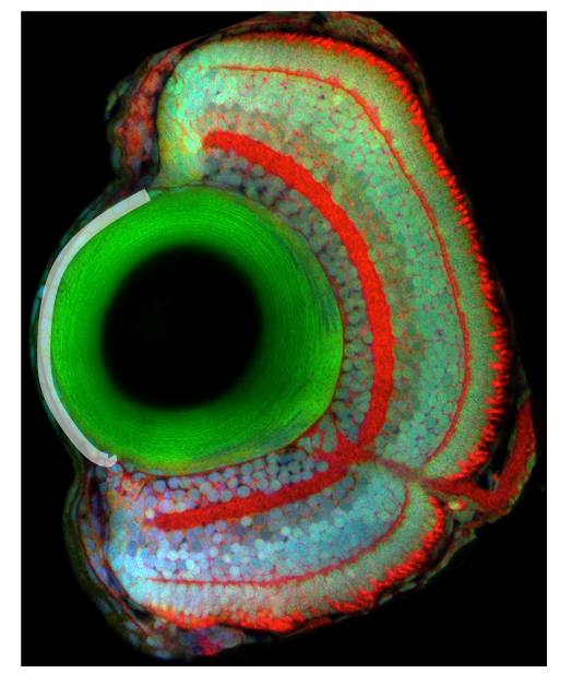
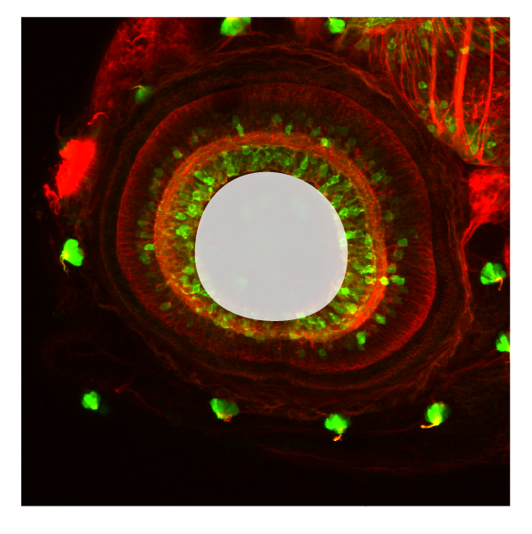
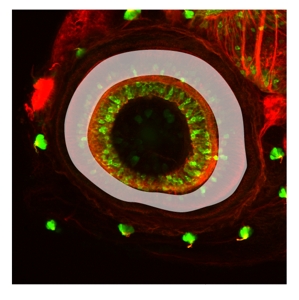
The cornea develops from the ectoderm overlying the lens. Corneal epithelial layers proliferate and generate multiple layers. the corneas main function is to refract or bend light. It also acts as a protective outer layer to the eye.


Ontology
 

is part of: eye

has parts:


Transgenic Lines/Antibodies that label this brain region


Key Publications

lens

lens

Schematic showing the position of the lens in sagittal, horizontal and coronal sections through the zebrafish brain.  Based on the anatomical segmentation of 3 day old zebrafish larval brain by Thomas Müller, Olaf Ronneberger, Wolfgang Driever and c…

Schematic showing the position of the lens in sagittal, horizontal and coronal sections through the zebrafish brain.
Based on the anatomical segmentation of 3 day old zebrafish larval brain by Thomas Müller, Olaf Ronneberger, Wolfgang Driever and colleagues. For details see Ronneberger et al., Nat. Meth. 2012 and http://vibez.informatik.uni-freiburg.de

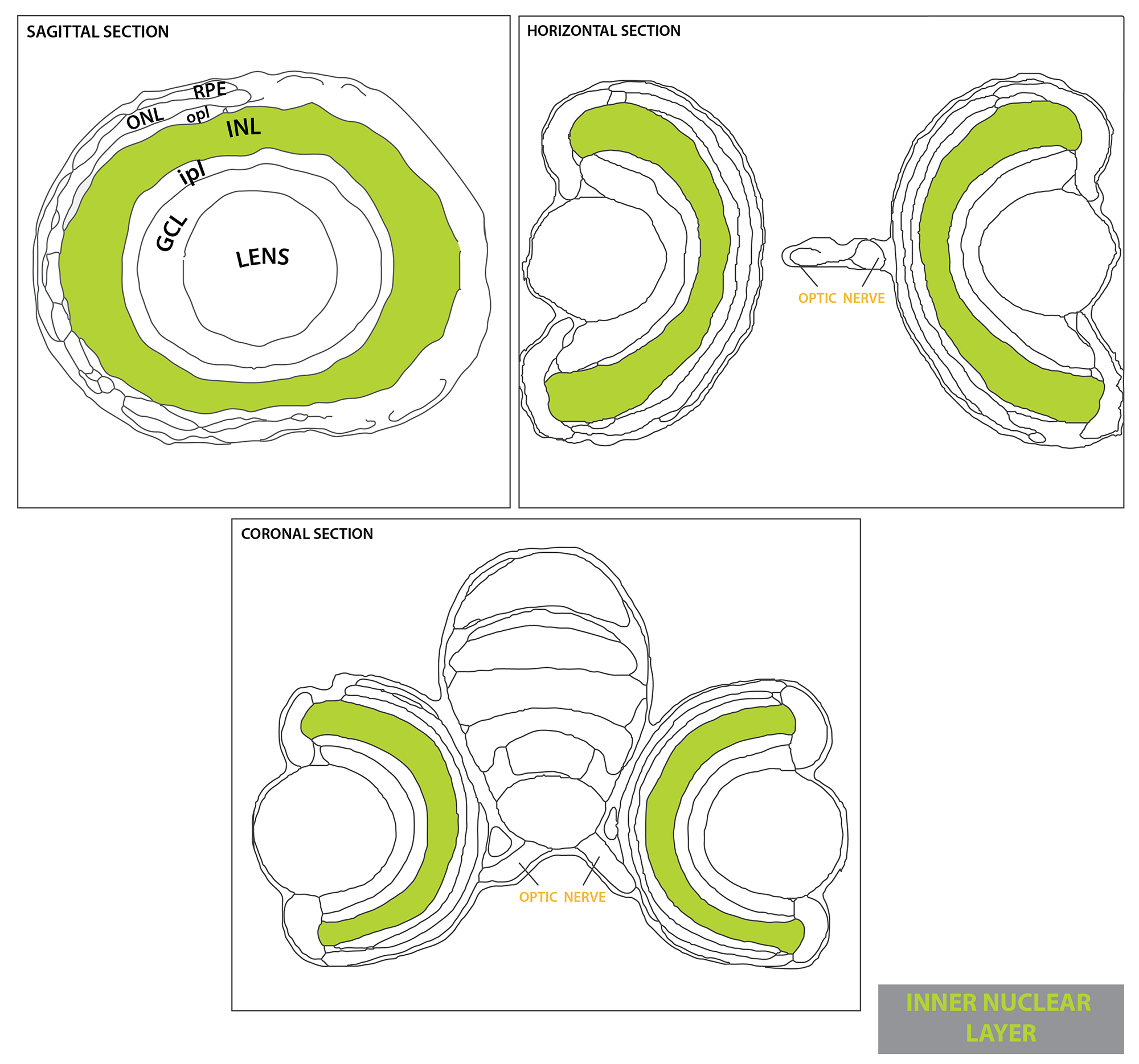
Abbreviations: GCL, ganglion cell layer; INL, inner nuclear layer ; ipl, inner plexiform layer ; ONL, outer nuclear layer ; opl, outer plexiform layer; RPE, retinal pigmented epithelium.

Description

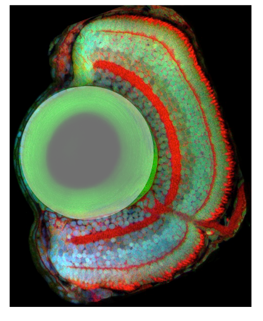
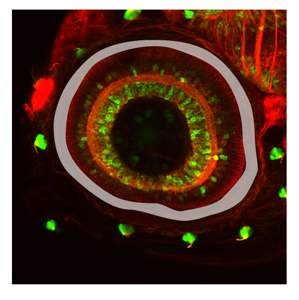
Upon neural retina specification, signals from the neural retina induce lens fate in the overlying surface ectoderm. The lens placode then invaginates to give rise to the lens vesicle, which buds out at around 24hpf. The lens vesicle keeps growing throughout adulthood and fibre cells differentiation is maintained from a pool of undifferentiated proliferating epithelial cells located at the most anterior region of the vesicle.


Ontology
 

is part of: eye

has parts:


Transgenic Lines/Antibodies that label this brain region


Key Publications

ciliary marginal zone

ciliary marginal zone

Schematic showing the position of the ? in sagittal, horizontal and coronal sections through the zebrafish brain.  Based on the anatomical segmentation of 3 day old zebrafish larval brain by Thomas Müller, Olaf Ronneberger, Wolfgang Driever and coll…

Schematic showing the position of the ? in sagittal, horizontal and coronal sections through the zebrafish brain.
Based on the anatomical segmentation of 3 day old zebrafish larval brain by Thomas Müller, Olaf Ronneberger, Wolfgang Driever and colleagues. For details see Ronneberger et al., Nat. Meth. 2012 and http://vibez.informatik.uni-freiburg.de

Abbreviations: CMZ, ciliary marginal zone; GCL, ganglion cell layer; INL, inner nuclear layer ; ipl, inner plexiform layer ; ONL, outer nuclear layer ; opl, outer plexiform layer; RPE, retinal pigmented epithelium.

Description

In the zebrafish eye, all the stages of progression from stem cell to differentiated neuron are found near the margin of the eye in a region termed the ciliary marginal zone (CMZ).  This type of retinal stem cell niche is found in all non-mammalian vertebrates and contain perpetually self-renewing, proliferative neuroepithelial cells that are spatially ordered with respect to cellular development and differentiation.  The youngest and least determined cells are most peripheral, proliferative retinoblasts are located in the middle, and the quiescent, differentiating cells are most central (Fig 1).  Cell behaviour of neuronal precursors within the CMZ is likely influenced by environmental signals emmanating from the surrounding tissues, including the RPE, lens, and mature retina.


Development of the CMZ:

The ciliary marginal zone is derived from the mitotic neural retina, but little is known about how progenitor cells choose to form the CMZ instead of differentiate into neurons.

Kara Cerveny

Kara Cerveny


Ontology
 

is part of: retina

has parts:


Transgenic Lines/Antibodies that label this brain region


Key Publications

retinal ganglion cell layer

retinal ganglion cell layer

GCL-03.png

Description


The layer of the nerual retina that contains Retinal Ganglion Cells (RGCs)

Ganglion cells are the final output neurons of the vertebrate retina.  The ganglion cell axons form the nerve fiber layer of the inner retina as they coalesce to form the optic nerve head, which is composed of ganglion cell axons and glia that migrate into the optic nerve as well as astrocytes that are derived from the optic stalk.

Retinal Cell Types found in the Retinal Ganglion Cell Layer


Ontology
 

is part of: neural retina

has parts:


Transgenic Lines/Antibodies that label this brain region


Key Publications

retinal inner plexiform layer

retinal inner plexiform layer

ipl-03.png

Description

Within the inner plexiform layer are localized synapses between the amacrine and bipolar interneurons and the retinal ganglion cells. Connections between cells that are depolarized upon an increase in illumination (ON) occupy approximately the inner half of the IPL, whereas synapses of cells hyperpolarized (OFF) by light are confined to the outer half of the IPL. 


Ontology
 

is part of: neural retina

has parts:


Transgenic Lines/Antibodies that label this brain region


Key Publications

retinal outer plexiform layer

retinal outer plexiform layer

Schematic showing the position of the outer plexiform layer in sagittal, horizontal and coronal sections through the zebrafish brain.  Based on the anatomical segmentation of 3 day old zebrafish larval brain by Thomas Müller, Olaf Ronneberger, Wolfg…

Schematic showing the position of the outer plexiform layer in sagittal, horizontal and coronal sections through the zebrafish brain.
Based on the anatomical segmentation of 3 day old zebrafish larval brain by Thomas Müller, Olaf Ronneberger, Wolfgang Driever and colleagues. For details see Ronneberger et al., Nat. Meth. 2012 and http://vibez.informatik.uni-freiburg.de

Abbreviations: GCL, ganglion cell layer; INL, inner nuclear layer ; ipl, inner plexiform layer ; ONL, outer nuclear layer ; opl, outer plexiform layer; RPE, retinal pigmented epithelium.

Description

In the outer plexiform layer, photoreceptors form chemical synapses with horizontal cells and bipolar cells.  


Ontology
 

is part of: neural retina

has parts:


Transgenic Lines/Antibodies that label this brain region


Key Publications

Retina outer nuclear layer

Retina outer nuclear layer

ONL-03.png

Description

Layer of the neural retina that contains the photoreceptor cells. Photoreceptor cells span the outer nuclear layer as well as the outer and inner segment layers. These light sensing cells of the retina, the cone and rod photoreceptors, are located in the apical-most layer of the retina, and display a stereotypical subcellular organization with their nuclei basal to the inner segment (cell body or soma) and outer segment, which abuts the retinal pigmented epithelium (RPE) and is full of membrane invaginations packed with light-sensitive cell-specific opsins.  All photoreceptors have opsin-containing outersegments, which are replaced on a daily basis.  The older membrane and protein debris is shed from the distal tip of the outersegment and phagocytosed by the adjacent RPE cells.  Because zebrafish are diurnal, their retinae contains a large number of bright-light sensitive cone subtypes in addition to dim-light sensitive rod photoreceptors, all of which are organized into regular mosaic patterns that can best be visualized in tangential sections.  Below is more detailed information about the distinct types of photoreceptors found in zebrafish.
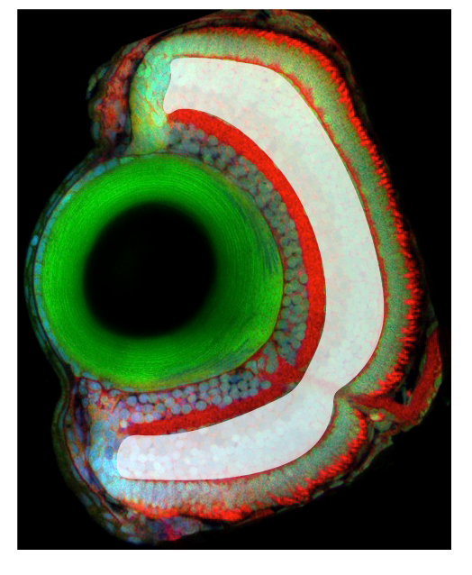
retinal cell types found in the outer nuclear layer


Ontology
 

is part of: neural retina

has parts: photoreceptor cell


Transgenic Lines/Antibodies that label this brain region


Key Publications

retinal pigmented epithelium

retinal pigmented epithelium

RPE-03.png

Description

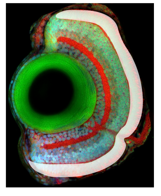
The retinal pigmented epithelium (RPE), like the neural retina, is derived from the neural ectoderm, and through complex morphogenetic movementsis during development it becomes tightly apposed to the photoreceptor cell layer of the neural retina.  This tissue is required for the function and survival of photoreceptor cells; numerous mutations affecting vacuole transport in the RPE result in non-cell autonomous photoreceptor cell degeneration.  The RPE is a typical epithelial monolayer of tightly packed cuboidal cells forming part of the blood brain barrier between the vascular choroid and the neural retina.  The cells contain large numbers of melanosomes packed with melanin granules.  The apical microvilli of the RPE interdigitate with the photoreceptor outer segments, and the basal surface rests on Bruch’s membrane, the basement membrane between the RPE and capillaries of the choroid. 


Ontology
 

is part of: retina

has parts:


Transgenic Lines/Antibodies that label this brain region


Key Publications